This protocol provides instructions for intranasal administration of IAV and downstream analysis of mouse lung damage and associated transcriptional changes to lung cell types.
Method Article
Modeling Dysplastic and Functional Lung Alveolar Repair after Influenza Infection
In This Article
Summary
Abstract
Viral infection causes both acute and long-term damage to the lung alveolus, the specialized tissue structure responsible for gas exchange between the cardiovascular system and the external environment. Influenza A virus (IAV) infection in mice represents a translational model for the response of the human lung to viral infection and induces both transient and persistent cell state changes in the alveolus. In some cases, aberrant cell states induced by viral injury that are not resolved over time may permanently impair the critical gas exchange function of the lung. This article demonstrates methods for intranasal infection of mice with A/PR/8/34 IAV and characterization of transient and long-lasting alterations to the cellular composition and tissue structure of damaged lungs. This model enables a detailed investigation into the molecular and cellular mechanisms underlying lung repair and chronic dysfunction. This approach also offers a platform for evaluating therapeutic interventions aimed at promoting effective lung regeneration and restoring respiratory function after viral injury.
Introduction
The lungs play a crucial role in supplying oxygen and nutrients to local tissues. Endothelial cells (ECs) that populate small capillary blood vessels of the lungs are essential for the proper functioning of the lung tissue, maintaining vascular integrity and efficient gas exchange1. ECs form the vascular niche and reciprocally crosstalk with other cells such as epithelial cells, immune cells, and mesenchymal cells. This contributes to the regulation of lung development and of tissue repair in response to injury or disease2.
H1N1 Influenza A virus infection is a worldwide problem3. Patients with H1N1 infection are often diagnosed with exudative diffuse alveolar damage (DAD)4. Infection with H1N1 can lead to serious lung damage and severe consequences, including pneumonia, severe acute lung injury (ALI), and acute respiratory distress syndrome (ARDS)5.The damage mainly arises from the virus infecting lung epithelial cells, which prompts an excessive immune response resulting in inflammation, tissue damage, and fluid buildup in the lungs. The epithelial cells lining airways and alveoli, which are responsible for gas exchange, are thus directly harmed by the H1N1 virus, leading to disruption of the normal structure of the lung6,7. It has become clear that other cell types, although not directly infected by IAV, can become damaged in response to infection8,9. Tissue damage after influenza infection, therefore, affects all cellular compartments of the lung, and it has recently been shown that IAV infection causes both transient and persistent changes to cell state in multiple cellular compartments9. This cell plasticity in response to disease can be helpful or harmful to successful tissue repair and warrants further study.
The most adopted experimental model for studying influenza and its pathogenesis is intranasal infection of mice with influenza virus A/PR/8/3410. It is an important technique permitting the study of disease severity, mortality, and viral infectivity. Intranasal infection of mice mimics the natural infection route in humans and allows the study of both adaptive and innate immune responses against the virus. In 1934, a specific strain of the Influenza A virus "A/PR/8/34" was isolated from a patient in Puerto Rico11. This strain of influenza virus has now been mouse-adapted and no longer productively infects humans. Because of the similarities in mouse and human immune systems, mice are the most popular animal model for IAV research12. In this method, the A/PR/8/34 H1N1 strain is directly administered to mice through the nostrils, mimicking a natural infection13. It is an easy, cost-effective, and convenient method to study the effects of viral infection on host lung tissue in wild-type (WT) as well as knockout (KO) conditions. Alternative techniques have advantages and disadvantages compared to the intranasal model. Intratracheal (IT) inoculation allows accurate delivery of the virus into the lungs, but this technique does not fully mimic infection by inhalation and does not provide information on toxicity in the upper respiratory tract. IT inoculation also requires great skill to reduce the risk of tracheal injury13,14. Aerosol inhalation (AR) inoculation mimics the natural route of infection and creates a more uniform lung pathology compared to intranasal infection, but it can be difficult to maintain uniform virus concentration in the aerosol and to achieve accurate delivery of viral aerosol. Safety is also an important concern of the AR inoculation method10. Therefore, the intranasal inoculation technique of IAV administration is a crucial approach to pulmonary-focused research on the downstream effects of viral infection in tissue injury and repair. Unlike chemical or mechanical models, it provides a more realistic and less invasive way to study influenza-induced lung damage and host-pathogen interactions.
Here, we establish a reproducible method for intranasal infection of mice with Influenza A virus (A/PR/8/34) and for characterizing both the short-term and long-term cellular and structural changes in the lung alveolus that result from viral injury. Studies using this method can aid in the discovery of mechanisms of lung tissue damage and repair to identify novel therapeutic targets for lung diseases.
Protocol
Working with the influenza A virus in an animal model is considered biosafety level 2 (BSL2) and animal biosafety level 2 (ABSL2). All animal procedures used to generate this data were approved by the Animal Care and Use Committee of the Center for Cancer Research, National Cancer Institute, National Institutes of Health (Protocol #24-452). This protocol is also approved by the Institutional Biosafety Committee of the Center for Cancer Research, National Cancer Institute, National Institutes of Health. Before beginning this protocol, it is recommended that users seek approval from their institutional animal care and use committee and institutional biosafety committee.
1. Preparation of working stock
NOTE: All work with the influenza virus should be performed on ice in a biosafety cabinet. Eject tips and anything else that contacts the virus into a container of 10% bleach. Ice buckets, pipettes, and tip boxes should be sprayed with 10% bleach solution rather than immersed. Incubate at room temperature for at least 30 min. Dispose of solid waste in a biohazard waste container and discard liquid waste down the sink, flushing with water.
- Thaw frozen stock (0.5 mL) of virus on ice in a biosafety cabinet.
- Set up a waste container containing 10% bleach in tap water. Place two plastic or cardboard freezer boxes that can hold up to 100 sample tubes in a -80 °C freezer to pre-chill.
- Pre-label 160 1.5 mL tubes for aliquots of virus. Screw-top tubes are preferable to flip-cap tubes to avoid creating aerosols when opening viral stock tubes for use. Place tubes on ice to pre-chill before making aliquots of virus.
- Make 3 µL aliquots of virus using a calibrated P20 pipette and tips, only placing virus into prechilled tubes and keeping aliquots on ice at all times.
- After making aliquots, store them in a prechilled freezer box labeled with "A/PR/8/34 Influenza Virus (IAV)", the catalog number, the lot number, and expiration date.
NOTE: Different lots of viruses have different infectivity and should not be mixed. - Place the box in a -80 °C freezer. Make sure to keep everything as cold as possible, as the viral titer will decrease with each freeze/thaw cycle.
2. Diluting aliquots of virus for use in intranasal infection experiments
- Thaw one 3 µL aliquot of viral stock on ice in the biosafety cabinet.
- Make a 1:100 dilution (dilution A) by transferring 2 µL of the 3 µL aliquot to a prechilled 1.5 mL tube containing 198 µL of cold D-PBS.
- Continue to dilute the viral stock until it reaches the appropriate concentration for 0.5-1 LD50 in the strain background of mice used. Keep everything on ice and use prechilled tubes to avoid over-warming the virus.
NOTE: For example, for VR-95PQ, lot 70063082, infect mice with 12,600 to 31,500 CEID50/µL virus (scaled by body weight) at a 1:500,000 dilution. - From dilution A (1:100), make a 1:50,000 dilution (dilution B) by adding 10 µL of dilution A to 4990 µL of cold D-PBS in a prechilled 15 mL tube. Mix well by pipetting up and down using a 5 mL serological pipette.
- From dilution B, make a 1:500,000 dilution (dilution C) by adding 200 µL of dilution B to 1.8 mL of D-PBS in a prechilled 5 mL tube. Mix well by pipetting up and down.
NOTE: To determine the ideal dilution of virus to be used in infecting the animals when purchasing a new lot, it is possible to estimate using the titer of the virus indicated on the product sheet and adjusting the dilution based on the titer of the previous lot. The best way to determine the ideal dilution is to empirically titer the new lot of virus using wild-type C57BL/6 mice. Plan for a range of dilutions spanning from 1:50,000 to 1:500,000 and order 1-2 cages of C57BL/6 female mice per dilution to test. Infect mice at different concentrations and monitor their weight loss. To test viral titers, the study proposes using only female mice to reduce variability that may be caused by sex or weight differences between mice. Female mice often become sicker than male mice, possibly due to their smaller size, or due to sex-specific differences in response to influenza infection, as occurs in other forms of mouse lung injury15. Although this was not studied in detail, female mice were used in the titer experiments because if the titer was determined using male mice, female mice might die at the determined dose. However, it was found that male mice get appreciably sick and have noticeable lung injuries when infected with a viral titer, which was determined using only female mice. An ideal dilution if using mixed-background or outbred mice will be "1 LD50," the dilution at which 50% of the wild-type C57BL/6 mice die. Outbred mice are more resistant to infection16 and will be injured and recover well at this dilution. If mice for the experiment will be on an inbred C57BL/6 background, the ideal dose will be half of this "0.5 LD50." Because the goal of these experiments is to study injury and repair, it is ideal to define a dose at which mice get sick and lose 20-25% of their body weight, but most mice ultimately recover (Figure 1). If all mice in the study are approximately the same weight (~25 g), it is possible to administer the influenza virus at 50 µL of the appropriate dilution to each mouse. However, mice range in weight from 15-35 g. Therefore, administer 2 µL of viral dilution per gram of body weight (for example, a 15 g mouse would receive 30 µL influenza, while a 35 g mouse would receive 70 µL). This results in a more consistent level of infection and tissue damage across differently sized animals. This information is needed to plan for the volume of dilution C.
3. Transportation of viral aliquots to animal facilities
- Place the 5 mL tube with the final viral dilution into the ice and label the container with a biohazard sticker. Line a plastic sample carrier box with paper towels to absorb possible spills.
- Place materials needed for intranasal infection inside the biohazard labeled box: a P200 pipette, an unopened box of filtered P200 pipette tips, a pen, and a 50 mL conical tube filled with fresh 10% bleach for viral waste. As an infection control, fill a 1.5 mL tube with the same cold D-PBS used to dilute the IAV.
4. Administration of IAV to mice
NOTE: Working with the influenza virus in the animal facility requires an ABSL2 safety level facility. Standard PPE includes scrubs, a disposable lab coat, two hair bonnets, double nitrile gloves, double shoe covers, protective eyewear (safety glasses), and a mask. The diluted influenza virus should be kept on ice throughout the administration protocol to avoid loss of infectivity.
- All work with viruses must take place inside a biosafety cabinet. Although the A/PR/8/34 influenza virus is a mouse-adapted strain, influenza viruses are prone to mutation, and therefore, treat the virus as though it is potentially infectious to humans.
- Administer isoflurane anesthesia to animals one at a time at a flow rate of 2.5-3% on the vaporizer machine. Move animals from an anesthesia box to a nose cone prior to administering influenza.
- When a mouse is under anesthesia, check its breathing and do a toe pinch to see if it reacts. Mice are ready for IAV infection when they display peripheral spinal reflexes (such as limb withdrawal) without central awareness (as indicated by a mouse moving its head and looking around).
NOTE: A sub-surgical plane of anesthesia is preferred (neither fully awake nor at a surgical plane) for intranasal administration. At a surgical plane, mice have an increased risk of mortality due to aspiration of liquid into the lungs, while insufficient anesthesia leads to expulsion of the inoculum and inconsistent dosing. This intermediate plane, where mice may respond to a toe pinch, optimizes safety and infection efficiency. - Using a P200 pipette set to 50 µL (or 2 µL/g body weight; see NOTE below step 2.5), aspirate diluted IAV (or PBS control) from the vial on ice and set the pipette down on the tip box or paper towel where the tip will not contact gloves or the surface of the biosafety cabinet.
- Pick up the anesthetized mouse by scruffing and hold the mouse facing upright at a 45° angle. Scruff the mouse tightly enough that the trachea is elongated for better inhalation, but not so tightly that breathing is obstructed. Ensure that the mouse's breath is shallow but consistent, without gasping.
- Slowly pipette the IAV dilution into the mouse's nostrils. This can be done successfully using either of two methods as described in steps 4.6.1 and 4.6.2.
- Drop the viral dilution into the nostrils dropwise, alternating between nostrils so that the virus enters both sides of the nose.
- Create a "bubble" of virus spanning both nostrils, maintained by consistently and slowly pipetting as the mouse inhales the liquid.
NOTE: This study consistently used the bubble instillation method, which was more controlled and reproducible. This method allows easy delivery of the virus suspension to the nostrils under anesthesia and facilitates natural breathing. The dropwise method is equally valid, but whichever method is used, consistency in method should be applied across experiments to ensure less variability in results.
- Hold the pipette tip close to the nose, but do not touch the nose. If the mouse is waking up, administer half of the dose and return the mouse to the isoflurane nose cone before administering the second half of the dose.
- If the mouse is awake enough to shake its head, cough, or spit out the virus, more anesthesia is necessary to avoid causing aerosolized virus and to ensure a good infection. If the mouse is not awake, administer the entire dose. If the mouse was placed back on the nose cone, repeat step 4.6 to administer the remaining IAV.
- Eject the tip into a waste container containing fresh 10% bleach. Use a fresh tip for each cage of mice.
- After each IAV administration, hold the mouse upright for 1-2 min to keep the airway open and make sure that the flu has been adequately delivered to the lungs. If the flu virus has been successfully delivered to the mouse lungs, crackles can be felt by placing one finger over each side of the mouse's chest.
- After the mouse has started to recover, place the mouse back into the cage to recover fully. Mice will recover quickly from isoflurane but need to be monitored until they are ambulatory.
- Record events such as crackles in the chest and deep breathing that indicate IAV has been inhaled well, or bubbles and nasal spraying that indicate some of it has been expelled from the mouse's nostrils. Keep track of this information in an injury log spreadsheet (Supplementary File 1) and lab notebook.
- Repeat steps 4.2-4.12 for each mouse to be infected.
5. Transportation back to the lab
- Place 50 mL conical containing bleach and tips back into the plastic sample carrier box, along with the leftover IAV dilution (if any) and the lab items used for flu infection.
- Once back in the lab, incubate anything that has come into contact with the virus with 10% bleach. Spray ice buckets, pipets, and tip boxes with 10% bleach solution rather than immersing. Incubate at room temperature for at least 30 min. Dispose of solid waste in the biohazard waste container and discard liquid waste down the sink, flushing with water.
6. Monitoring of mice and additional experimental procedures
- Monitor the body condition score (BCS)17 and body weight of all control (PBS) and IAV-infected mice daily after influenza infection, starting either the day after infection (day 1) or 3 days after infection (day 4). IAV-infected mice are unlikely to lose substantial weight until day 4 following infection (Figure 1).
NOTE: Mice that lose greater than 30% of their body weight or reach a body condition score of 1 should be euthanized. - If desired, administer EdU to mice by intraperitoneal (i.p.) injection or in the drinking water to monitor cell proliferation after influenza infection.
NOTE: Many cell types in the mouse lung proliferate after IAV-induced damage9. It is possible to administer other drugs, special food, or special water as desired for specific experimental purposes. Keep in mind that mice do not eat or drink as much as usual during their recovery from influenza, so an injection-based administration method may be more desirable; on the other hand, it can be difficult to inject mice with a BCS of 2-2.5.
7. Harvesting of lung tissue for downstream approaches
- Harvest lung tissue for downstream applications, including immunofluorescence analysis, flow cytometry, or single-cell RNA sequencing in the mouse facility or in the laboratory in a biosafety cabinet.
- Euthanize mice one at a time using CO2. Do not cervically dislocate to avoid dislocating the trachea. Bilateral thoracotomy is usually an approved secondary euthanasia method.
NOTE: This study follows the institution's approved animal care guidelines and CO2 euthanasia protocol and has consistently used this method in previous influenza studies without introducing significant variability in lung injury or histopathological outcomes. Ketamine/xylazine can also be used to euthanize mice to avoid damage caused by CO2 inhalation. However, CO2 inhalation is an acceptable method if the acquisition of ketamine/xylazine is too costly or if obtaining a DEA license is too difficult. - Dissect open the chest cavity and expose the rib cage and the trachea.
- Cut open the diaphragm and dissect it away from the rib cage carefully so as not to puncture the lungs. Cut along the sides of the rib cage and remove the rib cage at the top. Cut the clavicles and dissect the bone and soft tissue away from the front of the trachea.
- Dissect the muscle and tissue surrounding the trachea until the cartilage rings are visible, but do not puncture the trachea. Using curved forceps, push the esophagus out from behind the trachea and dissect it away from the trachea.
- Thread a piece of suture behind the trachea and tie a loose suture knot (double loop one end around one forceps with another, grab the loose end of the suture, and pull the loose end through the loops).
- Perfuse the circulatory system with D-PBS. Clip a large vessel (under the forelimb or at the bottom of the diaphragm) and perfuse D-PBS through the right ventricle (bottom of the heart, facing up) by pushing slowly on the end of the syringe. If the needle is not visible through the heart wall, it is likely in the left ventricle. This removes blood from the lungs.
NOTE: Do not push bubbles. If there are any bubbles in the line, expel them before pushing D-PBS with the syringe. If endothelial cell death is a concern, it is more gentle to use gravity perfusion rather than pushing PBS by hand. - If collecting tissue for histology or immunofluorescence only, proceed to step 7.9; if collecting for single-cell suspension only, cut off all lung lobes and place them in PBS on ice, then proceed to step 7.11.
- Lung inflation for downstream immunofluorescence or histology:
- Inflate the lungs in 2% paraformaldehyde (PFA) and dehydrate through ethanol for paraffin embedding or inflate in 2% PFA and dehydrate through sucrose for cryosectioning.
- For RNA detection techniques such as RNAscope or hybridization chain reaction (HCR), fix the lungs in 4% PFA. For best fixation, inflate the lungs in 2% ultra-low melting agarose and immediately section on a vibrating microtome into 2% or 4% PFA in a tissue culture plate for downstream immunofluorescence.
- Lung inflation by hand:
- Inflate the lungs by hand with a syringe and 21-23 G needle with or without tubing or using gravity by affixing a tube of PFA to a ring stand 30 cm above the bench surface.
- After isolating the trachea, insert the needle into the top of the trachea, bevel up, without piercing the bottom of the trachea, and tighten the suture around the trachea and needle prior to inserting liquid.
NOTE: Using a sharp needle does carry a slight risk of tracheal puncture compared to a blunt-tip needle, and the researchers must take great care during the procedure to avoid any damage. If a sharp needle is puncturing the trachea, a blunt-tip needle should be used.
- To create a single-cell suspension for downstream flow cytometry or single-cell sequencing analysis, remove lung tissue from D-PBS and mince lung tissue with scissors, then chop with a razor blade for 2-3 min to achieve even finer tissue pieces.
- Dissociate in digest buffer containing 480 U/mL collagenase I, 100 µL/mL dispase, and 2 µL/mL DNase in D-PBS, at 37 °C for 30-35 min. Filter through 100 µm and 40 µm filters, lyse any remaining red blood cells using Ammonium-Chloride-Potassium (ACK) lysis buffer, and resuspend in 1% BSA in PBS for downstream processing and analysis.
NOTE: To detect viral infection and clearance after infection in the lungs, it is possible to evaluate viral burden using qRT-PCR or TCID50 assay in lung homogenates to measure viral replication. Neuraminidase and capsid staining can also be used to detect viral infection. Viral clearance should occur by days 8-9 following infection18,19, while repair and regeneration of lung tissue continues for several weeks9.
Results
Compared to uninfected, PBS-treated mice, IAV-infected mice will demonstrate heterogeneous tissue damage (Figure 1A). Tissue damage can be quantified using a k-means clustering algorithm in MATLAB20 (Figure 1B). IAV-infected mice will begin to lose weight between 3-4 days following infection. At an ideal titer, mice will lose between 20-30% of their initial body weight, with the maximum weight loss occurring at 8-10 days post infection (dpi) (Figure 1C). By 11-12 dpi, mice should have begun to gain weight and should return to within 10-15% of their initial body weight by 14 dpi. For studies of regeneration, it is useful if most mice in the experiment lose weight and experience tissue damage but do not die. However, due to the heterogeneity in tissue damage (Figure 1A,B) and the possible variability in intranasal administration, it is normal for several mice in an experiment not to lose weight or experience tissue damage and for several mice to lose >30% body weight, resulting in euthanasia (Figure 1D). Experiments that endeavor to determine a difference in survival and weight loss between, for example, mice with KO of a particular gene and their WT controls may need to adjust the titer and dosage of IAV and will need to perform a power calculation to determine the sample size needed to detect such a difference in these two parameters.
In addition to changes to overall tissue structure and mouse health, transcriptional changes to all lung cellular compartments occur over the course of the tissue's response to damage. This includes both transient cell states that ultimately resolve as the tissue is repaired, including transitional or intermediate epithelial cell states and myeloid immune cell states, and persistent cell states that do not contribute to functional repair, including Krt5+ epithelial cells and TrkB+ endothelial cells9. For example, in the capillary endothelium of the mouse lung, a transient interferon-stimulated endothelial cell state arises at 6 dpi, characterized by high expression of interferon-stimulated genes (Figure 2A). This state resolves by 11-19 dpi (Figure 2A). An injury-induced capillary endothelial (iCAP) state arises at 11 dpi, characterized by high expression of Ntrk2 (TRKB), Sparcl1, and Bnip3 (Figure 2B). Unlike the interferon-stimulated endothelial cell state, the iCAP endothelial cell state then persists in regions of severely damaged tissue and is still present at a year post-infection (Figure 2C). Transient alveolar epithelial cell states, including alveolar transitional cells and immature alveolar type I epithelial (AT1) cell states, also arise after infection (Figure 2D). Finally, infection results in loss of many endogenous alveolar macrophages (aMAC_a) and their reconstitution from both inflammatory monocytes (iMON) and endogenous aMACs. Alveolar macrophages at 1 year post-infection have different transcriptional states (aMAC_a and aMAC_b) depending on their origin (Figure 2E)9. Transient and persistent cell states that arise following acute lung injury warrant further study, as their abnormal behavior within the niche may contribute either productively or negatively to successful tissue repair and subsequent tissue function. Single-cell RNA sequencing data have been previously published9 and are deposited in the Gene Expression Omnibus as GEO: GSE262927.
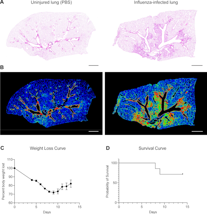
Figure 1: Monitoring lung injury and animal health. (A) Hematoxylin and eosin (HE) staining of lung tissue sections from a mock-infected (PBS-treated) lung on the left and an IAV-infected lung on the right. Compared to the uninjured lung, the IAV-infected lung shows heterogeneous tissue damage. Scale bars, 1 mm. (B) Using a k-means clustering algorithm in MATLAB20, tissue damage can be characterized as severe (red), damaged (green), or normal (blue). It is important to note that in the PBS-treated lung lobe on the left, airway structures are classified as "severe" due to the density and staining intensity of airway epithelial cells. However, it is still possible to easily discern the difference between PBS-treated and influenza-infected lungs given this baseline. Scale bars, 1 mm. (C) Example weight loss curve from a representative IAV infection experiment in which mouse lungs were collected at 13 days post-infection (dpi). The curve contains data from six wild-type mice, and dots represent the mean, while error bars represent the standard error of the mean. (D) Example survival curve from a representative IAV infection experiment in which mouse lungs were collected at 14 days post-infection (dpi). The curve contains data from seven wild-type mice. Please click here to view a larger version of this figure.
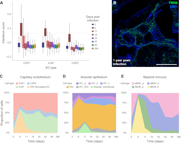
Figure 2: Monitoring capillary endothelial cell state changes after IAV infection. (A) An interferon-stimulated EC state arises at 6 dpi that is resolved between 11-19 dpi. Reanalysis of published single-cell RNA sequencing data from mouse lungs at several different time points following influenza infection shows that the interferon module score in all capillary endothelial cell types is highest at 6 dpi. Data was reanalyzed using R, and the interferon module score is based on the expression of 87 interferon-stimulated genes within each cell type. CAP1, capillary type 1 endothelial cell; iCAP, injury-induced endothelial cell; CAP2, capillary type 2 endothelial cell; EC, endothelial cell. (B) In contrast, an injury-induced EC state characterized by expression of Ntrk2 (TRKB) arises at 11 dpi and persists up to a year post-infection. Scale bar, 500 µm. (C) Transcriptional data from single-cell RNA sequencing of lung capillary endothelial cells after influenza infection demonstrate the dynamic nature of capillary endothelial cell state during tissue repair. (D) Alveolar epithelial cells also experience cell plasticity after influenza infection. An alveolar transitional cell type arises between 6-11 dpi and is resolved by 19 dpi. An immature AT1 cell state (AT1_c) arises at 11 dpi and persists until 90 dpi. (E) After infection, many existing alveolar macrophages (aMAC_a) are lost, and an influx of inflammatory monocytes (iMON) is seen at 6 dpi. The alveolar macrophage population is reconstituted both by existing aMACs to regenerate the aMAC_a population, and by inflammatory monocytes, generating the aMAC_b population. Although both represent an alveolar macrophage state, the resulting aMACs can be distinguished transcriptionally. Graphs in (C-E) indicate the percentage of cells in each population at each time point in the longitudinal influenza infection dataset and were generated using R. Please click here to view a larger version of this figure.
Supplementary File 1: Sample injury log, weight log, and body condition score (BCS) log for monitoring animals after influenza infection. Please click here to download this File.
Discussion
Here, we have reported a convenient method of transferring the influenza virus into mice to initiate rapid and effective damage to mouse lungs and study mechanisms of both successful regeneration and dysplastic repair. We demonstrate that intranasal inoculation of A/PR/8/34 IAV can be a stress-free technique. It takes less than 2 minutes per mouse to complete inoculation after isoflurane anesthesia. It is a simple and noninvasive technique of viral inoculation that follows the natural route of infection and allows the study of the response of both the tissue and the immune system in a more physiologically relevant manner. Virus inoculated through the mouse nostrils can induce both mucosal and systemic immunity21. More importantly, the damage caused by viral infection is clearly visible in lung tissue slices stained with hematoxylin and eosin (HE) staining22 (Figure 1). A complete timeline of expected transcriptional changes in lung cell types in each compartment of the lung has been previously published9. Here, we have reanalyzed a subset of this data to demonstrate that in the capillary endothelial, alveolar epithelial, and myeloid immune compartments of the mouse lungs, transient and persistent transcriptional changes occur over the course of the response to influenza infection from uninfected mice up to a year post-infection (Figure 2).
There are several critical steps in this protocol, and appropriate troubleshooting of these steps will produce a successful experiment. A/PR/8/34 influenza virus is a mouse-adapted strain of influenza that does not productively infect humans23, but it is always important to follow all indicated safety procedures during the experiment to avoid any possibility of infection of laboratory personnel. Isoflurane anesthesia administration is a critical step to ensure the successful delivery of the virus, as mice will not inhale the virus well when they are awake. Check the breathing and monitor the mouse head movement during the procedure to ensure the virus is inoculated into both nostrils. If mice are not fully anesthetized, bubbles can be generated, which both limit inhalation of the virus and generate aerosols. Both outcomes should be avoided, and mice should be placed back on the isoflurane nose cone if any sneezing or bubbles at the nostrils are observed. Post-infection monitoring, such as measuring weight loss, body condition score, and health status, as well as providing a recovery food supplement, is also an important component of this experiment. If it is observed that mice do not lose adequate weight or do not have the expected amount of tissue damage (Figure 1), it is possible that the dose is too low; on the other hand, if many mice reach the 30% body weight loss endpoint, it is possible that the dose is too high. In either case, it is best to determine the proper dose through an empirical titer of the lot of virus using a cohort of C57BL/6 mice as we describe here.
Since influenza infection continues to be prevalent worldwide, causes severe respiratory disease, and can be fatal, understanding the process of viral infectivity and the cellular response to infection and tissue injury in the lung is of prime importance. The intranasal infection method described in this study may therefore help in the identification of important regulatory mechanisms and the development of novel therapeutic approaches for lung diseases. In addition, future applications of the technique could use a different strain of mouse-adapted influenza virus, such as X31, to determine the effects on lung tissue structure and function in the case of repetitive respiratory infections. As many people are subject to multiple respiratory infections in their lifetime, such an extension of this model could have important implications for the study of lung tissue regeneration and, therefore, for public health.
Disclosures
The authors have nothing to disclose.
Acknowledgements
This research was supported by the Intramural Research Program of the US National Institutes of Health (NIH), National Cancer Institute, Center for Cancer Research.
Materials
| Name | Company | Catalog Number | Comments |
|---|---|---|---|
| 1.5 mL tube | Fisher Scientific | 02-682-557 | |
| 100 µm cell strainer | Falcon | 352360 | |
| 15 mL conical tube | Falcon | 352196 | |
| 1x PBS | Gibc0 | 10010-023 | |
| 40 µm cell strainer | Falcon | 352340 | |
| 5 mL tube | Globe | 6101C | |
| 50 mL conical tube | Falcon | 352098 | |
| A/PR/8/34 Influenza Virus stock | ATCC | VR-95PQ | Different lots have different viral titer |
| ACK Lysis Buffer | Quality Biological | 118-156-101 | |
| Anti-TRKB Antibody | R&D Systems | AF1494 | |
| Bleach | Clorox | 10% Bleach | |
| Bovine Serum Albumin | Sigma Aldrich | A8022 | |
| CO2 Anesthesia Induction Chamber | Any | N/A | |
| Collagenase Type I | Gibco | 17100017 | |
| DAPI | Milipore Sigma | 28718-90-3 | |
| Dispase | Corning | CB-40235 | |
| Dissection tools: scissors, curved forceps, straight forceps | Any | N/A | |
| Dnase | Sigma Aldrich | 4716728001 | |
| D-PBS | Corning | 21-031-CM | |
| Ethanol | Pharmco | 111000200 | |
| Glass vials | DWK Life Sciences | 986562 | |
| Ice bucket | Any | N/A | |
| Isoflurane | Covetrus | 29404 | |
| Needles, 21–23 G | Any | N/A | |
| P20/P200 pipette | Pipetman | F144056M/F144058M, | |
| P20/P200 pipette tips | Thomas Scientific | 1159M43/1159M40 | |
| Paraformaldehyde (PFA) | Thermo Scientific | J19943-k2 | 2%PFA |
| Pipetman | Any | N/A | |
| Plastic or cardboard freezer boxes | Any | N/A | |
| Razor blade | Any | N/A | |
| Ring Stand | Any | N/A | |
| Serological pipets, 5 mL | Falcon | 357543 | |
| Slide Top Induction Mouse Isoflurane Chamber | Somni Scientific | ||
| Suture | Fisher Scientific | NC9422522 | |
| Syringe tubing and luer-locks | Any | N/A | |
| Syringe, 10 mL | Any | N/A | |
| Table Top Anesthesia Machine Isoflurane | Ohmeda | Ohmeda Isotec- 4 | Flow rate should be 2.5–3% |
| Ultra-low melting agarose | Sigma Aldrich | A5030-1G | |
| Weighing balance | OHAUS | CS200 |
References
- Mammoto, A., Mammoto, T. Vascular niche in lung alveolar development, homeostasis, and regeneration. Front Bioeng Biotechnol. 7, 318(2019).
- Tsikis, S. T., Hirsch, T. I., Fligor, S. C., Quigley, M., Puder, M. Targeting the lung endothelial niche to promote angiogenesis and regeneration: a review of applications. Front Mol Biosci. 9, 1093369(2022).
- Torrego, A., Pajares, V., Mola, A., Lerma, E., Franquet, T. Influenza A (H1N1) organising pneumonia: figure 1. BMJ Case Rep. 2010, bcr1220092531(2010).
- Murota, M., et al. Influenza H1N1 virus-associated pneumonia often resembles rapidly progressive interstitial lung disease seen in collagen vascular diseases and COVID-19 pneumonia; CT-pathologic correlation in 24 patients. Eur J Radiol Open. 7, 100297(2020).
- Niethamer, T. K., et al. Defining the role of pulmonary endothelial cell heterogeneity in the response to acute lung injury. eLife. 9, e53072(2020).
- Klomp, M., Ghosh, S., Mohammed, S., Khan, M. N. From virus to inflammation, how influenza promotes lung damage. J Leukoc Biol. 110 (1), 115-122 (2021).
- Newton, A. H., Cardani, A., Braciale, T. J. The host immune response in respiratory virus infection: balancing virus clearance and immunopathology. Semin Immunopathol. 38 (4), 471-482 (2016).
- Jones, D. L., et al. An injury-induced mesenchymal-epithelial cell niche coordinates regenerative responses in the lung. Science. 386 (6727), eado5561(2024).
- Niethamer, T. K., et al. Longitudinal single-cell profiles of lung regeneration after viral infection reveal persistent injury-associated cell states. Cell Stem Cell. 32 (2), 302-321.e6 (2025).
- Belser, J. A., Gustin, K. M., Katz, J. M., Maines, T. R., Tumpey, T. M. Comparison of traditional intranasal and aerosol inhalation inoculation of mice with influenza A viruses. Virology. 481, 107-112 (2015).
- Oltean, T., Maelfait, J., Saelens, X., Vandenabeele, P. Need for standardization of influenza A virus-induced cell death in vivo to improve consistency of inter-laboratory research findings. Cell Death Discov. 10 (1), 247(2024).
- Nguyen, T. -Q., Rollon, R., Choi, Y. -K. Animal models for influenza research: strengths and weaknesses. Viruses. 13 (6), 1011(2021).
- Jin, X. -Y., et al. Comparative pathogenicity of influenza virus-induced pneumonia mouse model following intranasal and aerosolized intratracheal inoculation. Virol J. 21 (1), 240(2024).
- Revelli, D. A., Boylan, J. A., Gherardini, F. C. A non-invasive intratracheal inoculation method for the study of pulmonary melioidosis. Front Cell Infect Microbiol. 2, 164(2012).
- Cantu, A., Gutierrez, M. C., Dong, X., Leek, C., Anguera, M., Lingappan, K. Modulation of recovery from neonatal hyperoxic lung injury by sex as a biological variable. Redox Biol. 68, 102933(2023).
- Sunagar, R., Kumar, S., Namjoshi, P., Rosa, S. J., Hazlett, K. R. O., Gosselin, E. J. Evaluation of an outbred mouse model for Francisella tularensis vaccine development and testing. PLoS One. 13 (12), e0207587(2018).
- Cowley, P. M., Roberts, C. R., Baker, A. J. Monitoring the health status of mice with bleomycin-induced lung injury by using body condition scoring. Comp Med. 69 (2), 95-102 (2019).
- Gonzalez, A. J., Ijezie, E. C., Balemba, O. B., Miura, T. A. Attenuation of influenza A virus disease severity by viral coinfection in a mouse model. J Virol. 92 (23), e00881-e00918 (2018).
- Lv, J., et al. Kinetics of pulmonary immune cells, antibody responses and their correlations with the viral clearance of influenza A fatal infection in mice. Virol J. 11, 57(2014).
- Liberti, D. C., et al. Alveolar epithelial cell fate is maintained in a spatially restricted manner to promote lung regeneration after acute injury. Cell Rep. 35 (6), 109092(2021).
- Cipolla, D., et al. Inhalable and nasal biologics: analytical, formulation, development, and regulatory considerations. J Aerosol Med Pulm Drug Deliv. 2024, (2025).
- Cardiff, R. D., Miller, C. H., Munn, R. J. Manual hematoxylin and eosin staining of mouse tissue sections. Cold Spring Harb Protoc. 2014 (6), (2014).
- Bouvier, N. M., Lowen, A. C. Animal models for influenza virus pathogenesis and transmission. Viruses. 2 (8), 1530-1563 (2010).
Reprints and Permissions
