This protocol describes the surgical technique for harvesting a human perforator flap model based on the deep inferior epigastric artery pedicle, intended for experimental research.
Method Article
An Experimental Human DIEP Flap Model to Investigate Preservation Strategies for Vascularized Composite Allografts and Free Flaps
In This Article
Summary
Abstract
Recently, preservation approaches such as cytoprotective agents injection, ex vivo machine perfusion, and supercooling have emerged as strategies to enhance long-term preservation of both standard and marginal organs by mitigating ischemic and hypoxic injury. Although encouraging, its application in the field of vascularized composite allotransplantation (VCA) remains largely confined to preclinical research. To date, most studies investigating VCA perfusion strategies have relied on animal models, particularly swine or rodent composites. While these models provide valuable mechanistic insights, their anatomical, immunological, and physiological differences limit reproducibility and translational relevance to human applications.
In this protocol, each surgical step required for the procurement of a human deep inferior epigastric perforator (DIEP) flap for preservation studies is described in detail. The perforator is transected above the fascia without any subfascial dissection, yielding a short yet sufficient pedicle for catheterization. This model takes advantage of discarded tissue from standard abdominoplasty procedures, posing no additional risk to the patient. Critical steps are outlined to ensure a functional flap is harvested without prolonging operative time or compromising patient safety. Functional imaging is subsequently performed to confirm flap viability prior to experimental use, and sequential biopsies may be performed to follow tissue integrity. This model is particularly suited for research involving muscle-sparing VCA procedures -- such as partial facial transplantation -- and may also have relevance for the study of autologous free flap preservation.
Introduction
Vascularized composite allotransplantation (VCA) represents a promising reconstructive solution for patients with complex tissue defect, particularly in the face and upper limbs1. However, VCA is highly susceptible to ischemia-reperfusion injury, which can compromise graft viability and long-term outcomes2. In this context, preservation approaches such as cytoprotective agents injection, ex vivo machine perfusion, and supercooling have emerged as strategies to extend preservation time, optimize graft quality, and enable viability assessment prior to transplantation3,4,5,6,7. While normothermic ex vivo perfusion of solid organs has been successfully translated from an experimental laboratory technique into clinical practice over the past decade8, with promising results, it remains largely confined to the research setting in the field of VCA9.
To date, most studies investigating VCA perfusion protocols have relied on animal models, particularly swine or rodent composites10. While these models offer valuable insights, they inherently limit reproducibility and translational relevance due to anatomical, immunological, and physiological differences11,12. Human tissue models provide more relevant insights, but fresh tissues from brain-dead donors are extremely difficult to access for research purposes, requiring complex logistics, strict regulatory approval, and raising ethical concerns13,14. An ideal alternative would be a non-cadaveric, ethically acceptable, human-based flap model that does not carry the complexity or risk of a full VCA procedure and is entirely safe for the patient. Importantly, it should also be easily accessible to facilitate reproducible, scalable research.
Herein, a human deep inferior epigastric perforator (DIEP) flap model for VCA preservation research is described, using discarded tissue from abdominoplasty procedures without any additional risk to the patient.
Protocol
All patients received standard humane care in accordance with the Helsinki Declaration, and the protocol was approved by the institutional ethics committee (project No. IRB00014528_2025_32). The patient provided informed consent for the use of discarded tissue for research purposes.
1. Preoperative care
NOTE: These cutaneous perforator flaps, excluding fascia and muscle, are derived from surgical waste in patients, most often post-bariatric, and can only be harvested in cases of prior dermolipectomy with predictable umbilical transposition (i.e., a significant abdominal pannus). Planned liposuction during the procedure is an exclusion criterion. Concomitant diastasis repair is not a contraindication, as this procedure occurs after flap harvesting. The abdominal pannus generally provides at least partial coverage of the pubic area. Patients must have no history of thromboembolic events, and the body mass index (BMI) must be below 30 kg/m², as higher values are associated with obesity and an unfavorable risk-benefit ratio for elective non-vital procedures under general anesthesia. Previous abdominal surgery, except liposuction, is usually not a contraindication, provided that a perforator is identified by Doppler examination or directly visualized intraoperatively. This research procedure should also be avoided in true clinical unilateral DIEP free-flap reconstructions, which are already lengthy and complex operations where additional preoperative steps would be irrational and increase procedural risk.
- Perform the preoperative dermolipectomy marking with the patient in the standing position with a dermographic pen. Identify the midline from the pubic symphysis to the xiphoid process, and draw a transverse line approximately 7 cm above the vulvar commissure. Extend this line laterally about 7 cm on each side, gently curving it upward to join just below the anterior superior iliac spines.
- Identify the perforators around the umbilicus using a handheld acoustic Doppler probe (8-10 MHz) with the patient in the supine position.
NOTE: Apply a small amount of sterile ultrasound gel to the skin to ensure proper acoustic coupling. Hold the probe at a 45° angle with light contact pressure to avoid dampening the signal. A strong, triphasic pulsatile sound indicates an arterial perforator. Mark each perforator on the skin with a sterile marker to establish the perforator map, as routinely done prior to DIEP flap harvest. - Using a dermographic pen, outline a skin paddle in an elliptical shape of the desired dimensions, including the perforator along its medial border and extending on both sides of the umbilicus (Figure 1).
- Prepare and drape the surgical field from the xiphoid process to the upper third of the thighs, including the pubic region, using sterile surgical drapes as routinely performed in standard abdominal surgery.
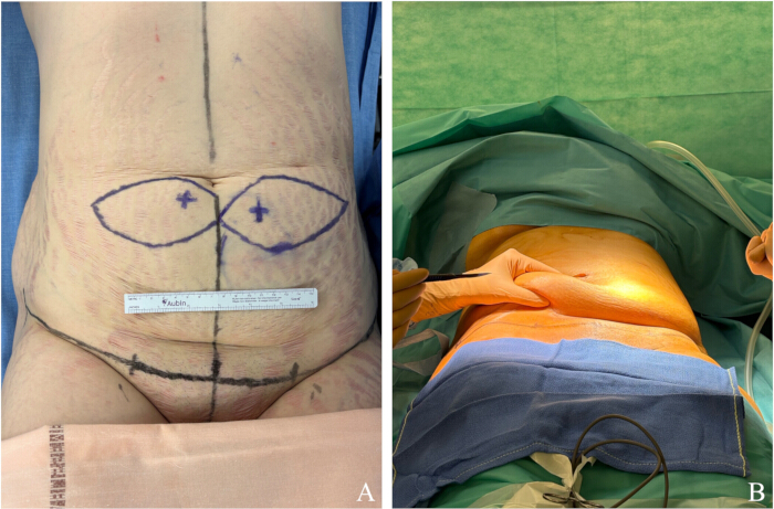
Figure 1: Preoperative evaluation before post-bariatric abdominal dermolipectomy with DIEP flap harvest. (A) Preoperative marking performed using an acoustic Doppler and a dermographic pen to identify and outline the paraumbilical perforators on each side of the midline. (B) The surgeon demonstrates the laxity of the abdominal skin and subcutaneous tissue to be resected, highlighting the adipocutaneous panniculus before incision along the horizontal marking line. Please click here to view a larger version of this figure.
2. Flap procurement
- Detach the umbilicus from the abdominal wall through a circumferential incision down to the hypodermis using a No. 15 scalpel blade.
- Place a long, loose, non-absorbable suture on either side of the umbilical margin.
- Dissect the umbilicus vertically using Mayo scissors, from the superficial to the deep plane, until it is completely freed while preserving its umbilical pedicle.
NOTE: Assistant participation is critical at this step. Gillies retractors should be used to expose the umbilicus, while applying gentle longitudinal traction on the long non-absorbable suture to facilitate dissection. The fat surrounding the umbilical pedicle forms a firm cord that is easily palpable during umbilical isolation, allowing precise identification and preservation of the pedicle. - Make a low transverse skin incision along the preoperative marking, approximately 7 cm above the vulvar commissure, gently curving toward the anterior superior iliac spines on each side, using a No. 15 or No. 21 scalpel blade.
- Raise the superior abdominoplasty flap in the subcutaneous plane, starting from the lower incision and progressing cranially toward the umbilicus. Use a fine-tip monopolar electrocautery set to approximately 50-70 °C and 80 W in coagulation mode to separate the anterior rectus fascia from the overlying subcutaneous tissue and skin while maintaining meticulous hemostasis throughout the dissection.
- As the dissection approaches the previously isolated umbilical pedicle, reduce the electrocautery temperature to approximately 20-30 °C, keeping the power set at 80 W in coagulation mode, and continue with fine dissection using Metzenbaum scissors to avoid pedicle injury.
- Identify and carefully isolate the two dominant paraumbilical perforators arising from the deep inferior epigastric system. Dissect each perforator circumferentially under direct vision using Stevens or small Metzenbaum scissors, preserving their vascular pedicles. Do not extend the dissection above the level of the anterior rectus aponeurosis (Figure 2).
NOTE: Perforators are selected based on their size; those appearing larger and pulsatile under direct vision are preferred. Dissection around the umbilicus must be meticulous to avoid damaging these perforators. - Divide the adipocutaneous panniculus longitudinally along the midline, from the center of the lower pubic incision up to the umbilicus, using a No. 15 blade followed by monopolar electrocautery set at approximately 50-70 °C.
- Continue the dissection cranially up to the xiphoid region and along the lateral costal margins with electrocautery set at approximately 50-70 °C, keeping the flap pedicles intact and not divided (Figure 3).
NOTE: This step 9 can also be delayed for practical reasons if the pedicle makes the supraumbilical dissection difficult and may instead be performed after pedicle division at Step 12, depending on intraoperative conditions.. - Control minor bleeding with medium-intensity coagulation, using a standard monopolar electrocautery set to 50-60 °C and 80 W in coagulation mode to maintain precise hemostasis.
- Readjust the outline of the skin paddle with a dermographic pen, based on the initial markings, to ensure that the perforator is included along the medial edge of the flap on each side of the adipocutaneous panniculus that was divided longitudinally at step 8 (Figure 4).
NOTE: To avoid bias due to differences in surface area exposed to the test solution, both flaps (if two are harvested) must be of identical size. The flap thickness is determined by the subcutaneous tissue of the flap and cannot be surgically reduced without risking compromise of flap integrity. - Ligate the deep inferior epigastric artery perforator pedicle using resorbable 3-0 sutures or automatic clips and transect the perforator above the fascia, with no subfascial dissection. Record the time of the warm ischemia.
NOTE: An approximate 10-min period of flap ischemia is observed before complete detachment and treatment with a cytoprotective agent injection. - Advance the supraumbilical skin and subcutaneous fat downward, and secure the undermined abdominal flap to the inferior incision margin at the midline using a non-absorbable suture, leaving one end of the knot long.
NOTE: Place the patient in a semi-flexed position during this step. Downward traction on the upper abdominal flap helps determine the amount of excess skin to be resected. - Use the long end of the midline non-absorbable suture connecting the supraumbilical and pubic areas as a guide to draw, with a dermographic pen, the resection line on the excess adipocutaneous panniculus, extending laterally toward each anterior superior iliac spine, as typically performed during abdominoplasty. The DIEP flaps are located below this marked resection line (Supplementary Figure 1).
- Incise the flap along the preoperative markings using a No. 15 scalpel blade. Harvest the perforator flap following the pre-established design, using monopolar electrocautery set to approximately 50-60 °C and 80 W in coagulation mode (Figure 5).
NOTE: This is one of the most delicate steps, as the perforator must be included within the flap. Assistance is recommended to provide gentle traction, while the operator maintains continuous visual control of the perforator throughout the dissection. Cold-knife dissection is not advised, as it may compromise flap integrity and lead to vascular leakage. - Completely detach the perforator flap from the surrounding discarded tissue.
NOTE: If two flaps are harvested and subjected to different treatments, they can be distinguished by placing a suture on one of them. - Excise the redundant dermo-adipose tissue corresponding to the excess skin and fat removed during the abdominoplasty.
NOTE: If the perforator flap is harvested after excision of the excess dermo-adipose tissue for practical reasons, use a sterile, single-use, battery-operated disposable cautery pen (large tip, approximately 1,200 °C) to complete the detachment. A return electrode cannot be used in this situation, as the tissue is fully separated from the patient. After harvesting the DIEP flaps, the surgical team hands the flap to the research team waiting in the operating room, then proceeds with the abdominoplasty by dissecting along the pre-marked resection lines to detach and remove the redundant adipocutaneous panniculus.
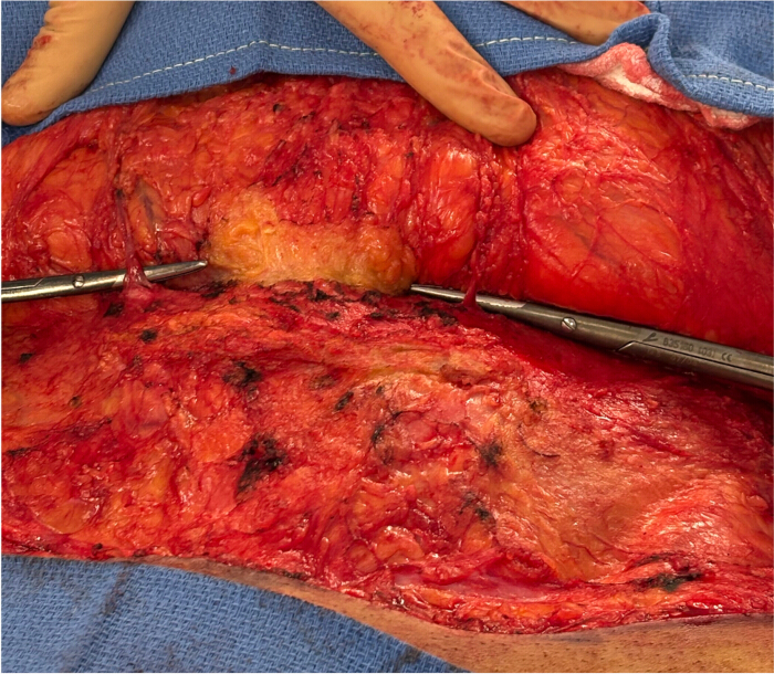
Figure 2: Intraoperative view of the abdominal dermolipectomy procedure with skeletonization of the deep inferior epigastric perforator pedicles. The image shows the operative field before supraumbilical dissection and longitudinal infraumbilical division. The two dominant bilateral paraumbilical deep inferior epigastric perforators are exposed and highlighted with surgical instruments. Please click here to view a larger version of this figure.
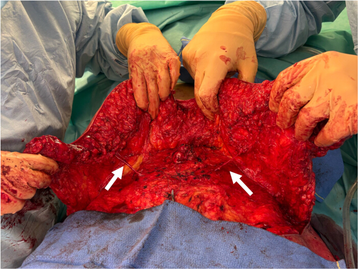
Figure 3: Intraoperative view after supraumbilical dissection showing the bilateral deep inferior epigastric perforators. The perforators are not ligated, and their subfascial course and length can be better appreciated under gentle traction (white arrows). Please click here to view a larger version of this figure.
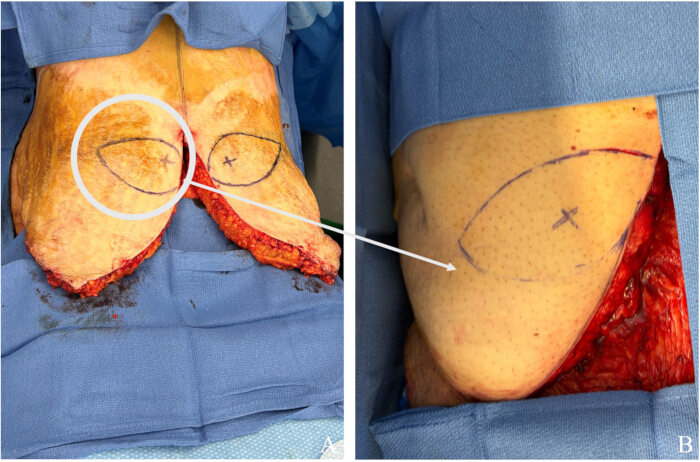
Figure 4: Intraoperative view showing the DIEP flaps outlined on the skin in an elliptical shape before ligation. (A) The skin paddle is adjusted and outlined in an elliptical shape using a dermographic pen (white circle) to ensure inclusion of the perforator along the medial edge of the flap on each side of the adipocutaneous panniculus. (B) Enlarged view showing the DIEP flap outline and the perforator marked with an "x" according to preoperative Doppler mapping (white arrow). Please click here to view a larger version of this figure.
Supplementary Figure 1: Downward traction on the upper abdominal flap helps determine the amount of redundant skin to be resected. The long end of the midline non-absorbable suture connecting the supraumbilical and pubic regions is used as a guide. Using a dermographic pen, draw the resection line along the course of the suture (white arrows) on the redundant adipocutaneous panniculus. Extend this line laterally toward each anterior superior iliac spine. The DIEP flaps are located below this marked resection line. Please click here to download this File.
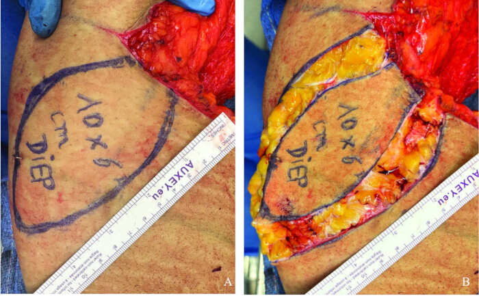
Figure 5: Intraoperative view of the deep inferior epigastric artery perforator (DIEP) flaps during harvest, following pedicle ligation. (A) The DIEP flap, measuring approximately 10 cm × 6 cm, is outlined with a surgical marker after localization of the perforator. (B) Skin incision and flap harvest are performed along the pre-established markings. The initial incision is made with a No. 15 blade, followed by dissection using a monopolar electrocautery set at approximately 50-60 °C, and 80 W in coagulation mode, when the excess cutaneous-adipose tissue remains attached to the patient. When the tissue has been completely detached, a sterile, single-use, battery-operated disposable cautery pen (large tip, approximately 1,200 °C) is used to complete the separation. Please click here to view a larger version of this figure.
3. Preparation of the flap
- Dissect the vascular pedicle under magnification using microsurgical instruments, and identify both the artery and the vein (Figure 6). Gently open the arterial lumen with a microvascular dilator.
NOTE: Use a dedicated sterile table equipped with microsurgical instruments to prepare the flap. Trim 2-3 mm from the arterial end to obtain a clean, regular edge suitable for catheterization. - Weigh each flap at the time of harvest to allow later quantification of edema formation.
- Catheterize the artery using an 18-24 G cannula and secure it in the lumen with 5-0 silk suture ligation (Figure 7).
NOTE: For this experiment, the flaps were catheterized using a 24-G cannula, as the study involved the evaluation of cytoprotective agents through a single injection. For ex vivo machine perfusion or extracorporeal membrane oxygenation (ECMO) applications, cannulation should be performed with a larger 18-20-G cannula. In case of catheter occlusion during ex vivo perfusion, flush gently with heparinized saline or replace the cannula to restore flow. Maintaining low perfusion pressure (<25 mmHg) helps prevent recurrence. - Inject contrast agent intra-arterially through the catheter into the perforator artery, and evaluate the flap under fluoroscopy (Figure 8).
NOTE: The contrast agent should render the small capillaries uniformly radiopaque throughout the flap, indicating an adequate perfusion territory supplied by the selected pedicle. At this stage, the presence of non-perfused areas may render the model unsuitable for further research use or require excision of the distal part of the flap. The harvested flaps can subsequently be used for experimental protocols, placed on a perfusion machine, ECMO device, or injected with a test solution as cytoprotective agents. Punch biopsies (ranging from 1 mm to 8 mm in diameter) can be performed at different time points without compromising flap integrity, allowing kinetic assessment of histological changes within the tissues.
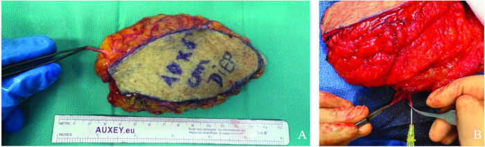
Figure 6: Post-harvest view of a research DIEP flap. (A) The harvested DIEP flap is shown with the vascular pedicle dissected and isolated using microsurgical forceps. The arterial end is trimmed by a few millimeters to obtain a clean cut, and the lumen is gently opened with a micro-dilator forceps. (B) Close-up view showing microsurgical forceps securing the dominant perforator artery, with a 24-G catheter inserted into the lumen for contrast agent injection. Please click here to view a larger version of this figure.
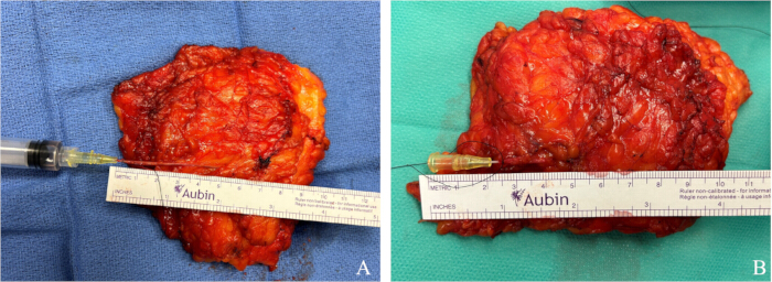
Figure 7: Catheterization of the DIEP flap pedicle. (A) The catheter is secured in place with a 5-0 silk suture tied gently to avoid obstructing the lumen of the pedicle. (B) View of the catheter positioned without the syringe attached, showing the extent of catheter insertion into the vascular lumen. Please click here to view a larger version of this figure.
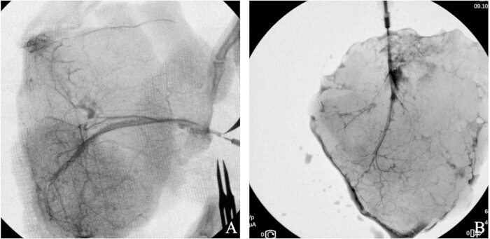
Figure 8: Post-harvest fluoroscopy of the DIEP flap demonstrating the vascular anatomy. (A,B) Contrast agent is injected through a 24-G catheter into the dominant perforator artery, depicting the branching pattern and the vascularized area, confirming flap viability on two different flaps. Please click here to view a larger version of this figure.
Results
Eleven flaps were harvested from six female patients included in this study. Two senior plastic surgeons, experienced in flap surgery and abdominoplasty, performed the dissections. In one patient, the flap was harvested unilaterally due to a dissection issue. The harvested flaps had a mean weight of 198.6 ± 24.4 g (n = 11) and an average size of 10 cm × 6 cm, which corresponded to the sample requirements for the preservation study in which these flaps were used. The pedicle length, measured under gentle tension, averaged 3.85 ± 0.74 cm, and the external vessel diameter, measured with a caliper, was 1.2 ± 0.3 mm. The catheter was observed to be inserted to an average depth of 1.75 cm (n = 2). All flaps demonstrated good perfusion on arteriography, with homogeneous and complete vascular filling throughout the flap, as assessed similarly in previous studies15.
The total mean operative time for standard abdominoplasty with umbilical transposition in a case series of six patients was 123 ± 24 min. There was no significant difference in operative time among these six procedures (p = 0.61). To assess any difference compared with the standard procedure without research flap harvest, the records of six patients who underwent similar surgery at the University Hospital were retrospectively reviewed (abdominoplasty with abdominal transposition). The mean operative time was 131 ± 21 min. There was no statistically significant difference in operative time compared with the six standard abdominoplasty procedures without flap harvest (p = 0.47, two-tailed Student's t-test).
No postoperative abdominal wall deficits or infections were observed in any of the six patients included in the study, with a mean follow-up of 6.5 ± 5.8 months (minimum: 2 weeks; maximum: 14 months) (Table 1).
In this protocol, the flaps were used for cytoprotective agent injection studies, and catheterization via the arterial pedicle was performed using either a 22- or 24-G catheter without technical difficulty, as in this protocol. Venous catheterization is not required in this type of study, consistent with static cold storage and supercooling protocols. The outcome of the catheterized flap depends on the protocol used. In this study, eleven DIEP flaps were included in a protocol designed to evaluate the effect of a cytoprotective agent, an inhibitor of eIF5A hypusination. After catheterization, the flaps were wrapped in sterile surgical towels, placed in sterile vacuum-sealed bags, and labeled. In the experience we conducted on these flaps, two experimental groups were defined: a control group perfused with University of Wisconsin preservation solution, and a treatment group receiving the cytoprotective agent administered intra-arterially through the vascular pedicle. All flaps were stored under static cold conditions at 4 °C in sealed bags immersed in ice water within an insulated container. Two 0.8-mm punch biopsies were taken from identical locations on both treated and control flaps at predefined time points: baseline (t = 0, before preservation), and after 1, 2, 4, 6, 8, 12, 24, 36, and 48 hours of cold ischemia. Tissue samples were fixed overnight in 10% neutral buffered formalin and embedded in paraffin. Four-micrometer sections were then deparaffinized in xylene, rehydrated through graded alcohols, and washed in phosphate-buffered saline (PBS). Sections were stained with hematoxylin and eosin for microscopic analysis. Histological evaluation was performed in a blinded fashion to compare treated and control flaps and to assess time-dependent ischemic changes over a 48-h period (Figure 9).
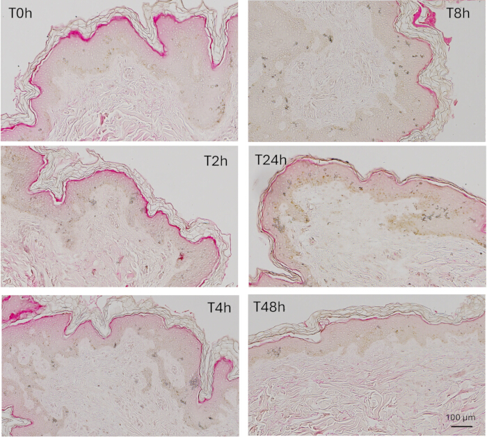
Figure 9: Representative hematoxylin and eosin (HE) image. Representative histology of flap biopsy cross-sections stained with HE from 0-48 h after surgical resection, following treatment with a single cytoprotective agent at the time of harvest. Scale bar = 100 µm. Please click here to view a larger version of this figure.
| Parameter | n | Mean ± SD | Range (Min–Max) | Unit | Notes |
| Flap weight | 11 | 198.6 ± 24.4 | 148–218 | g | Average flap size 10 × 6 cm |
| Pedicle length | 11 | 3.85 ± 0.74 | 2.1–4.6 | cm | Measured under gentle tension |
| External vessel diameter | 11 | 1.2 ± 0.3 | 0.9–1.5 | mm | Measured with caliper |
| Catheter insertion depth | 2 | 1.75 ± 0.21 | 1.6–1.9 | cm | Average depth for 24-G catheter |
| Operative time | 11 | 123 ± 24 | 98–165 | min | No significant difference (p = 0.61) |
| Post-operative follow-up | 11 | 6.5 ± 5.8 | 0.5–14 | months | No wall deficit or infection observed |
Table 1: Main characteristics of the harvested DIEP research flaps. Summary of morphometric and operative parameters from eleven research DIEP flaps harvested during abdominoplasty procedures. Measurements include flap weight, pedicle length, external vessel diameter, and catheter insertion depth. Operative time and postoperative follow-up were recorded to assess surgical safety and reproducibility. No postoperative abdominal wall complications or infections were observed.
Discussion
Human models help bridge findings from animal studies to clinical practice, improving the relevance and predictivity of preclinical research16,17,18. They must be carefully designed to ensure safety and ethical compliance, often limiting the scope and invasiveness of experiments17. Across the four harvested flaps, no additional dissection was necessary, and there was no significant prolongation of operative time in any of the cases. The relatively large diameter of the vessels of this flap (1.2 mm mean) makes cannulation easy in comparison with animal models. Clinical observation of flap perfusion is also relatively easier through the abdominal skin. Unlike the flaps commonly used in clinical practice (DIEP and transverse rectus abdominis myocutaneous [TRAM]), this experimental model does not involve any incision of the rectus abdominis fascia or muscle harvesting. Consequently, although the described flap pedicle is shorter, its length remains adequate for experimental preservation studies (Figure 10).
Regarding the final step and flap cannulation, it depends on the preservation studies. For cytoprotective agent injection studies, catheterization via the arterial pedicle can be performed using either a 22- or 24-G catheter without technical difficulty, as in this protocol. For ex vivo machine perfusion studies, a systematic literature review highlights that, in large animal studies using grafts of comparable size to the one presented here, arterial catheterization is typically performed with 18-20 G catheters and venous catheterization with 16-18 G catheters5. The specific perfusion system and catheter size depend on the animal species and the graft type. For ECMO studies, Ozturk et al. developed a human extracorporeal free flap perfusion model using a pediatric ECMO device to perfuse a DIEP flap comparable to the one described in this study. Their setup included a centrifugal pump head connected to a pediatric hollow-fiber oxygenator with a countercurrent heat exchanger and a 150-mL cardiotomy reservoir with a filter, all integrated into a standard pediatric ECMO circuit. The tubing diameter was adjusted to the appropriate range (16-20 G), confirming that the catheters are insertable into such flaps, which typically range from 16 to 20 G19.
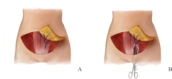
Figure 10: Schematic representation of the harvested deep inferior epigastric artery perforator (DIEP) flap for research versus clinical purposes, illustrating differences in harvest technique according to the intended use of the flap. (A) Research DIEP (preservation study): the flap is harvested without muscle or fascia, and the pedicle dissection remains suprafascial to avoid harming the patient. (B) Clinical DIEP (free flap reconstruction): the flap is also harvested without muscle or fascia, but a subfascial dissection is performed to obtain additional pedicle length for subsequent microsurgical anastomosis. Please click here to view a larger version of this figure.
To date, this model has rarely been described19,20. The most relevant report briefly outlined the use of a deep inferior epigastric artery perforator flap model for extracorporeal perfusion with an extracorporeal membrane oxygenation device, achieving a mean perfusion duration of 6 days. Yet, it requires careful preparation and an optimized strategy to ensure both patient safety and model integrity. In their description, Ozturk et al. folded the flap onto itself after excision, shaped it into a tube, and sutured it, a method that may compromise anatomical fidelity and perfusion assessment19. Since the deep inferior epigastric arteries are bilateral, harvesting two smaller flaps of the same size can be preferred, with a contralateral flap that can be used as a reliable control from the same abdominoplasty specimen and during the same procedure.
The use of arteriography or intra-arterial fluorescein in a research setting can be useful to ensure the validity of the model and confirm homogeneous perfusion throughout the entire flap. Indeed, evaluating flap preservation often relies on biopsies, and if these are taken from poorly perfused areas, the results may be misleading. Arteriography can also assist in guiding the resection, helping to optimize the harvesting procedure.
There is currently no consensus on the optimal temperature for ex vivo machine perfusion (EVMP), for either solid organ transplant21 or VCA9. This model is particularly relevant for subnormothermic and normothermic perfusion studies, where maintaining an optimal balance between metabolic demand and preservation time is critical. Conversely, hypothermic perfusion is of limited relevance for muscle-free VCAs and cutaneous flaps, as their inherently low metabolic activity reduces the benefits of such cooling strategies22. To a lesser extent, it can also support decellularization and recellularization research, as it provides fresher tissue than any cadaveric source could offer23,24. Moreover, the proposed model is relatively simple to perform for a plastic surgeon, whereas VCA models based on cadaveric or brain-dead donors are technically demanding and require extensive specialized training25,26,27,28.
Doppler ultrasound is recommended, as it is non-irradiating, inexpensive, and provides rapid localization of perforators for an experienced operator29. However, its use is not mandatory, since under direct vision, the perforator is usually located within a few centimeters of the umbilicus, as consistently reported in multiple anatomical studies on DIEP flaps29. Preoperative CT angiography is not justified in the setting of abdominoplasty-derived research flaps, as it is both irradiating and costly. In this study, fluoroscopic assessment was performed for all 11 harvested flaps prior to cytoprotective treatment, consistently demonstrating homogeneous vascular opacification without evidence of misinterpretation or tissue injury. For small flaps of approximately 10 cm × 6 cm, omitting fluoroscopy can reasonably save time before initiating the preservation protocol, provided the procedure is performed by an experienced surgeon or when fluoroscopy is not readily available. Perfusion assessment relies primarily on the surgeon's experience, with homogeneous vessel opacification confirming adequate flap perfusion. For larger flaps, fluoroscopy remains recommended to ensure complete vascular integrity. Although not performed in the present study, indocyanine-green angiography may also be used for perfusion assessment; however, it does not provide a significant time advantage compared with fluoroscopy.
While the procedure is technically straightforward, it presents several practical limitations that require anticipation and close coordination between the surgical and research teams. If the operating surgeon is not comfortable harvesting the flap while it remains attached to the patient, preferring instead the conventional abdominoplasty approach based on bilateral panniculus excess measurements, the excision can be performed in the usual manner, and the discarded tissue subsequently transferred to the research team. This workflow requires close collaboration between the surgical and research teams. The research team must be prepared to handle the flap immediately after excision to prevent prolonged ischemia, with all experimental materials and instruments ready in advance. The operating surgeon should have prior experience in abdominal wall surgery, typically in digestive or plastic surgery, and be familiar with the basic principles of flap physiology, particularly hemodynamics. Similarly, the research team should be trained in primary manipulation of microsurgical instruments to ensure proper handling of the specimen; even minor trauma from sharp microforceps can damage the pedicle or compromise perfusion30. The use of monopolar electrocautery carries a risk of thermal injury, particularly when operating near the perforator pedicle. To minimize this risk, coagulation should be performed at moderate power, with continuous saline irrigation to prevent heat accumulation, and scissors should be used for dissection when working in close proximity to the pedicle. Proper placement of the return electrode is essential to avoid skin burns, as in any surgical procedure31. Fluoroscopy for contrast injection must follow radiation safety standards, including the use of lead aprons and minimizing exposure through low-dose settings and beam collimation. In our series, one case of intraoperative perforator identification failure occurred (no suitable perforator was found on one side of the umbilicus, and the others were accidentally transected). No cannulation leaks were observed with the 22-24-G injection protocol, and incomplete opacification on fluoroscopy was not encountered. The model itself also presents significant drawbacks. Unlike most VCAs, it does not include bone or muscle components but consists solely of vascularized soft tissue. As such, it does not allow for the study of muscle viability or the early ischemic damage typically observed in muscle compartments during VCA. Nevertheless, it is worth noting that many partial face transplants have been performed without muscle32,33,34, particularly when the defect involves only the skin, subcutaneous tissue, and occasionally portions of facial bone or mucosa. Additionally, although anatomically realistic, this model does not reproduce the systemic physiological and inflammatory milieu associated with brain-dead organ donors, which may influence immune activation and endothelial injury in VCA settings25,35. The relatively short pedicle length, while sufficient for perfusion and injection studies, may limit compatibility with certain ex vivo perfusion devices compared with larger animal models. Inter-donor variability in flap thickness, vascular anatomy, and perfusion pattern also represents an inherent limitation, potentially affecting reproducibility in experimental applications. This can be mitigated by using pairwise contralateral control flaps when available and by standardizing perfusion solutions, flow rates, and environmental conditions. Finally, the representative data presented here are based on relatively small flaps (10 cm × 6 cm) and have not been tested for reproducibility in larger specimens. Further validation would be required to confirm whether fluoroscopy or other perfusion assessment methods can be safely omitted in larger flap models.
This model may also be beneficial for research on autologous flap preservation36. While the preservation of autologous tissue is generally considered less critical, it can become relevant in specific situations, such as saving time for free flap rescue or complex logistical scenarios37,38,39.
Disclosures
The authors have nothing to disclose.
Materials
| Name | Company | Catalog Number | Comments |
|---|---|---|---|
| 1 mL and 5 mL Luer-Lok syringe | Becton Dickinson | https://www.bd.com/en-us/products-and-solutions/products/product-page.309646 | |
| 11.5” Medium and Small Premium Surgiclip II Auto Suture vessel clip applier | Covidien | N/A | |
| 22–24 G vessel cannula blunt tip | Medtronic Inc | N/A | |
| 5-0 silk suture | N/A | Any compatible suture | |
| A single-use, battery-powered disposable cautery pen | Helpmedical, Europe | N/A | 1200 °C, large-tip |
| Adson Forceps | MPM | 106-2112A | |
| Bipolar Coagulation Forceps | Olsen | 20-1320I | |
| Disposable Scalpel #15 & #21 | Sklar | N/A | |
| Eosin | Microm Microtech | U/C0363 | |
| Faraboeuf retractors | Medicta Instruments | N/A | |
| Fine needle cautery | Cormedica | N/A | |
| OEC MiniView Max | GE Healthcare | N/A | Fluoroscopy system |
| Forceps Dilators | WPI | 15910 | |
| Hematoxylin | Microm Microtech | U/C0303 | |
| Iohexol 300 mg I/mL | GE Healthcare Canada Inc. | N/A | |
| Micro scissors | WPI | 504492 | |
| Valleylab Monopolar Diathermy | Medtronic Inc | ||
| Non-ionic iodinated contrast medium, Omnipaque 300 mg I/mL | GE Healthcare | N/A | |
| Portable acoustic Doppler, 8 MHz | Parks Medical Electronics | https://www.parksmed.com/ | |
| Saline solution 0.9% | GenDepot | S0600-101 | |
| Standard plastic surgery instrument set | Aesculap | N/A | including scalpel handle No. 3, Metzenbaum and Mayo scissors, Adson and Gillies forceps, dissecting scissors, hemostats, and needle holders. |
| Strabismus scissors | Surtex | 102-4109 | |
| Surgical marking pen | Cardinal health | 212PR | |
| Sutures Ethilon 4.0 and 3.0 | Ethicon | 1667G | |
| Tissue Forceps | MPM | 106-0511 |
References
- Lupon, E., et al. vascularized composite allografts in france: An update. Ann Chir Plast Esthet. , (2024).
- Messner, F., Grahammer, J., Hautz, T., Brandacher, G., Schneeberger, S. Ischemia/reperfusion injury in vascularized tissue allotransplantation: Tissue damage and clinical relevance. Curr Opin Organ Transplant. 21 (5), 503-509 (2016).
- Berkane, Y., et al. advances and perspectives in vascularized composite allotransplantation preservation. Bull Acad Natl Med. 208, 1299-1308 (2024).
- Baker, C. E., et al. Pretreatments of ex vivo vascularized composite allografts: A scoping review. Ann Plast Surg. , (2025).
- Njessi, P., et al. Preservation strategies for vascularized composite allotransplantation: An updated systematic review of a rapidly expanding field. bioRxiv. , (2025).
- Cougnon, M., et al. Inhibition of eif5a hypusination enhances antioxidant defense to prevent kidney ischemia/reperfusion injury. Redox Biol. 86, 103814(2025).
- Le Meur, Y., et al. First-in-human use of a marine oxygen carrier (m101) for organ preservation: A safety and proof-of-principle study. Am J Transplant. 20 (6), 1729-1738 (2020).
- Nicholson, M. L., Hosgood, S. A. Renal transplantation after ex vivo normothermic perfusion: The first clinical study. Am J Transplant. 13 (5), 1246-1252 (2013).
- Muss, T. E., et al. Ex-vivo perfusion of limb vascularized composite allotransplants: A systematic review of published protocols. Transpl Int. 38, 14132(2025).
- Muss, T. E., et al. A guide to the implementation and design of ex vivo perfusion machines for vascularized composite allotransplantation. Plast Reconstr Surg Glob Open. 12 (11), e6271(2024).
- Mcgonigle, P., Ruggeri, B. Animal models of human disease: Challenges in enabling translation. Biochem Pharmacol. 87 (1), 162-171 (2014).
- Pound, P., Ritskes-Hoitinga, M. Is it possible to overcome issues of external validity in preclinical animal research? Why most animal models are bound to fail. J Transl Med. 16 (1), 304(2018).
- Pirnay, J. P., et al. Access to human tissues for research and product development: From eu regulation to alarming legal developments in belgium. EMBO Rep. 16 (5), 557-562 (2015).
- Charmode, S., Ratanpara, L., Sheikh, N., Ravi, K. S., Mehra, S. Legal frameworks upholding deceased individuals' rights and enabling the use of cadavers in anatomy education and research: A systematic review. Cureus. 16 (4), e58473(2024).
- Lupon, E., et al. Engineering Vascularized Composite Allografts Using Natural Scaffolds: A Systematic Review. Tissue Eng Part B Rev. 28 (3), 677-693 (2022).
- Van Lier, D., Geven, C., Leijte, G. P., Pickkers, P. Experimental human endotoxemia as a model of systemic inflammation. Biochimie. 159, 99-106 (2019).
- Olesen, A. E., Andresen, T., Staahl, C., Drewes, A. M. Human experimental pain models for assessing the therapeutic efficacy of analgesic drugs. Pharmacol Rev. 64 (3), 722-779 (2012).
- Littman, B. H., Williams, S. A. The ultimate model organism: Progress in experimental medicine. Nat Rev Drug Discov. 4 (8), 631-638 (2005).
- Ozturk, M. B., et al. Extracorporeal free flap perfusion using extracorporeal membrane oxygenation device: An experimental model. Ann Plast Surg. 83 (6), 702-708 (2019).
- Kreidstein, M. L., Pang, C. Y., Levine, R. H., Knowlton, R. J. The isolated perfused human skin flap: Design, perfusion technique, metabolism, and vascular reactivity. Plast Reconstr Surg. 87 (4), 741-749 (1991).
- Parente, A., et al. Machine perfusion techniques for liver transplantation - a meta-analysis of the first seven randomized-controlled trials. J Hepatol. 79 (5), 1201-1213 (2023).
- Drivas, E. M., et al. Reply: A guide to the implementation and design of ex vivo perfusion machines for vascularized composite allotransplantation. Plast Reconstr Surg Glob Open. 13 (5), e6763(2025).
- Lupon, E., et al. A new method for preparation of decellularized human scaffolds for facial reconstruction. Curr Issues Mol Biol. 47 (4), (2025).
- Lupon, E., et al. Optimized decellularization of a porcine fasciocutaneaous flap. Bioengineering (Basel). 11 (4), (2024).
- Moris, D., Cendales, L. C. Sensitization and desensitization in vascularized composite allotransplantation. Front Immunol. 12, 682180(2021).
- Perraudin, T., et al. Vascularized composite hand allograft procurement and preparation for distal and proximal forearm allotransplantation: A stepwise approach. J Vis Exp. (219), e67767(2025).
- Perraudin, T., et al. Vascularized composite upper limb allograft harvesting for proximal arm allotransplantation. J Vis Exp. (220), e68170(2025).
- Barbat, P., et al. Procurement for a vascularized and reinnervated abdominal wall allotransplantation. J Vis Exp. (221), e68418(2025).
- Lupon, E. Perforator identification for propeller flap harvest: Technical insights from a case serie. Int J Surg Case Rep. 135, 111921(2025).
- Villavisanis, D. F., et al. Assisting in microsurgery: Operative and technical considerations. J Hand Surg Glob Online. 5 (3), 358-362 (2023).
- Saaiq, M., Zaib, S., Ahmad, S. Electrocautery burns: Experience with three cases and review of literature. Ann Burns Fire Disasters. 25 (4), 203-206 (2012).
- Pomahac, B., Diaz-Siso, J. R., Bueno, E. M. Evolution of indications for facial transplantation. J Plast Reconstr Aesthet Surg. 64 (11), 1410-1416 (2011).
- Barret, J. P. From partial to full-face transplantation: Total ablation and restoration, a change in the reconstructive paradigm. Int J Surg. 12 (2), 109-112 (2014).
- Dubernard, J. M., et al. Outcomes 18 months after the first human partial face transplantation. N Engl J Med. 357 (24), 2451-2460 (2007).
- Barklin, A. Systemic inflammation in the brain-dead organ donor. Acta Anaesthesiol Scand. 53 (4), 425-435 (2009).
- Slater, N. J., et al. Ex-vivo oxygenated perfusion of free flaps during ischemia time: A feasibility study in a porcine model and preliminary results. J Surg Res. 205 (2), 292-295 (2016).
- Lupon, E., et al. Microsurgery in low- and middle-income countries: Results of 20 years of experience in cambodia. J Plast Reconstr Aesthet Surg. 98, 161-169 (2024).
- Berkane, Y., et al. Reconstruction of a complex foot defect with a chimeric triple-component osteocutaneous scip-siea free flap: A case report and literature review. Arch Plast Surg. 52 (5), 310-316 (2025).
- Albadia, R., et al. Management of a large abdominal dermatofibrosarcoma protuberans requiring a life-threatening excision: A case report. Int J Surg Case Rep. 133, 111579(2025).
Reprints and Permissions
