The surgical protocol describes a laparoscopic partial splenectomy protocol in a 30-year-old female with a splenic hemangioma. The minimally invasive, organ-preserving procedure resulted in minimal blood loss, no complications, and preserved residual spleen perfusion, offering a reproducible approach for benign splenic lesions.
Method Article
How to Preserve the Spleen: Laparoscopic Partial Splenectomy as a Treatment for Splenic Hemangioma
* These authors contributed equally
In This Article
Summary
Abstract
Advances in understanding splenic function and the implementation of precision medicine and enhanced recovery after surgery (ERAS) have established laparoscopic partial splenectomy (LPS) as the preferred surgical approach for benign splenic diseases, hematologic diseases involving the spleen, and minor splenic trauma. This technique not only preserves splenic function but also offers advantages, including safety, reduced postoperative pain, and accelerated recovery. The widespread adoption of LPS represents an evolving trend in splenic surgery. A case of a 30-year-old female patient with a splenic hemangioma is presented. Computed tomography (CT) angiography revealed a bifurcated splenic artery. This protocol demonstrates the key steps of a laparoscopic partial splenectomy, with a detailed focus on approach selection, vascular management, parenchymal transection, and wound closure, all performed uneventfully with minimal intraoperative blood loss. Follow-up ultrasonography demonstrated a well-perfused residual spleen. This case highlights the importance of precise preoperative vascular imaging and careful hilar dissection to ensure adequate perfusion of the residual spleen. The stepwise protocol presented here may serve as a practical reference for surgeons performing spleen-preserving laparoscopic procedures.
Introduction
Splenic hemangioma is a common benign tumor of the spleen, predominantly of the cavernous type, followed by capillary hemangioma1. It has a female predominance, potentially associated with hormonal changes during pregnancy or oral contraceptive use2. Splenic hemangiomas exhibit slow growth, yet 2%–10% have a predisposition to spontaneous rupture, potentially leading to massive intra-abdominal hemorrhage and hemorrhagic shock1. Consequently, precise diagnosis and appropriate management are essential to reduce the risk of rupture and prevent serious complications. With advancements in laparoscopic techniques, laparoscopic splenectomy has undergone significant technical refinement3,4,5.
Compared with open surgery, laparoscopic splenectomy is associated with reduced surgical trauma, less postoperative pain, and faster recovery4,5. LPS is primarily indicated for benign focal lesions confined to the upper or lower splenic pole with well-defined vascular boundaries, select hematologic disorders requiring splenic preservation (e.g., hereditary spherocytosis), and localized splenic trauma, whereas contraindications include lesions highly suspicious for malignancy, diffuse or centrally located growth (particularly with hilum involvement that compromises safe vascular dissection), and complex vascular anatomy or tumor blood supply identified on preoperative three-dimensional CT angiography that would preclude preservation of a sufficient functional splenic volume (typically >25%–30%)4,6,7,8. In these scenarios, total splenectomy is recommended as a more anatomically definitive and procedurally controlled alternative9,10.
Historically, total splenectomy was the standard approach despite the high risk of overwhelming post-splenectomy infection (OPSI)11. Greater recognition of splenic immune function has shifted the clinical preference toward partial splenectomy, particularly for lesions localized to the poles. In patients with a Type I (distributed) arterial pattern and limited intersegmental anastomoses, ligating a segmental branch creates a distinct ischemic demarcation line, providing the anatomical basis for precise, parenchyma-preserving resection6. Therefore, laparoscopic partial splenectomy is a viable surgical option for benign splenic tumors located at the superior or inferior poles of the spleen7,8. Several studies have reported favorable outcomes of laparoscopic partial splenectomy compared with total splenectomy9,10.
This protocol presents a case of a 30-year-old female patient in whom a splenic space-occupying lesion was incidentally discovered during routine examination six months prior. CT findings suggested a diagnosis of splenic hemangioma at the inferior pole of the spleen, measuring approximately 3.5 × 3.5 × 3.0 cm (Figure 1). Given the documented interval enlargement of the lesion, surgical intervention was recommended after multidisciplinary evaluation, and informed consent was obtained. CT angiography indicated a bifurcated splenic artery (Figure 2). After completing the preoperative evaluation, a laparoscopic partial splenectomy was performed on the patient. Intraoperative blood loss was 300 mL (hemodynamically stable), and postoperative pathology confirmed splenic cavernous hemangioma.
Protocol
Informed consent was obtained from the patient. All procedures performed were in accordance with the ethical standards of the People's Hospital of Ningxia Hui Autonomous Region committee.
1. Pre-surgical steps
- Anesthesia and positioning
- Administer general anesthesia with endotracheal intubation and position the patient supine.
- Surgeon positioning
- Position the surgeon and camera operator on the patient's right side.
- Position the assistant on the patient's left side.
- Trocar placement (A five-port approach)
- Place a 12 mm trocar left paramedian to the umbilicus as the observation port.
- Place a 5 mm trocar below the xiphoid process, slightly left.
- Position a 12 mm trocar at the midpoint between the xiphoid and umbilicus, with left deviation.
- Place a 12 mm trocar near the umbilical level along the left midclavicular line as the main operating port.
- Place a 5 mm trocar near the umbilical level along the left anterior axillary line as an auxiliary port.
2. Exposure of the surgical field
- Retraction of the left hepatic lobe and gastric body
- Utilize purse-string sutures to retract the left hepatic lobe.
- Suture the purse-string suture into the resilient tissue at the edge of the left hepatic lobe to avoid laceration of the liver parenchyma.
- Utilize a urinary catheter to constrain the gastric body. Achieve optimal exposure of the surgical field (Figure 3).
- Suspension of the gastric body for deep exposure
- Incise the hepatogastric ligament at the inferior border of the left lateral liver lobe.
- Suspend the gastric body using a self-adjustable band fashioned from a urinary catheter, looped around the stomach and tightened to achieve upward traction.
- Introduce a straight needle with suture through a selected point on the right upper abdominal wall into the peritoneal cavity.
- Externalize the suture tail through a corresponding point on the left upper abdominal wall.
- Secure the suspension suture by fixing the externalized suture to the hepatic side of the divided hepatogastric ligament using a clip.
- Place a small gauze roll under the suture on the abdominal wall to facilitate external adjustment and fixation.
- Gather the two suture ends externally.
- Apply appropriate tension to achieve adequate upward traction.
- Tie the suture ends securely to the skin.
NOTE: This maneuver provides optimal exposure of the superior pancreatic border, portal vein, and splenic vein while avoiding excessive traction that could cause iatrogenic liver injury.
3. Mobilization and control of the splenic artery
- Dissection of the splenic artery
- Dissect the main trunk of the splenic artery at the upper border of the pancreatic tail.
- Open the retroperitoneum at the avascular area superior to the pancreas with blunt dissection.
- Identify and isolate the splenic artery (located posterior-superior to the splenic vein) by carefully freeing it from adjacent tissues.
- Placement of vascular control
- Place a blocking band (vascular loop) around the splenic artery (Figure 4) to establish immediate proximal control in case of hemorrhage.
4. Hilar dissection and identification of target vessels
- Dissection of the splenic hilum
- Dissect the splenic hilum to identify the course and distribution of the splenic vessels.
NOTE: Keep all dissections immediately adjacent to the splenic capsule to preserve the pancreatic tail.
- Dissect the splenic hilum to identify the course and distribution of the splenic vessels.
- Establishment of the initial ischemic line
- To establish the initial ischemic line, first control and divide the lower-pole vascular pedicle to the target segment.
- A clear ischemic line will become evident within 3–5 min of vascular division, appearing as a dusky purple demarcation against the normal red-brown parenchyma.
- Division of target branches
- Meticulously dissect the target branch arteries and veins along the splenic parenchyma and divide them separately (Figure 5).
5. Parenchymal transection
- Transection along the ischemic line
- Transect the splenic parenchyma approximately 1 cm inside the ischemic demarcation line using an ultrasonic scalpel (Figure 6 and Figure 7).
- Advance the ultrasonic dissector with its active blade facing the resection side.
- Technique for hemostasis during transection
- Employ a deliberate, slow-paced coagulation-before-transection sequence to achieve reliable hemostasis, allowing gradual sealing of smaller vessels before division.
- Management of larger vessels
- Ligate larger vessels encountered within the splenic parenchyma for secure hemostasis (Figure 8).
6. Hemostasis of the resection surface
- Bipolar electrocoagulation
- Achieve complete hemostasis of the splenic resection surface using bipolar electrocoagulation (Figure 9).
- Apply bipolar electrocautery in a focal and precise manner to individual bleeding points.
CAUTION: Avoid prolonged or broad-area coagulation to prevent eschar formation and delayed hemorrhage, ensuring a dry resection surface with minimal thermal damage to the preserved parenchyma.
7. Drain placement
- Place a latex drainage tube with a specific size of 9.3 mm (18F) in the splenic fossa.
- Confirm proper positioning before concluding the procedure.
8. Postoperative management
- Monitoring
- Monitor vital signs and drain output closely to detect early complications.
- The patient had an estimated blood loss of 300 mL during the 230 min procedure.
- Remove the drain on postoperative day (POD) 4 following an uncomplicated course.
- Thromboprophylaxis
- Administer low-molecular-weight heparin subcutaneously beginning 24–48 h postoperatively for thromboprophylaxis.
- Platelet monitoring
- Track platelet counts serially.
- Platelet count on POD 1 was 170 × 109/L.
- Platelet count on POD 4 was 187 × 109/L.
- Values remained physiologic.
- Diet advancement
- Advance diet as tolerated (achieved on POD 2).
- Postoperative imaging
- Confirm uncomplicated recovery and residual splenic perfusion with cross-sectional imaging (CT on POD 6, Figure 10), which permitted discharge.
- Histopathological confirmation
- Histopathological confirmation ultimately revealed a cavernous hemangioma of the spleen (Figure 11).
Results
Preoperative contrast-enhanced CT revealed a well-defined lesion located at the inferior pole of the spleen (Figure 1). Intraoperatively, the splenic artery and its branches were successfully identified and selectively ligated (Figure 2).
Following vascular control, parenchymal transection was performed. The total operative time was 230 min, with an estimated intraoperative blood loss of 300 mL. The patient resumed oral intake on postoperative day (POD) 2, and the abdominal drain was removed on POD 4. Postoperative CT scan performed on POD 6 confirmed adequate perfusion of the remnant spleen (Figure 10). The patient was discharged on the same day. Histopathological examination of the resected specimen confirmed the diagnosis of a splenic cavernous hemangioma (Figure 11).
Suboptimal outcomes to be aware of in this procedure include an indistinct ischemic demarcation line following arterial ligation, which may complicate parenchymal transection; increased intraoperative bleeding requiring transfusion; or postoperative partial splenic infarction due to inadequate collateral perfusion, which may be detected on follow-up imaging.
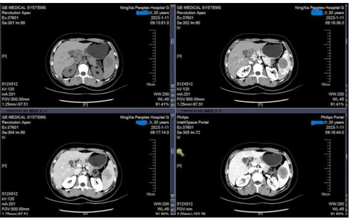
Figure 1: Preoperative CT shows a 3.5 × 3.5 × 3.0 cm hemangioma in the inferior pole of the spleen. Preoperative contrast-enhanced CT image demonstrating a 3.5 cm × 3.5 cm, well-defined lesion at the inferior pole of the spleen. Imaging characteristics were consistent with splenic hemangioma. Please click here to view a larger version of this figure.
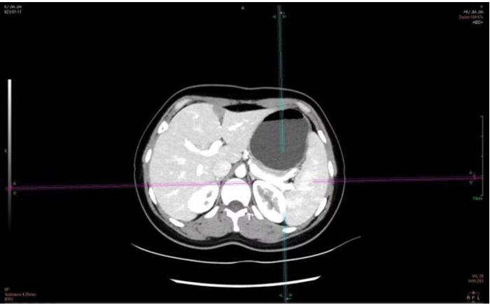
Figure 2: Preoperative CT reconstruction shows a two-branch pattern of the splenic artery. Preoperative contrast-enhanced CT image demonstrating a clear bifurcation of the splenic artery before entering the splenic hilum. This anatomical variation provides a favorable surgical plane for laparoscopic partial splenectomy. Please click here to view a larger version of this figure.
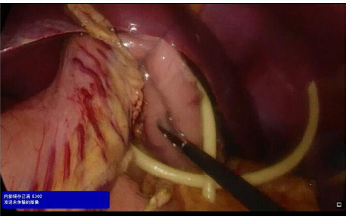
Figure 3: Suspension of the liver and stomach. Intraoperative photograph showing the liver and stomach being retracted to expose the gastrospheric ligament and the splenic hilum. This maneuver provides a clear visualization of the surgical field and facilitates access to the splenic pedicle. Please click here to view a larger version of this figure.
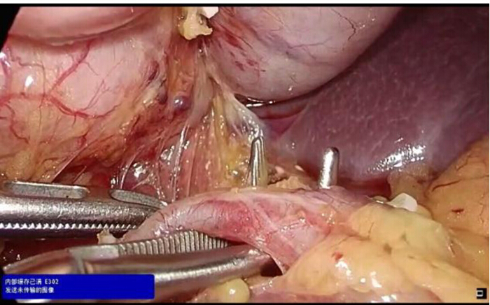
Figure 4: Mobilization of the splenic artery for preemptive clamping. Intraoperative image showing the splenic artery being carefully dissected at the upper border of the pancreatic tail and prepared for temporary clamping if needed. This maneuver provides the option for proximal vascular control during challenging dissection or in case of unexpected bleeding, thereby reducing the risk of conversion to open surgery and enhancing the safety of the procedure. Please click here to view a larger version of this figure.
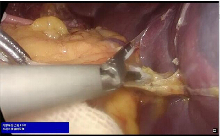
Figure 5: Sequential dissection of branching vessels adherent to the splenic capsule. Intraoperative image demonstrating the meticulous dissection of small vascular branches adhering to the splenic capsule using a laparoscopic energy device. This step-by-step approach ensures safe mobilization of the spleen while preserving capsular integrity and minimizing intraoperative bleeding. Please click here to view a larger version of this figure.
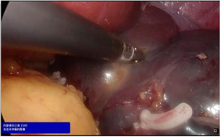
Figure 6: Demarcation of the ischemic line on the visceral surface. Intraoperative image showing a clear color change on the splenic surface following selective vascular clamping, with the ischemic area (dusky purple) sharply demarcated from the perfused area (pink). This ischemic line precisely defines the planned resection plane and guides the parenchymal transection during partial splenectomy. Please click here to view a larger version of this figure.
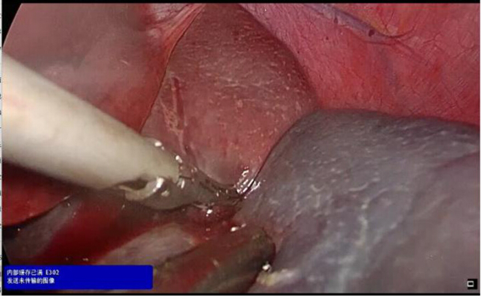
Figure 7: Centrifugal splitting of splenic parenchyma along the ischemic line. Intraoperative image demonstrating the parenchymal transection performed in a centrifugal direction following the previously demarcated ischemic line. The splenic tissue is divided using an energy device, progressing from the hilum toward the periphery. This technique ensures precise resection along the avascular plane while preserving the integrity of the remaining splenic parenchyma. Please click here to view a larger version of this figure.
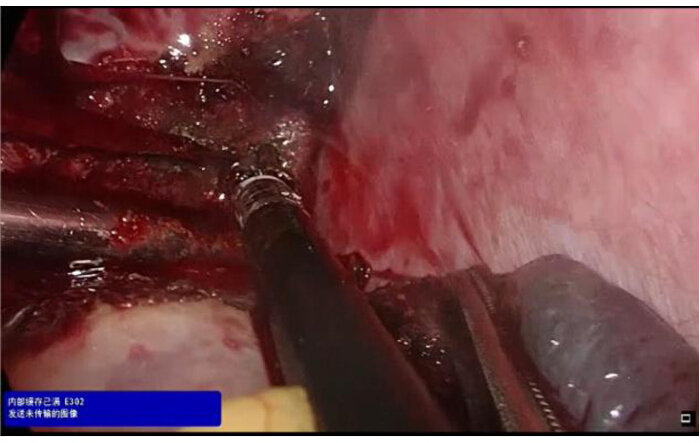
Figure 8: Bipolar hemostasis of the transection surface. Intraoperative image showing the use of bipolar forceps to achieve meticulous hemostasis on the splenic parenchymal surface following transection. The residual spleen is inspected carefully, and any bleeding points along the cut edge are coagulated under direct visualization. Please click here to view a larger version of this figure.
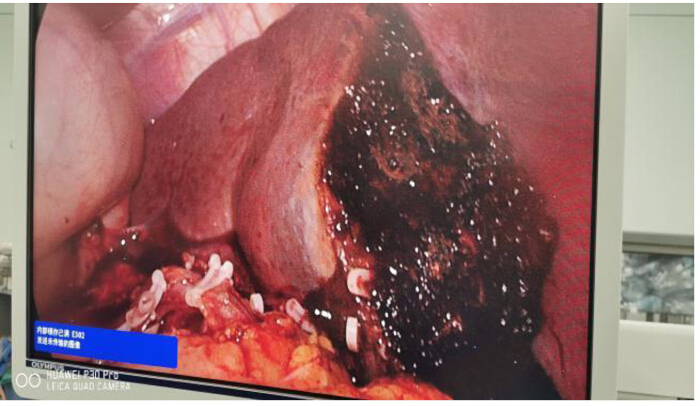
Figure 9: Postoperative appearance of the splenic transection surface. Intraoperative image showing the remaining splenic parenchyma after completion of laparoscopic partial splenectomy. The transection surface demonstrates effective hemostasis with no active bleeding, and the preserved splenic tissue appears well perfused with normal coloration. Please click here to view a larger version of this figure.
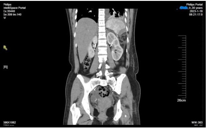
Figure 10: Postoperative CT scan. Postoperative computed tomography image showing the residual spleen following laparoscopic partial splenectomy, with the patient in an upright position. The preserved splenic parenchyma demonstrates good perfusion and adequate volume, with no evidence of ischemia, infarction, or fluid collection in the splenic fossa. Please click here to view a larger version of this figure.
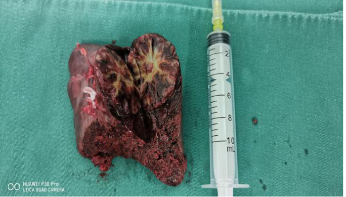
Figure 11: Postoperative pathological specimen. Gross photograph of the resected splenic specimen showing the cut surface following partial splenectomy. The transection margin is clearly visible, and the lesion (splenic hemangioma) is well demarcated from the surrounding normal splenic parenchyma. The resection margin appears intact and negative for tumor involvement. Please click here to view a larger version of this figure.
Discussion
As an indispensable organ, the spleen participates in vital functions such as regulating circulating blood volume, filtering blood, producing various immunoglobulins, and modulating the endocrine system11. Historically, due to limited understanding of splenic function and anatomy, clinicians frequently performed total splenectomy for benign splenic tumors and splenic trauma. Postoperative patients face significantly increased risks of thrombocytosis, pylephlebitis, intra-abdominal abscesses, OPSI, thromboembolism, and pulmonary hypertension12,13,14. With advances in laparoscopic techniques and improved understanding of spleen physiology, an increasing number of scholars recognize the importance of preserving splenic function when treating splenic diseases. Consequently, spleen-preserving strategies have gained prominence, with laparoscopic partial splenectomy emerging as the optimal choice for selected patients with benign conditions confined to the splenic poles15,16. The patient had no significant medical history, and preoperative assessment identified the lesion as a benign tumor located at the inferior pole of the spleen; therefore, the surgical approach selected was laparoscopic partial splenectomy.
The segmental blood supply of the spleen provides the anatomical basis for laparoscopic partial splenectomy. Splenic arterial branching patterns are classified into two main types: magistral (concentrated, 30%) and distributed (dispersed, 70%). Splenic lobar arteries (primary branches of the splenic artery) typically exhibit three branching patterns: Type I (3%–5.7%), Type II (76.9%–98%), and Type III (20%–23.8%). The predominance of Type II branching in splenic arteries has reached domestic and international consensus17,18, and was intraoperatively confirmed in this patient. Previous work established the fundamental framework for splenic vascular anatomy and segmental nomenclature17. The segmental blood supply of the spleen enables surgeons to ligate the splenic lobar arteries supplying the affected region; subsequently, partial splenectomy can be safely performed along the ischemic demarcation line formed on the splenic surface6,17. In most cases, the ischemic demarcation line largely coincides with the plane of splenic notches. Therefore, during partial splenectomy, dissection along the plane connecting the splenic hilum and the corresponding splenic notch minimizes intraoperative blood loss.
Precise preoperative imaging (CT angiography) to identify vascular anatomy, such as the bifurcated splenic artery in this case, is essential for surgical planning. Intraoperatively, the meticulous dissection of the splenic hilum and selective division of target vessels are paramount to establishing a clear ischemic demarcation line. As shown in Figure 6 and Figure 7, transecting the parenchyma approximately 1 cm inside this line using an ultrasonic scalpel with a slow-paced "coagulate-before-transect" technique ensures reliable hemostasis. Furthermore, achieving complete hemostasis on the resection surface with focal bipolar electrocautery (Figure 9) is critical to preventing postoperative bleeding. In certain anatomical situations, technical modifications may be required. If intraoperative bleeding occurs from the parenchyma, temporary clamping of the main splenic artery using the pre-placed vessel loop can provide immediate control. When the ischemic line is unclear after pedicle division, re-evaluation of the hilar anatomy for missed feeding vessels is necessary. For larger or deeper resection planes, hemostatic agents or reinforced suturing may serve as adjuncts to bipolar coagulation.
The crucial aspect of laparoscopic partial splenectomy lies in the precise dissection of splenic vessels and their branches. Particularly for patients with anatomical variations, preoperative angiography should be performed to delineate the vascular anatomy19. Preoperative CT angiography was performed to clarify the branching pattern of the splenic vessels. To minimize adjacent organ injury, the gastro-splenic and spleno-colic ligaments are divided by adhering closely to the splenic surface—a key maneuver for protecting the stomach, colon, and pancreatic tail. For managing the bulk of the splenic artery, temporary occlusion using bulldog clamps or urinary catheters is recommended. This maneuver provides the option for proximal vascular control during challenging dissection or in case of unexpected bleeding, thereby reducing the risk of conversion to open surgery and enhancing the safety of the procedure. Studies confirm the safety of main splenic artery occlusion for up to 1 h during surgery20,21, which enables emergency hemostasis in cases of unexpected bleeding. Furthermore, when managing the nutrient vessels supplying the spleen segment scheduled for resection, dissection close to the splenic parenchyma was performed to minimize potential pancreatic injury.
Beyond splenic hemangioma, laparoscopic partial splenectomy is indicated for benign space-occupying lesions confined to one pole of the spleen, such as hemangiomas, nonparasitic splenic cysts, and abscesses, as well as selected hematologic diseases and partial splenic trauma22,23. For patients with Grade I–III splenic injuries who are hemodynamically stable, laparoscopic partial splenectomy may be considered. Hematologic diseases determined through a Multi-Disciplinary Team (MDT) discussion, such as hereditary spherocytosis, are also appropriate indications. Other conditions, including unexplained splenomegaly, may warrant a partial splenectomy to obtain a pathological diagnosis. Iatrogenic splenic injuries occurring during abdominal surgery may likewise undergo partial splenectomy as appropriate24. Comprehensive preoperative assessment should be performed, with MDT discussion involving the hematology department when necessary.
Although the present report provides a detailed technical description, several limitations should be acknowledged. As a single-case report, the findings may not be generalizable to broader patient populations. No comparative or control data were available, as the study was designed as a technical demonstration rather than a comparative trial. Although short-term follow-up imaging confirmed preserved splenic perfusion, long-term outcomes regarding splenic function, potential complications, and recurrence remain to be established. Furthermore, the procedure was performed by a surgical team with extensive experience in laparoscopic splenic surgery, and the results may not be directly transferable to less experienced settings. Despite these limitations, this report provides a detailed technical framework that may serve as a useful reference for surgeons performing laparoscopic partial splenectomy.
Given the spleen's critical roles in immune, hematologic, and endocrine systems, partial splenectomy holds greater clinical significance than total splenectomy. When strict indications are followed, laparoscopic partial splenectomy is safe and feasible for benign splenic diseases. This approach preserves splenic tissue and function while providing favorable clinical outcomes. The step-by-step framework may serve as an educational tool for surgeons adopting this technique and promote the broader application of spleen-preserving surgery.
Disclosures
All authors have disclosed any conflicts of interest.
Materials
| Name | Company | Catalog Number | Comments |
|---|---|---|---|
| 3D laparoscope | KARL STORZ SE & Co. KG | 26605BA | Visualization system |
| Absorbable ligation clips | Hangzhou Sunstone Technology Co.,Ltd | K12 | Vessel ligation |
| Laparoscopic instruments | Tonglu Youshi Medical Equipment Co., Ltd | Includes graspers, scissors, etc. | |
| Latex T-shape Catheter | Zhenjiang Star Enterprise Co.,Ltd | 16 Fr | Drainage catheter |
| Reusable Laparoscopic Bipolar Electrosurgical Forceps | Hangzhou Kangji Medical Instrument Co., Ltd. | Electrosurgical instrument | |
| Ultrasound knife | Johnson & Johnson | Energy device for tissue dissection |
References
- Vancauwenberghe, T., Snoeckx, A., Vanbeckevoort, D., et al. Imaging of the spleen: what the clinician needs to know. Singap Med J. 56 (3), 133-144 (2015).
- Chatzoulis, G., Kaltsas, A., Daliakopoulos, S., et al. Co-existence of a giant splenic hemangioma and multiple hepatic hemangiomas and the potential association with the use of oral contraceptives: a case report. J Med Case Rep. 2, 147(2008).
- Yano, H., Imasato, M., Monden, T., et al. Hand-assisted laparoscopic splenectomy for splenic vascular tumors: report of two cases. Surg Laparosc Endosc Percutan Tech. 13 (4), 286-289 (2003).
- Han, X. L., Zhao, Y. P., Chen, G., et al. Laparoscopic partial splenectomy for splenic hemangioma: experience of a single center in six cases. Chin Med J (Engl). 128 (5), 694-697 (2015).
- Makrin, V., Avital, S., White, I., et al. Laparoscopic splenectomy for solitary splenic tumors. Surg Endosc. 22 (9), 2009-2012 (2008).
- Zheng, C. H., Xu, M., Huang, C. M., et al. Anatomy and influence of the splenic artery in laparoscopic spleen-preserving splenic lymphadenectomy. World J Gastroenterol. 21 (27), 8389-8397 (2015).
- Chen, J., Yu, S., Xu, L. Laparoscopic partial splenectomy: a safe and feasible treatment for splenic benign lesions. Surg Laparosc Endosc Percutan Tech. 28 (5), 287-290 (2018).
- Wang, X., Wang, M., Zhang, H., et al. Laparoscopic partial splenectomy is safe and effective in patients with focal benign splenic lesion. Surg Endosc. 28 (12), 3273-3278 (2014).
- Cai, H., An, Y., Wu, D., et al. Laparoscopic partial splenectomy: a preferred method for select patients. J Laparoendosc Adv Surg Tech A. 26 (12), 1010-1014 (2016).
- Wang, L., Xu, J., Li, F., et al. Partial splenectomy is superior to total splenectomy for selected patients with hemangiomas or cysts. World J Surg. 41 (5), 1281-1286 (2017).
- Vasilescu, C., Stanciulea, O., Tudor, S., et al. Laparoscopic subtotal splenectomy in hereditary spherocytosis: to preserve the upper or the lower pole of the spleen. Surg Endosc. 20 (5), 748-752 (2006).
- Lee, D. H., Barmparas, G., Fierro, N., et al. Splenectomy is associated with a higher risk for venous thromboembolism: a prospective cohort study. Int J Surg. 24, 27-32 (2015).
- Mouttalib, S., Rice, H. E., Snyder, D., et al. Evaluation of partial and total splenectomy in children with sickle cell disease using an internet-based registry. Pediatr Blood Cancer. 59 (1), 100-104 (2012).
- Buesing, K. L., Tracy, E. T., Kiernan, C., et al. Partial splenectomy for hereditary spherocytosis: a multi-institutional review. J Pediatr Surg. 46 (1), 178-183 (2011).
- Li, Q., Liu, Z., Hu, M., et al. Laparoscopic partial splenectomy of benign tumors assisted by microwave ablation. J Cancer Res Ther. 16 (5), 1002-1006 (2020).
- Utria, A. F., Goffredo, P., Keck, K., et al. Laparoscopic splenectomy: has it become the standard surgical approach in pediatric patients. J Surg Res. 240, 109-114 (2019).
- Redmond, H. P., Redmond, J. M., Rooney, B. P., et al. Surgical anatomy of the human spleen. Br J Surg. 76 (2), 198-201 (1989).
- Covantsev, S., Alieva, F., Mulaeva, K., et al. Morphological evaluation of the splenic artery, its anatomical variations and irrigation territory. Life (Basel). 13 (1), 195(2023).
- Hansen, M., Moller, A. Splenic cysts. Pediatr Ann. 45 (7), e251-e256 (2016).
- Ouyang, G., Li, Y., Cai, Y., et al. Laparoscopic partial splenectomy with temporary occlusion of the trunk of the splenic artery in fifty-one cases: experience at a single center. Surg Endosc. 35 (1), 367-373 (2021).
- Teperman, S. H., Whitehouse, B. S., Sammartano, R. J., et al. Bloodless splenic surgery: the safe warm-ischemic time. J Pediatr Surg. 29 (1), 88-92 (1994).
- Salinas, S. A., Hernandez, M. I., Virseda, R. J., et al. Problem-based learning in urology training. Actas Urol Esp. 29 (1), 8-15 (2005).
- de la Villeon, B., Zarzavadjian, L. B. A., Vuarnesson, H., et al. Laparoscopic partial splenectomy: a technical tip. Surg Endosc. 29 (1), 94-99 (2015).
- Rosman, C., Broens, P., Trzpis, M., et al. A long-term follow-up study of subtotal splenectomy in children with hereditary spherocytosis. Pediatr Blood Cancer. 64 (10), (2017).