This experimental protocol demonstrates a deep cervical lymph duct-cannulated rat model that enables direct and quantitative assessment of lymphatic drainage from the head region. The model facilitates detailed analysis of central nervous system (CNS)-derived solutes, immune cell populations, and the distribution and clearance of CNS-targeted therapeutics.
A subscription to JoVE is required to view this content. Sign in or start your free trial.
Method Article
Cervical Lymph Duct-Cannulated Rat Model for Assessing Lymphatic Transport from the Head and Brain
In This Article
Summary
Abstract
Lymph flows from the central nervous system (CNS) via a series of lymphatic vessels (LVs) and lymph nodes that converge at the cervical lymph ducts. In addition to maintaining fluid balance, these lymphatic ducts play a key role in transporting a wide array of substances, including endogenous metabolites, signaling molecules, immune cells, and small and macromolecular drugs. This is critical for physiological homeostasis and immune function in both intracranial (e.g., brain) and extracranial regions (e.g., nasal and oral cavities). Impaired lymphatic drainage from the brain has increasingly been linked to a range of neurological and neurodegenerative disorders. Cervical lymph duct cannulation enables the collection of lymph draining the head and brain, allowing the measurement of the concentration and transport rate of various substances via the lymphatic system. Changes in these factors in response to different challenges (e.g., drugs, stress, trauma) and diseases (e.g., stroke, infection, Alzheimer's disease) can also be determined. Here, we describe an anesthetized, deep cervical lymph duct cannulated rat model that enables lymph collection for several hours following surgery. The method may be combined with imaging and multi-omics technologies for the measurement of a wide range of parameters of interest in the lymph. This facilitates fundamental physiological and pathophysiological research of the head and neck region, as well as pharmacokinetic/pharmacodynamic studies of drugs, particularly for the treatment of CNS diseases.
Introduction
The lymphatic system, which is distributed throughout the body, is essential for maintaining fluid homeostasis, regulating immune responses, and facilitating the transport of nutrients and signaling molecules1. The peripheral lymphatics begin in the lymphatic capillaries, which collect interstitial fluid and solutes and drain into larger collecting lymphatic vessels (LVs) that possess a surrounding smooth muscle layer; these vessels then deliver lymph to local lymph nodes for immune processing2,3. The lymphatic network draining the head region is important for the homeostasis of both intracranial (e.g., brain) and extracranial (e.g., nasal and oral cavities) organs. The structure and organization of the extracranial lymphatic network are believed to resemble those of peripheral lymphatics throughout the body, whereas the intracranial lymphatic system exhibits unique anatomical and functional features (see Figure 1 for a detailed schematic representation in rat models)4,5. Although typical LVs are absent within the brain parenchyma, lymphatic-like functions are mediated by the specialized glial-dependent lymphatic transport (glymphatic) system that facilitates the exchange between cerebrospinal fluid (CSF) and interstitial fluid (ISF)6. While the glymphatic system enables the mixing of ISF within the brain parenchyma with CSF, the meningeal, perivascular, and nasopharyngeal LVs transport solutes from the CSF away from the cranial cavity. Specifically, the meningeal lymphatic pathway directs CSF from the subarachnoid space into meningeal LVs, linking intracranial fluid dynamics with peripheral lymphatic clearance7. In parallel, perineural pathways enable the efflux of CSF and associated solutes along cranial and spinal nerve sheaths, most prominently via the olfactory nerves into the nasopharyngeal lymphatic plexus5,8. Concurrent work of these systems maintains fluid homeostasis within the brain6,7,8. Recent advances in understanding the CNS-draining lymphatic system have provided new insights into brain physiology and pathophysiology. These findings may offer new avenues for therapeutic intervention in CNS diseases9.
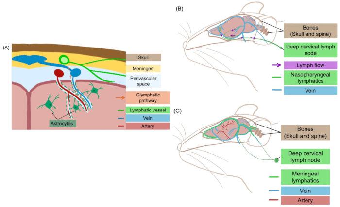
Figure 1: Schematic representation of CNS lymphatic clearance pathways. These pathways consist of (A) the glymphatic route via perivascular spaces; (B) the nasopharyngeal lymphatic route, and (C) the meningeal LVs. Please click here to view a larger version of this figure.
The lymphatic networks draining from the head and CNS converge into the cervical lymph ducts located in the neck, where lymph is filtered through the cervical lymph nodes before entering the systemic circulation. The cervical lymph nodes are classified into superficial (sCLNs) and deep cervical lymph nodes (dCLNs) according to their anatomical locations within the neck. Alterations in the composition of lymph fluid within the cervical lymph ducts are thought to reflect underlying pathophysiological variations in the brain, with Alzheimer's disease (AD), Parkinson's disease (PD), traumatic brain injury (TBI), and post-stroke cognitive impairment (PSCI) being representative examples9,10,11,12,13. Notably, in AD, impaired drainage of amyloid-β (Aβ) and phosphorylated tau proteins (P-tau) through these lymphatic pathways has been implicated as a contributing factor in disease progression9,10,14. By directly connecting lymph ducts to adjacent veins to reroute lymphatic drainage, lymphatic venous anastomosis (LVA) is being investigated in both animals and humans as a therapeutic strategy to enhance lymphatic outflow from the CNS, thereby facilitating Aβ clearance from the brain and potentially delaying the progression of AD6,15,16. The importance of this pathway is further illustrated by observations in mice subjected to surgical removal of the cervical lymph nodes (lymphadenectomy), resulting in reduced cervical lymphatic clearance. Impaired clearance leads to toxic accumulation of Aβ and P-tau, with subsequent activation of pathological kinase signaling pathways. These changes are associated with a progressive worsening of AD-like tauopathy and an increased risk of anxiety- and depression-like behaviours14,16. Furthermore, CSF-based studies suggest that CNS tumors, such as brain lymphomas, can physically obstruct lymphatic clearance through the deep cervical lymph ducts while concurrently elevating oncoprotein concentrations in the CSF9,17,18. Evidence from CNS inflammation-based studies further indicates that cells originating in the meninges can migrate via the meningeal LVs to the cervical lymph nodes8,19. These cells consist primarily of lymphocytes, with smaller populations of dendritic cells, macrophages, and endothelial cells19,20. During inflammation or following CNS injury or surgery, lymphatic immune cell trafficking becomes evidently enhanced, accompanied by elevated levels of immune biomarkers such as cytokines, chemokines, and other inflammatory mediators21.
In addition to its patho-physiological roles, the cervical lymphatic network is increasingly acknowledged as an important conduit for drug disposition. Previous animal studies have demonstrated that several drug formulations designed for CNS administration can undergo clearance from the brain via the cervical lymph ducts and lymph nodes (to varying extents), exemplified by intracerebrally administered nanoparticle-based agents (e.g., lamotrigine, talazoparib) and biologics (e.g., monoclonal antibodies, radiolabeled tracer proteins)22,23,24,25. In humans, MRI imaging studies have demonstrated that certain molecules, such as gadobutrol, undergo active transport from the glymphatic system to the cervical lymph ducts26.
As aforementioned, cervical lymph nodes, both sCLNs and dCLNs, drain lymph from distinct regions of the head; however, the specific cranial regions drained by each node remain unclear27,28. Overlapping drainage areas, inter-individual anatomical variability, and potential interspecies structural differences further complicate the description of cervical lymphatic drainage patterns in both animals and humans29. Within the complex cervical lymphatic network, the deep cervical lymph ducts have been identified as major outflow routes for CSF, located downstream of the nasopharyngeal and meningeal LVs8. Compared with the superficial cervical lymph ducts, which form fine and variable networks in the subcutaneous tissue and differ greatly in size and branching between individual rats, the deep cervical lymph duct offers a more accessible and practical route for collecting CNS-derived lymph in small animal models20,30. The current protocol is therefore focused on establishing a reliable method for collecting lymph from the deep cervical lymph duct.
Only a limited number of studies conducted in the past decade have described the collection of cervical lymph fluids to study CNS diseases or analyze drug distribution. A previous study described measurement of drug concentrations in cervical lymph by flushing or squeezing fluid from isolated tissue sections (such as those adjacent to a single dCLN)31. However, experimental protocols for the continuous collection of lymph from the cervical lymph ducts in small laboratory animals, particularly rats, remain underdeveloped. This protocol establishes a readily accessible and reproducible method for cannulating the (rat) cervical lymph duct to enable continuous lymph collection and to investigate CNS lymphatic transport, immune responses, and pharmacokinetics. Its key advantage lies in the ability to quantify lymph flow and composition over defined time intervals, thereby enabling the quantitative measurement of drugs, lipids, fatty acids, and cellular constituents. Particularly, this model captures lymphatic transport processes that are not adequately represented by plasma/CSF sampling or imaging techniques, offering novel insights into CNS drug disposition, lipid and nutrient transport, and immune signalling20.
Access restricted. Please log in or start a trial to view this content.
Protocol
The procedure(s) described in this protocol were approved by the university animal ethics committee and were performed in accordance with the experimental ethical guidelines of China Pharmaceutical University. Prior to commencing any animal-related procedures, obtain necessary approvals from the relevant institutional or organizational ethics committee. Male Sprague-Dawley (SD) rats weighing 220 to 250 g are used as the experimental animals. Animals are sourced from authorized experimental animal suppliers (refer to Table of Materials) and housed in dedicated cages with free access to food and water until the commencement of the experiment. In this protocol, cannulation of the left deep cervical lymph duct is described as a representative example. Currently, there is limited evidence describing anatomical differences between the left and right deep cervical lymph ducts. Based on observations from previously completed experiments, no significant differences have been noted between the two sides, including in lymph flow rates.
1. Safety precautions and procedures
- Perform all procedures in accordance with institutional biosafety guidelines, and all personnel should receive adequate training in microsurgical techniques, experimental animal handling, and chemical safety prior to conducting this protocol.
- Wear appropriate personal protective equipment (PPE), including a laboratory coat, surgical gloves, and protective goggles, throughout the procedure. As a volatile anesthetic (isoflurane) is used and may cause central nervous system depression and cardiovascular toxicity in humans following prolonged exposure, perform the procedure in a well-ventilated environment. Food and drink must not be brought into the laboratory, and ensure that flowing water is readily accessible at all times.
- Handle all sharp items, including syringe needles, razor blades, and micro-scissors, with care and dispose of them immediately after use in designated sharps containers; under no circumstances should disposable sharp items be reused.
- Treat all biological samples (e.g., collected lymph) and animal body fluids (e.g., blood) as potentially hazardous, and handle all collection equipment carefully while keeping samples away from the surgical area. In the event of an accidental needle-stick injury or skin contact with hazardous substances, immediately wash the affected area with soap and water and report the incident in accordance with institutional safety protocols.
2. Preparation on the day before the surgery
NOTE: This is a terminal procedure in rats; therefore, strict aseptic technique and postoperative analgesia are not required. However, the experimental area should be maintained in a clean and hygienic condition throughout the procedure.
- Fast or feed the rats according to the requirements of the specific experiment. This protocol does not mandate a specific fasting or fed state.
- Prepare an anticoagulant solution by diluting heparin or ethylenediaminetetraacetic acid (EDTA) to a final concentration of 10 IU/mL. Pre-load this mixture (approximately 0.7 mL) into a 1 mL sterile insulin syringe fitted with a 30 G needle, to pre-coat the inner surface of the polyethylene (PE) cannula (used for lymph duct cannulation). Store this prepared mixture under sterile conditions until use.
- Prepare cannulas for lymph sample collection by assembling a PE cannula in a head-to-tail configuration, using 0.5 mm outer diameter (OD), 0.3 mm inner diameter (ID) tubing for both terminal sections and 0.6 mm OD, 0.5 mm ID tubing for the middle section.
- Cut the cannula segments to lengths of 2 cm for the proximal portion connecting to the syringe needle, 40 cm for the middle section, and 8 cm for the distal portion intended for cannulation.
- Cut and bevel the distal end of the cannula assembly at an angle of approximately 45° in order to facilitate smooth insertion into the cervical lymph duct. Refer to Figure 2 for a detailed illustration of the cannula assembly.
NOTE: This bevel facilitates smooth insertion into the lymph duct and reduces the risk of blockage by increasing the effective cross-sectional area. Following successful cannulation, the proximal (2 cm) section is disconnected, and lymph is collected directly from the exposed proximal end of the middle section (as described in detail in Step 4.9 & 4.10).

Figure 2: Schematic diagram of the cannula assembly (not to scale). The diagram illustrates the dimensions of each component of the assembled PE tubing used to construct the cannula. From left to right, the assembled cannula will hereafter be referred to as the proximal end, the middle section, and the distal end, respectively. Please click here to view a larger version of this figure.
- Prepare the anesthesia inhalation system by pre-loading liquid isoflurane into the pressurized container equipped with a vaporizer. Prior to use, perform a test run (priming) of the vaporizer to confirm airtightness and to ensure stable and consistent gas flow.
- If required, prepare the test drug candidate in a suitable vehicle. Conduct stability studies to confirm formulation integrity during storage and throughout the administration period. Include additional agents (e.g., biological dyes such as Evans Blue) where applicable.
- Prepare the containers for lymph collection by pre-weighing sterile microcentrifuge tubes and recording their initial weights. Label each tube sequentially according to the planned collection time points (e.g., hourly intervals). Add concentrated heparin (or EDTA) to each tube and remove the solvent by nitrogen blowdown evaporation, leaving 2 to 5 IU of dried heparin (or EDTA) per tube to prevent uneven dilution of collected lymph.
3. Preparations immediately prior to surgery
- Verify the patency of each cannula by flushing with the prepared anticoagulant solution. Connect a syringe to the proximal end of the assembled cannula and gently release a small volume of solution. Ensure that the solution exits smoothly from the distal end without evidence of leakage. If leakage is detected, as typically indicated by fluid escaping at the connection point(s), discard the entire cannula assembly and reconstruct a new one according to Step 2.3.
NOTE: This step simultaneously pre-coats the inner surface of the cannula with anticoagulant (this step may also be conducted the day before surgery, so the cannula is precoated overnight). - Start administering inhaled isoflurane in a transparent anaesthesia induction chamber at a flow rate of 0.6 L/min under 101.3 kPa at 20 °C. Allow approximately 5 minutes for the rat to become fully anaesthetised before commencing the subsequent steps. Confirm an adequate depth of anaesthesia with a toe pinch test before proceeding.
- Remove the rat from the induction chamber and continue the anaesthesia via a nose-cone mask. Maintain anaesthesia throughout the procedure at a lower rate of 0.4 L/min.
- Continuously monitor the depth of anaesthesia throughout the procedure. If signs of distress or physiological instability are observed, immediately euthanise the rat.
- Position the rat supine on a heated surgical pad maintained at 37 °C. Tilt the head and extend the neck, using a small support (pillow) if necessary, to maximize exposure of the cervical region. Secure the head within the anesthesia mask to ensure continuous delivery of isoflurane and gently restrain the forelimbs using rubber bands or surgical tapes to minimize movement during the procedure.
- Rinse the fur over the cervical region with sterile normal saline to facilitate shaving. Thoroughly shave the entire surgical site (using a double-edged safety razor blade), typically the neck and clavicle region, to ensure complete removal of fur prior to incision (refer to Figure 3A for an example of the shaved neck area).
- Clean the entire shaved area with disinfectants, such as an iodine-containing agent.
- Mark the surgical site by drawing a 2.5 cm dotted line to confirm the length of the initial incision. Position the line slightly to the right of the midline of the rat's body. Place the upper end of the incision approximately 1 to 1.5 cm below the lower lip (refer to Figure 3B for an example of the incision site, with measurements aided by a ruler).
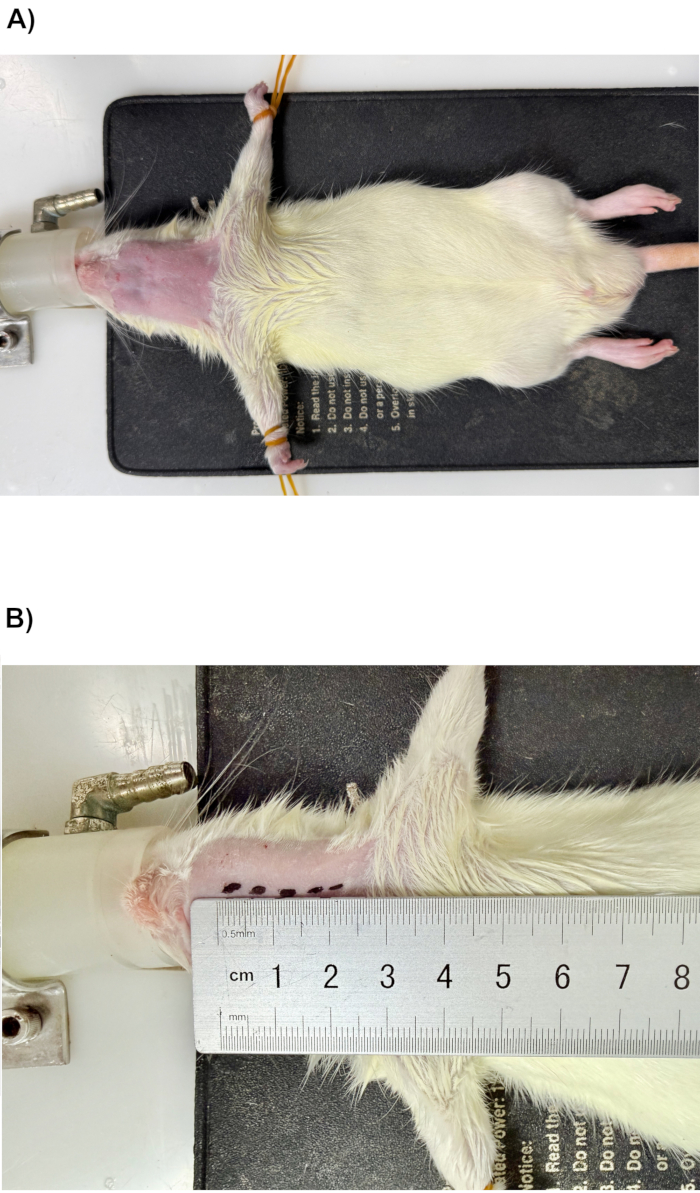
Figure 3: Preparation of the surgical site. (A) The neck region is shaved in preparation for incision; (B) approximate length and location of the incision, indicated by the 2.5 cm dotted line. Please click here to view a larger version of this figure.
4. Cannulation of the cervical lymph duct
- Position the rat with its head pointed towards the operator. Use a microscope for this step due to the relatively small diameter of the cervical lymph duct.
- Using a sterile scalpel, carefully incise the superficial layer of the cervical musculature along the aforementioned 2.5 cm-long mark, oriented nearly parallel to the midline (refer to Figure 4A for the opened superficial skin layer).
- Gently separate the remaining muscle layers, primarily the superficial cervical fascia, to expose the cervical artery, which is typically distinguishable by its red coloration.
- Retract the sternomastoid muscle and submandibular gland and carefully tilt them away from the surgical field to facilitate identification of the cervical lymph duct. Employ a tissue expander to maintain the surgical window and use a cotton swab to temporarily hold aside the muscle tissue while locating the lymph node and associated lymph duct. Ensure the tissue expander is in place throughout the entire cannulation process (refer to Figure 4B for the opened surgical window with the tissue expander in place).
- Locate the cervical lymph duct (preferably under a microscope): The duct appears as an opaque, colorless vessel with approximately 0.2 to 0.5 mm in diameter and runs immediately parallel to the carotid artery. One or more adjacent, flesh-colored dCLNs can often be observed along the course of the cervical lymph duct (refer to Figure 4C for a microscopic view of the exposed deep cervical lymph duct and an adjacent dCLN).
NOTE: The cervical lymph ducts typically have a color very similar to the surrounding tissues, and their identification often requires careful inspection, particularly in rats with more fat tissue. For surgical practice purposes (but not for studies on lymphatic transport of drugs or endogenous substances), Evans Blue may be subcutaneously injected under the neck at the beginning of the surgery so that the cervical lymph duct can be better visualized when the dye drains via the lymphatics. - Using a pair of blunt forceps, gently pass through the separated connective tissues and maintain the forceps in position to ensure the cervical lymph duct remains clearly visible within the surgical field.
- Carefully isolate the cervical lymph duct from the surrounding connective and adipose tissues by blunt dissection, taking particular care to avoid damage, as the vessel is extremely fragile.
- Use a pair of microscissors to create a small incision in the upper half of the exposed duct. Do not detach the duct completely; the two halves must remain intact.
- Check and make sure the prepared cannula pre-coated with anticoagulant solution (as described in Step 2.3) is free of air bubbles by performing a gentle flush. Immediately prior to insertion, carefully grasp the distal end of the cannula with forceps, orienting it so that the beveled face is directed upward. Then, gently advance the cannula approximately 2 mm into the duct through the incision, directing it toward the head (refer to Figure 4D for an illustration of successful cannulation). The actual depth of insertion may vary depending on the individual rat.
NOTE: Since the cannula insertion is relatively shallow in this procedure, an additional fixation point to the surrounding area can be helpful. This is achieved by creating a small secondary skin puncture (hole) approximately 1.5 cm from the surgical window using scissors. This opening will be further stabilized in subsequent steps (e.g., with suture or adhesive). Figure 4E demonstrates this step in the context of using tissue adhesive. The cannula should be passed through the hole to assist in securing its position. - Detach the insulin syringe along with the short (2 cm) proximal segment of the cannula from the rest of the cannula assembly. Lymph can then be collected directly from the exposed proximal end of the larger-diameter middle segment. Monitor the distal tip of this collecting segment for 1–2 min. Confirm successful cannulation by a visible, slow, and steady outflow of lymph fluid from the cannulated duct.
- Once cannulation is verified to be successful, secure the cannula by applying a small drop of cyanoacrylate tissue adhesive with a pipette at both the lymph duct incision site and the secondary fixation hole (refer to Figure 4E for a demonstration of cannula fixation). Take extra care to avoid occluding the vessel or impairing lymph flow.
- Observe the lymph flow for several minutes to confirm a steady outflow (a visible droplet typically forms at the end of the cannula within a minute). Once verified, carefully remove the tissue expander from the surgical window and reposition the submandibular gland to its original location.
- Attach a pre-labelled anticoagulant-coated microcentrifuge tube (see Step 2.6) to the distal end of the cannula to collect the free-flowing lymph.
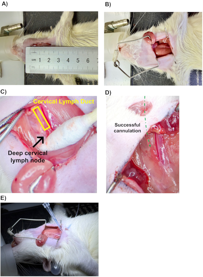
Figure 4: Cannulation of cervical lymph duct. (A) Incision site in the neck region; (B) surgical window opened with a tissue expander in place and the submandibular gland retracted to the right; (C) cervical lymph duct and one dCLN visualised under a microscope. (D) Successful cannulation, indicated by the green dotted line (microscopic view). (E) Tissue adhesive was applied to the incision site, and a secondary skin puncture was made using a pipette. Please click here to view a larger version of this figure.
5. Post-surgical period and drug administration
- Upon completion of cervical lymph duct cannulation, reposition all muscle layers to their original locations. Subsequently, close the skin incision with sutures and/or tissue adhesive; typically, a 2.5 cm long incision can be properly closed with 8 stitches (refer to Figure 5A for a surgical window closed with suture). Remove the constraining rubber band(s) or surgical tapes on the forelimbs upon completion of this step.
- Administer drugs into the CSF using an appropriate route, such as intra-cisterna magna (ICM), intra-cerebroventricular (ICV), or lumbar intrathecal (LIT) injection. Using ICM administration as an example, identify the injection site at the cisterna magna, located at the base of the skull and visible as the only soft depression on the dorsal surface. Shave the surrounding fur as required to facilitate visualization (refer to Figure 5B for a marked suitable area for ICM injection).
NOTE: To maintain intracranial pressure (ICP) during ICM injection, an equivalent volume of CSF is often withdrawn prior to drug administration. It is recommended to use a new, pre-filled syringe for injection. Bending the syringe needle can facilitate easier administration (refer to Figure 5C for a demonstration of ICM injection using a bent syringe needle). - Keep the rat supine on the heated surgical pad (maintained at 37 °C) to preserve body temperature after injection, and throughout the sample collection period.
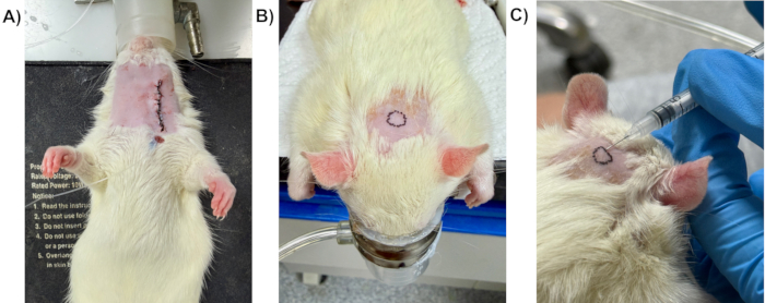
Figure 5: Post-surgery management. (A) Closed incision site using silk suture. (B) Approximate location of cisterna magna with surrounding fur shaved. (C) ICM administration of a drug candidate using an insulin syringe with a bent needle. Please click here to view a larger version of this figure.
6. Collection of lymph samples and analysis
- Collect lymph continuously into anticoagulant-coated microcentrifuge tubes, replacing the tubes at pre-determined intervals (typically hourly). Store the collected samples with icepacks using a cooler box. Refer to Figure 6 for a collected cervical lymph sample in a microcentrifuge tube.
- Upon completion of the lymph collection process, euthanize the rat humanely according to appropriate protocols.
- Determine lymph flow rate by measuring the volume of lymph collected during each planned interval (hourly).
- Measure the concentrations of drugs (or substances of interest) in the lymph using appropriate analytical methods, including HPLC, HPLC-MS, or commercially available assay kits, to characterize their clearance kinetics from the CNS via the lymphatic system.
- Calculate lymphatic drug mass transport by multiplying the measured drug concentrations by the corresponding lymph volumes.
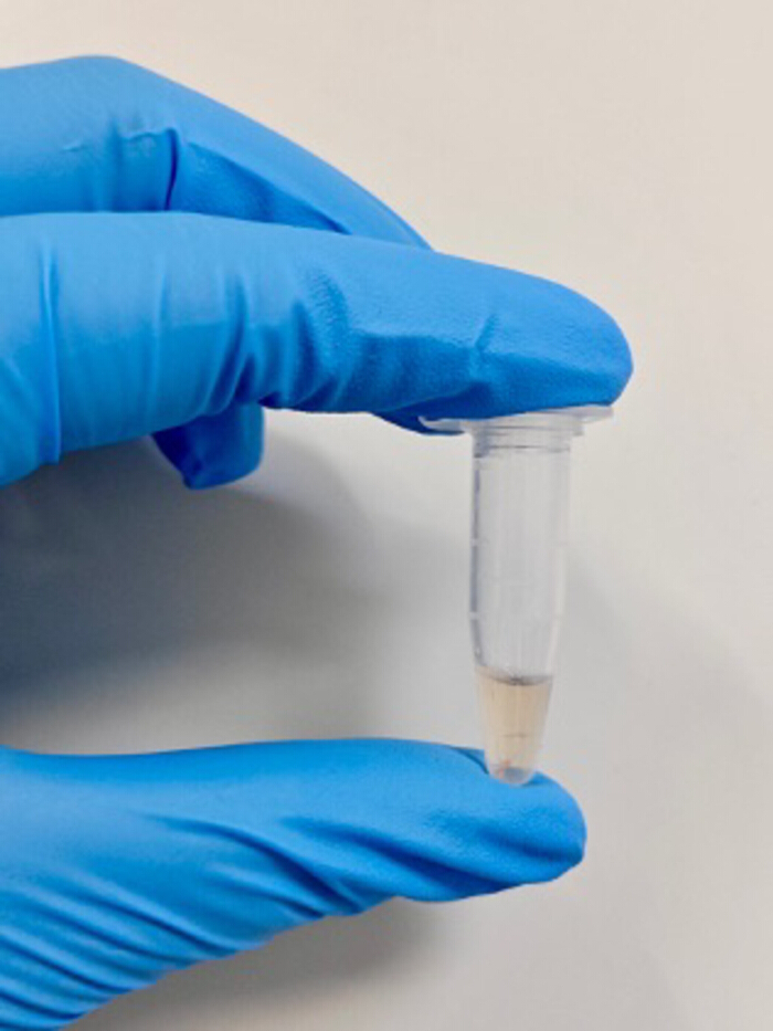
Figure 6: Lymph sample collection. Photograph of one collected sample (approx. 100 µL) in a microcentrifuge tube. Please click here to view a larger version of this figure.
7. Post-procedure handling and waste disposal
- Temporarily store euthanized rat(s) in designated medical waste containers within a dedicated cold storage area (e.g., a refrigerator) until they are collected and disposed of by a professional medical waste processing team.
- Collect all disposed sharp items in a designated sharps container, separate from all other waste.
- Dispose of all other non-sharp items used in the experiment (e.g., cotton swabs, rubber bands, PE cannulas) in medical waste bags, and do not dispose of them with household or general waste. Ensure that no sharp items are placed in this bag.
- Clean and disinfect all remaining reusable tools in accordance with laboratory protocols.
Access restricted. Please log in or start a trial to view this content.
Results
A representative dataset of hourly lymph flow rates (mL/h) was obtained using the cervical lymph cannulation model (refer to Figure 7 for the corresponding chart). In this experiment involving three (n = 3) study rats, lymph was collected continuously into microcentrifuge tubes, which were replaced hourly over a 6 h period.
Access restricted. Please log in or start a trial to view this content.
Discussion
This protocol establishes a reproducible method for cannulating the cervical lymph duct in rats, enabling continuous lymph collection over extended periods. With adequate microsurgical training, the procedure can be performed efficiently and yields consistent lymph samples under stable physiological conditions, maintained by continuous anesthesia, making it suitable for routine experimental use. Once mastered, the surgical procedure typically requires approximately 40 to 50 min to complete, enabling the processing of mul...
Access restricted. Please log in or start a trial to view this content.
Disclosures
The authors declare that they have no known competing financial interests or personal relationships that could have appeared to influence the work reported in this article.
Acknowledgements
The authors sincerely thank Mr. Zhenglin Hao and Ms. Jie Zhao at the Animal Experimental Centre of China Pharmaceutical University for their support in conducting animal experiments.
Access restricted. Please log in or start a trial to view this content.
Materials
| Name | Company | Catalog Number | Comments |
|---|---|---|---|
| 30 gauge needle | Yushou Health | Used for connecting the proximal end of cannula (0503) with the heparin syringe | |
| Anaesthesia dispenser with vaporiser | YYUN Instrument Company | ||
| Blunt forceps | RWD Life Science | F22002-10 | |
| Cotton swabs | Qingdao Shultz Biotechnology | MQ-02 | Used to hold muscle tissue in place temporarily |
| Heated surgical pad | Shanghai Yiheng Instruments | Dimension 15 × 20 cm | |
| Heparin sodium | Shanghai Aladdin Biochemical Technology | H123383 | Dilution required before use |
| Insulin syringe (1 mL) | Yushou Health | U100-1ML | Used for heparin pre-coating only |
| Isoflurane solution for inhalation | RWD Life Science | R510-22-10 | Used for anesthesia through vaporiser |
| Marker pens | Any brand | N/A | |
| Micro-centrifuge tube (1.5 mL) | Nantong Hairui Experimental Equipment | HR10003 | Used for collection of lymph |
| Micro-scissors | RWD Life Science | GD234BA0007925 | |
| Nylon surgical suture | Shanghai Yiheng Instruments | 4-0 35CM | |
| Pipette (volume adjustable, measuring range 5 to 50 μL) | Dragon Laboratory Instruments Limited | 7030301006 | Used for measuring tissue adhesive only |
| Pipette tips | Biosharp Life Sciences | BS-10-T | Used for transferring tissue adhesive only, no specific required volume |
| Polyeyhylene (PE) cannula 0.5 mm OD, 0.3 mm ID (0503) | AniLab Scientific Instruments(Ningbo) | PE-0503 | |
| Polyeyhylene (PE) cannula 0.6 mm OD, 0.5 mm ID (0605) | AniLab Scientific Instruments(Ningbo) | PE-0605 | |
| Razor blades | Gillette Shanghai | Q31/0115000301C004 | Used for fur shaving only |
| Rubber band | Any brand | N/A | Used for restraining forelimbs of rats |
| Ruler | Any brand | N/A | |
| Scapel blades (#23) | RWD Life Science | S31010-01 | |
| Sodium chloride (0.9%) solution for infusion | Shijiazhuang No. 4 Pharmaceutical | H20066533 (approval number) | Used for fur rinsing only |
| Sprague-Dawley Rats | Beijing Vital River Laboratory Animal Technology | 101 | Weight 220 to 250 grams, no age requirement |
| Surgical light | Beijing Heo Bio-Tech | XHA011-1 | |
| Surgical microscope | Shanghai Cewei Optoelectronic Technology | SMZ755T | Dissecting microscope without a mechanical stage, can be equipped with a retractable stand |
| Tissue adhesive | 3M Vetbond | 1469SB | Main ingredient cyanoacrylate |
| Tissue expander (Colibri) | RWD Life Science | R22029-04 | Expansion range 20mm/4cm |
References
- Hu, D., et al. Lymphatic system identification, pathophysiology and therapy in the cardiovascular diseases. J Mol Cell Cardiol. 133 (1), 99-111 (2019).
- Breslin, J. W., et al. Lymphatic vessel network structure and physiology. Compr Physiol. 9 (1), 207-299 (2018).
- Moore, J. E. Jr, Bertram, C. D. Lymphatic system flows. Annu Rev Fluid Mech. 50, 459-482 (2018).
- Dupont, G., et al. Our current understanding of the lymphatics of the brain and spinal cord. Clin Anat. 32 (1), 117-121 (2019).
- Aspelund, A., et al. A dural lymphatic vascular system that drains brain interstitial fluid and macromolecules. J Exp Med. 212 (7), 991-999 (2015).
- Xu, J., et al. The lymphatic system: a therapeutic target for central nervous system disorders. Neural Regen Res. 18 (6), 1249-1256 (2023).
- Raper, D., Louveau, A., Kipnis, J. How do meningeal lymphatic vessels drain the CNS. Trends Neurosci. 39 (9), 581-586 (2016).
- Louveau, A., et al. Understanding the functions and relationships of the glymphatic system and meningeal lymphatics. J Clin Invest. 127 (9), 3210-3219 (2017).
- Salvador, A. F. M., Abduljawad, N., Kipnis, J. Meningeal lymphatics in central nervous system diseases. Annu Rev Neurosci. 47 (1), 323-344 (2024).
- Guo, X., et al. Emerging roles of meningeal lymphatic vessels in Alzheimer's disease. J Alzheimers Dis. 94 (S1), S355-S366 (2023).
- Mestre, H., et al. Cerebrospinal fluid influx drives acute ischemic tissue swelling. Science. 367 (6483), eaax7171(2020).
- Da Mesquita, S., et al. Functional aspects of meningeal lymphatics in ageing and Alzheimer's disease. Nature. 560 (7717), 185-191 (2018).
- Zou, W., et al. Blocking meningeal lymphatic drainage aggravates Parkinson's disease-like pathology in mice overexpressing mutated α-synuclein. Transl Neurodegener. 8 (1), 7(2019).
- Wu, C., et al. Tauopathy after long-term cervical lymphadenectomy. Alzheimers Dement. 21 (4), e70136(2025).
- Fang, Y., et al. A new perspective on cerebrospinal fluid dynamics after subarachnoid hemorrhage: from normal physiology to pathophysiological changes. J Cereb Blood Flow Metab. 42 (4), 543-558 (2022).
- Yen, Y. H., Chen, M. W., Lim, J. X., Chew, K. Y. Exploring lymphovenous anastomosis for Alzheimer's disease: addressing brain lymphatic dysfunction, feasibility, and outcome metrics. Plast Reconstr Surg. , (2025).
- Rubenstein, J., Ferreri, A. J., Pittaluga, S. Primary lymphoma of the central nervous system: epidemiology, pathology and current approaches to diagnosis, prognosis and treatment. Leuk Lymphoma. 49 (1), 43-51 (2008).
- Camilleri-Broët, S., et al. A uniform activated B-cell-like immunophenotype might explain the poor prognosis of primary central nervous system lymphomas: analysis of 83 cases. Blood. 107 (1), 190-196 (2006).
- Papadopoulos, Z., Herz, J., Kipnis, J. Meningeal lymphatics: from anatomy to central nervous system immune surveillance. J Immunol. 204 (2), 286-293 (2020).
- Hoang, T. A., et al. Development and application of a novel cervical lymph collection method to assess lymphatic transport in rats. Front Pharmacol. 14 (1), 1111617(2023).
- Licastro, E., et al. Glymphatic and lymphatic communication with systemic responses during physiological and pathological conditions in the central nervous system. Commun Biol. 7 (1), 229(2024).
- Sarker, A., et al. Intrathecal [(64)Cu]Cu-albumin PET reveals age-related decline of lymphatic drainage of cerebrospinal fluid. Sci Rep. 13 (1), (2023).
- Khang, M., et al. Intrathecal delivery of nanoparticle PARP inhibitor to the cerebrospinal fluid for the treatment of metastatic medulloblastoma. Sci Transl Med. 15 (720), eadi1617(2023).
- Pizzo, M. E., et al. Intrathecal antibody distribution in the rat brain: surface diffusion, perivascular transport and osmotic enhancement of delivery. J Physiol. 596 (3), 445-475 (2018).
- Yan, J., et al. Lymphatic clearance is the main drainage route of lamotrigine-loaded micelles following delivery to the brain. J Pharm Pharmacol. 71 (10), 1488-1496 (2019).
- Ringstad, G., Eide, P. K. Glymphatic-lymphatic coupling: assessment of the evidence from magnetic resonance imaging of humans. Cell Mol Life Sci. 81 (1), 131(2024).
- Pan, W. R., Suami, H., Taylor, G. I. Lymphatic drainage of the superficial tissues of the head and neck: anatomical study and clinical implications. Plast Reconstr Surg. 121 (5), 1614-1624 (2008).
- Bou-Assaly, W. The forgotten lymph nodes: review of the superficial head and neck lymphatic system. Radiol Imaging. 1 (1), 9-13 (2016).
- Yağmurlu, K., et al. Anatomical features of the deep cervical lymphatic system and intrajugular lymphatic vessels in humans. Brain Sci. 10 (12), 953(2020).
- Suami, H., Scaglioni, M. F. Lymphatic territories (lymphosomes) in the rat: an anatomical study for future lymphatic research. Plast Reconstr Surg. 140 (5), 945-951 (2017).
- Zawieja, D. C., et al. Lymphatic cannulation for lymph sampling and molecular delivery. J Immunol. 203 (8), 2339-2350 (2019).
- Hoang, T. A., et al. Quantifying the lymphatic transport of model therapeutics from the brain in rats. Mol Pharm. 21 (5), 2473-2483 (2024).
- Hoang, T. A., et al. Acute neuroinflammation alters the transport of a model therapeutic protein from the brain into lymph and blood. Mol Pharm. 21 (10), 5138-5149 (2024).
- Maloveska, M., et al. Dynamics of Evans blue clearance from cerebrospinal fluid into meningeal lymphatic vessels and deep cervical lymph nodes. Neurol Res. 40 (5), 372-380 (2018).
- Trevaskis, N. L., et al. The mesenteric lymph duct cannulated rat model: application to the assessment of intestinal lymphatic drug transport. J Vis Exp. (97), e52389(2015).
- Gakuba, C., et al. General anesthesia inhibits the activity of the glymphatic system. Theranostics. 8 (3), 710-722 (2018).
- Miao, A., et al. Brain clearance is reduced during sleep and anesthesia. Nat Neurosci. 27 (6), 1046-1050 (2024).
- Bachmann, S. B., et al. Differential effects of anaesthesia on the contractility of lymphatic vessels in vivo. J Physiol. 597 (11), 2841-2852 (2019).
- Ruscic, K. J., Padera, T. P. Silence of the lymphs: some anaesthetic regimens inhibit lymphatic pumping. J Physiol. 597 (11), 2827-2828 (2019).
- Dahan, A., et al. The effect of general anesthesia on the intestinal lymphatic transport of lipophilic drugs: comparison between anesthetized and freely moving conscious rat models. Eur J Pharm Sci. 32 (4), 367-374 (2007).
- de Jong, W. H., et al. Long-term cannulation of the vena cava of rats for blood sampling: local and systemic effects observed by histopathology after six weeks of cannulation. Lab Anim. 35 (3), 243-248 (2001).
Access restricted. Please log in or start a trial to view this content.
Reprints and Permissions
