Integrating analytical approaches from established pressure-controlled perfusion methods optimized a simple, low-cost syringe-pump system for assessing outflow facility and ocular compliance in mouse eyes.
A subscription to JoVE is required to view this content. Sign in or start your free trial.
Method Article
Measuring Outflow Facility And Ocular Compliance In Ex Vivo Mouse Eyes Using A Syringe-pump System
* These authors contributed equally
In This Article
Summary
Abstract
Maintaining intraocular pressure (IOP) at a suitable and stable level is essential for ocular health. The biomechanical properties of the trabecular meshwork (TM), Schlemm’s canal (SC), and the entire corneoscleral shell play a crucial role in IOP homeostasis. Outflow facility (C) is a key parameter for evaluating the effectiveness of TM and SC in draining aqueous humor, while ocular compliance (ϕ) reflects the elasticity of the corneoscleral shell and transient outflow through TM and SC. Previously, a simple, cost-effective syringe-pump system was developed to assess C. However, using simple linear regression for in vivo data analysis has limitations in characterizing TM and SC function. In this study, the syringe-pump system was optimized by applying analytical approaches developed for established pressure-controlled ocular perfusion systems. The measured C and ϕ values in ex vivo eyes were compared with previously published perfusion-system values and were consistent with those reported ranges. In summary, this straightforward, low-cost syringe-pump system facilitates the evaluation of the biomechanical properties of TM and SC.
Introduction
Maintaining a suitable and stable level of intraocular pressure (IOP) is essential for ocular health1. Sustained elevation in IOP is the primary risk factor for glaucoma2,3. Long-term IOP fluctuations over months or years also contribute to vision loss4,5. Recent studies identified another form of IOP fluctuation, transient fluctuations caused by daily activities, which may also accelerate glaucoma progression6,7. For example, a 46-year-old man with a 20-year history of eyelid rubbing and a 52-year-old man who had rubbed his eyes for 10 years both exhibited accelerated optic disc damage8,9. These findings highlight the importance of timely, appropriate responses to the changes in IOP to minimize the risk of visual loss.
IOP is regulated by the balance between aqueous humor (AH) production and its drainage10. AH exits the eye primarily through the conventional and uveoscleral pathways11. In both normal and glaucomatous eyes, the conventional pathway comprising the trabecular meshwork (TM) and Schlemm’s canal (SC) is the principal source of outflow resistance that influences IOP homeostasis12,13,14. Therefore, the biomechanical properties of TM and SC are essential for the eye to adapt to changes in IOP15,16,17.
Recent technological advances have facilitated more accurate assessment of AH outflow. Invasive approaches, such as gravity-based perfusion and the syringe-pump system, have been developed to calculate outflow facility (C), which indicates how TM and SC regulate steady-state outflow18,19,20,21,22,23,24. Noninvasive methods, such as tonography25 and fluorophotometry26, have been used clinically to determine C in patients with glaucoma. However, most approaches are unable to assess TM and SC function specifically. For example, in the syringe-pump system, steady-state flow rate and pressure data are fitted using simple linear regression, and the slope is interpreted as a pressure-dependent C. However, this calculated C in live mice cannot accurately reflect AH outflow capacity through the conventional pathway because it is affected by AH secretion, uveoscleral outflow, and episcleral venous pressure, as described by the Goldmann equation.
To address this limitation, Millar et al. calculated AH inflow, conventional outflow, and uveoscleral outflow in the same mouse eye by multiple constant-flow measurements based on the assumption of absent AH inflow, no episcleral venous pressure, and unchanged uveoscleral outflow after euthanization22. In 2016, Sherwood et al. reported that uveoscleral outflow is 0 in ex vivo eyes at 0 mmHg, and then addressed the same question by introducing a power-law model using iPerfusion (a pressure-controlled perfusion system hereafter referred to as the perfusion system)27. The methodological advancements have clarified the distinct roles of TM and SC, enhancing understanding of their physiological functions in maintaining steady-state AH outflow.
In addition, the transient response of TM and SC to IOP fluctuations is essential for maintaining IOP homeostasis. To track this motion, Xin et al. developed phase-sensitive optical coherence tomography (OCT), which enables observation on a timescale of seconds28. Three-dimensional serial block-face scanning electron microscopy can examine this motion at an ultrastructural level29. However, image-based methods only estimate the biomechanical properties of TM and SC and do not yield actual values of transient AH drainage. In 2019, after developing iPerfusion, Sherwood et al. calculated ocular compliance (ϕ) as a key indicator of both corneoscleral elasticity and the transient response of TM and SC30. This advancement provides a quantitative parameter for assessing the transient behaviors of TM and SC in response to IOP fluctuations.
Based on these advances in assessing TM and SC function, this study improved the syringe-pump system by incorporating the power-law model for C calculation and integrating new methodologies to calculate ϕ. These measurements were compared with published data obtained using the perfusion system to evaluate whether the optimized syringe-pump system is suitable for assessing outflow facility and ocular compliance in ex vivo mouse eyes.
Access restricted. Please log in or start a trial to view this content.
Protocol
All experiments were conducted in accordance with the ARVO Statement for the Use of Animals in Ophthalmic and Vision Research and the laboratory animal care and use guidelines of Qingdao University Medical Center (QDU-AEC-2022069).
1. Animal selection and handling
- Use 5-month-old mice with a 50% C57BL/6 and 50% BALB/c genetic background. Do not consider sex in the experimental design.
- Maintain mice under a 12 h/12 h light–dark cycle at 23 ± 2 °C and humidity of 50 ± 5%.
2. IOP measurement
- Anesthetize mice using 2.5% isoflurane with 80% (vol/vol) oxygen in a sealed chamber for 3.5 min.
CAUTION: Isoflurane is a volatile anesthetic. Perform anesthesia in a well-ventilated area or an appropriate containment system. - Transfer mice to an operating platform. Maintain anesthesia using a mask for an additional 30 s.
- Perform all measurements between 9:00 and 12:00. Record three measurements per eye. Calculate the average value for each data point.
3. System setup (Figure 1)
- Connect a 100 µL syringe to a syringe pump.
- Connect the syringe to a flowthrough pressure sensor.
- Connect the sensor to a data acquisition system.
NOTE: Ensure that the pressure sensor and eye holder are aligned at the same height to avoid pressure artifacts. - Fill the system with 1× phosphate-buffered saline (PBS). Remove all air bubbles.
- Place the eye in a holder with a circular recess (4–6 mm diameter). Ensure the eye is stable during cannulation. Position the pressure sensor and eye holder at the same height.
NOTE: Misalignment may introduce pressure measurement errors. The STL design file for the customized eye holder is provided as Supplemental File 1.
4. Software operation
- Pressure sensor calibration
- Open the data acquisition software. Load the calibration setup file. Select the calibration function.
- Connect the system to a water reservoir at two defined heights.
- Input measured and actual pressures using the conversion:
1 cm H2O = 0.735 mmHg
- Pump setup
- Open the module settings. Load the regulator configuration file.
- Select the pump device. Input the maximum flow rate and the syringe diameter.
- Data recording
- Start recording.
- Stop recording after each pressure step.
- Input the next pressure value.
- Restart recording.
5. Needle preparation
- Pull a glass micropipette according to an established protocol8.
- Grind the tip to an inner diameter of 80–100 µm. Polish the needle tip.
CAUTION: Handle glass needles with care to avoid injury. - Mount the needle on a micromanipulator. Connect the needle to the pressure transducer.
- Fill the needle with PBS.
- Position the needle tip at the same height as the pressure sensor.
- Resistance measurement
- Apply a pressure of 1 mmHg. Record the flow rate.
- Calculate needle resistance:

- Use needles with resistance between 0.01 and 0.5 mmHg/(µL/min).
6. System facility and compliance
- Facility measurement
- Perfuse the system with PBS. Seal the system using a three-way valve.
- Apply pressure steps of 6, 8, 10, 12, 14, and 16 mmHg.
- Record P and Q for 10 min at each step. Calculate average values from the last 5 min.
- Perform linear regression. Interpret the slope as a system facility.
- Validation
- Repeat measurements 3x.
- Confirm that the system facility is less than 0.2 nL/min/mmHg.
- Compliance measurement
- Introduce a 35 cm silicone tube into the system.
- Measure compliance with and without Teflon tubing.
- Calculate volume change using a discrete-volume method14.
- Determine system compliance as 4 nL/mmHg.
- Sensitivity check
- Confirm that the detectable system compliance is greater than 9.9 nL/mmHg.
7. Enucleation
- Euthanize mice using CO2 at a flow rate of 0.7 L/min for 3 min.
CAUTION: CO2 euthanasia must follow institutional guidelines to ensure humane endpoints. - Dissect extraocular muscles and optic nerves using sterile instruments. Enucleate the eyeballs.
- Immediately mount one eye in the holder at 35.5 °C.
- Store the contralateral eye at 4 °C.
- Measure both eyes using the same system.
8. Cannulation
- Needle insertion
- Insert the needle through the central cornea at a 45° angle.
- Advance the needle to a depth of 0.3–0.5 mm into the anterior chamber.
- Validation
- Stabilize the system. Confirm pressure fluctuation ≤ 0.4 mmHg over the final 5 min.
9. Ocular Cr
- Stabilize the eye at 8 mmHg for 30 min.
- Apply pressure steps of 6, 8, 10, 12, 14, and 16 mmHg.
- Apply a final step at 8 mmHg.
- Data collection
- Set maximum flow rate to 4 µL/min.
- Record P and Q for 10 min at each step.
- Analysis
- Plot P–T and Q–T curves. Calculate mean values from the last 5 min.
- Fit data to the power-law model27:
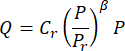
- Define Cr (after subtracting system facility) as ocular outflow facility.
- Set Pr = 8 mmHg.
- Define β as the nonlinear coefficient.
10. Ocular Φr
- Analyze P–T and Q–T curves during pressure increments. Identify the flow increase to Qmax and subsequent deceleration.
- Calculate the volume change between tp and tmax using the discrete-volume method30.
- Fit data to the modified Friedenwald equation30:
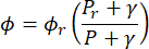
- Set Pr = 13 mmHg. Define Φr (after subtracting system compliance) as ocular compliance Φr,eye and γ as a material parameter.
Access restricted. Please log in or start a trial to view this content.
Results
The performance of the optimized syringe-pump system was evaluated by comparing C and ϕ in 5-month-old mice with previously published values from pressure-controlled ocular perfusion studies. The normality of datasets in Figure 2F was assessed using the Shapiro–Wilk test, and statistical comparisons were performed using a two-tailed paired Student’s t-test.
As shown in Figure 2A–C, the IOP, Cr,eye,...
Access restricted. Please log in or start a trial to view this content.
Discussion
This study presents an optimized syringe-pump system for estimating outflow facility and ocular compliance in mouse eyes, yielding values consistent with those reported using pressure-controlled ocular perfusion systems. The present approach improves the accuracy of syringe-based measurements while maintaining a simple and accessible experimental setup. This combination of methodological refinement and accessibility may facilitate broader adoption in laboratories where more advanced systems are not readily available.
...Access restricted. Please log in or start a trial to view this content.
Disclosures
The authors have no conflicts of interest to declare.
Acknowledgements
We thank Prof. Harald Stauss at the University of Iowa for developing the HemoLab software. This study was supported by the National Key Research and Development Program (2022YEF0132500), Qingdao Key Technology and Industrialization Project (23-1-4-xxgg-16-nsh), Taishan Scholar Youth Expert Program (tsqn202103055), Shandong Excellent Youth Science Fund (ZR2022YQ72), Ophthalmology Joint Project of Qingdao University.
Access restricted. Please log in or start a trial to view this content.
Materials
| Name | Company | Catalog Number | Comments |
|---|---|---|---|
| Reagent | 1× PBS Buffer | Thermo Fisher Scientific | 10010023 |
| Isoflurane | Shenzhen Rewoode Life Sciences Co., Ltd., China | R640 | |
| Material | 100 μL High Precision Syringe | Hamilton | 1710 |
| Borosilicate Glass | Sutter, Novato, CA | BF100-50-10 | |
| Disposable sterile syringe | Fenglin Medical Devices Co., Ltd. Jiangxi, China | 10 ml | |
| Pressure Sensor | IcuMedical | PX26-015G | |
| Silicone tube | Runze Fluid Co., Ltd, Nanjing, China | N/A | |
| Transparent PTFE tube | Runze Fluid Co., Ltd, Nanjing, China | N/A | |
| Equipment | Flaming/Brown pipette puller | Sutter, Novato, CA | P-97 |
| Manual Micromanipulator | World Precision Instruments (WPI) | M3301 | |
| Metal bath | Beaver Biology, Suzhou, China | 2016C | |
| MicroForge | Narishige Scientific Instrument Lab., Tokyo, Japan | MF830 | |
| Micropipette Grinder | MPInstrument Co., Ltd., Wuhan, China | KDG-02 | |
| PZMIII Stereo Zoom Binocular Microscope | World Precision Instruments (WPI) | PZMIII | |
| Syringe-Pump | World Precision Instruments (WPI) | AL-1000 |
Reprints and Permissions
This article has been published
Video Coming Soon