Metabolic reprogramming drives PDAC angiogenesis via HIF-1α, PKM2, KRAS, lactate, and lipid pathways. Compensatory mechanisms limit monotherapy, underscoring the need for rational combination strategies.
Review Article
Metabolic Regulation of Angiogenesis in Pancreatic Ductal Adenocarcinoma
In This Article
Summary
Abstract
Pancreatic ductal adenocarcinoma (PDAC) remains highly lethal, with a 5‑year survival of only 13%. Its aggressive biology and late diagnosis often lead to metastatic spread at presentations. This review examines how metabolic reprogramming regulates angiogenesis in PDAC and explores the therapeutic implications of this coupling. We systematically analyzed published studies on glucose, amino acid, and lipid metabolic alterations and their interconnection with neovascularization through hypoxia-inducible factor-1α (HIF-1α), oncogenic signaling, and metabolite-mediated pathways. Based on a synthesis of the current literature, metabolic-angiogenic coupling appears to operate through at least three interconnected mechanisms: the HIF‑1α/pyruvate kinase M2 (PKM2)-driven glycolytic-vascular endothelial growth factor (VEGF) axis; lactate acting as a signaling molecule that promotes M2‑polarized tumor‑associated macrophages and stabilizes HIF‑1α; and lipid metabolites--including eicosanoids and fatty acid‑binding protein 4 (FABP4)-that modulate endothelial function and survival. In addition, glutamine-derived intermediates support endothelial sprouting and extracellular matrix remodeling, while tryptophan and serine pathways influence the immune-angiogenic balance. Importantly, compensatory pathways (e.g., HIF-1α-independent angiogenesis via glycogen accumulation and protease-activated receptor‑2 activation) limit the efficacy of single‑agent interventions, highlighting the need for combination strategies. Understanding these metabolic-angiogenic networks provides a rationale for biomarker‑guided therapies that simultaneously target tumor metabolism and vascularization. Integrating metabolic and angiogenic biomarkers (e.g., VEGF, lactate dehydrogenase A) with imaging-based metabolic profiling may improve patient stratification and treatment monitoring in PDAC.
Introduction
Pancreatic ductal adenocarcinoma (PDAC) remains one of the deadliest solid malignancies. For patients with metastatic disease, median overall survival is approximately 6–11 months, and even those with locally advanced or resectable tumors face extremely poor long-term outcomes1,2. Over the past three decades, the 5-year survival rate has improved only modestly, from 4% to 13%, largely due to advances in surgical techniques and centralization of care rather than to breakthroughs in systemic therapy3,4. Epidemiologically, pancreatic cancer is projected to become the second leading cause of cancer-related death in the United States by 20305. In China, the disease ranks 8th in incidence and 6th in mortality6, and its incidence is rising among young adults7,8.
Despite this high burden, therapeutic progress has been frustratingly slow, primarily because of the tumor’s pronounced heterogeneity, early systemic dissemination, and incomplete understanding of the key drivers of its aggressive biology. Metabolic adaptation and vascular abnormalities within the tumor microenvironment are considered central to PDAC progression and treatment resistance, yet the causal relationships and signaling crosstalk between these two processes remain inadequately characterized. In particular, how cancer cells actively shape the angiogenic niche through metabolic reprogramming, whether metabolic intermediates act as direct signaling molecules on endothelial cells, and how these mechanisms collectively contribute to therapy evasion are not systematically understood9,10. Therefore, a comprehensive elucidation of the metabolic–angiogenic coupling network is of substantial theoretical and clinical importance for revealing PDAC progression mechanisms and for developing novel intervention strategies. Nevertheless, to date, no systematic review has synthesized the multifaceted mechanisms by which metabolic reprogramming governs angiogenesis in PDAC, nor has the translational potential of these findings been critically appraised. To address this gap, the present review aims to: synthesize the core mechanisms by which glucose, amino acid, and lipid reprogramming regulate angiogenesis in PDAC, with emphasis on the hub roles of hypoxia‑inducible factor‑1α (HIF-1α), oncogenic signals, and metabolic intermediates; critically evaluate the clinical utility of metabolic and angiogenic biomarkers based on available evidence; and discuss the prospects and challenges of combination therapies that simultaneously target metabolic and vascular pathways. By integrating these multi‑level data, we seek to provide a conceptual framework for precision diagnosis and treatment of PDAC. Importantly, while elucidating these coupling mechanisms may offer a theoretical basis for biomarker identification and targeted therapy development, the translational value of these findings awaits validation in well‑designed prospective clinical studies, and the strength of the evidence for each proposed application will be critically examined in the subsequent sections of this review.
Review and Perspective
Pancreatic ductal adenocarcinoma (PDAC) develops within a uniquely hostile microenvironment characterized by dense desmoplasia, profound hypoxia, and aberrant metabolic reprogramming. The tumor vasculature that emerges from this milieu is not merely a passive conduit but an active participant in disease progression, shaped by the convergence of oncogenic signaling, metabolic adaptation, and stromal crosstalk. As depicted in Figure 1, metabolic inputs from glucose, amino acid, and lipid pathways converge on central regulators--hypoxia-inducible factor-1α (HIF-1α), mutant KRAS (present in ~93% of PDAC), and mTOR complex 1 (mTORC1)—to coordinate transcriptional and post-translational angiogenic outputs. The resultant vasculature is characteristically hypovascular, leaky, and immature, comprising four distinct vascularization modes: sprouting, intussusceptive, and coalescent angiogenesis, alongside vasculogenic mimicry (VM). Understanding this mechanistic heterogeneity is essential for interpreting the failure of anti-angiogenic monotherapies and for designing rational combination strategies.
Basic overview of angiogenesis in pancreatic cancer
Modes of angiogenesis
Sprouting angiogenesis is the predominant mode of neovascularization in PDAC, triggered by hypoxia, tissue injury, or oncogenic signaling and regulated by multiple pro-angiogenic mediators11. Vascular endothelial growth factor (VEGF), secreted by tumor cells, constitutes the most critical regulatory factor. Upon binding to VEGFR2, VEGF activates the PI3K/AKT and MAPK/ERK pathways: PI3K phosphorylates PIP2 to generate PIP3, recruiting and activating AKT to promote endothelial cell (EC) proliferation and migration, while the MAPK/ERK cascade proceeds through RAS-RAF-MEK-ERK activation, with ERK translocating to the nucleus to modulate proliferation- and migration-associated gene expression. Preclinical studies demonstrate that VEGF inhibition significantly reduces sprouting and attenuates tumor growth in PDAC models12. Recent advances highlight that endothelial phenotypic plasticity critically modulates sprouting efficiency. Endothelial cells reversibly switch between quiescent “phalanx” cells, migratory “tip” cells, and proliferative “stalk” cells. Tip-stalk dynamics are metabolically specialized: tip cells preferentially rely on glycolysis to fuel migration, whereas stalk cells depend on fatty acid oxidation to sustain proliferation. Tumor-derived proteases such as ADAM9 further promote sprout formation through shedding of heparin-binding EGF-like growth factor. Collectively, these findings indicate that PDAC sprouting angiogenesis is governed not merely by VEGF, but by a complex interplay of metabolic, signaling, and stromal factors13,14,15,16.
Intussusceptive angiogenesis expands and branches vessels through remodeling of the existing vascular wall without reliance on endothelial cell proliferation or migration17. Within tumor tissue, rapid cancer cell proliferation increases local pressure, causing the vessel wall to invaginate into the lumen and form columnar or septal structures. These structures gradually widen and fuse, dividing the original lumen into multiple smaller channels, thereby increasing vessel surface area and volume. Three-dimensional imaging in PDAC mouse models indicates this mode is more common in central tumor regions, where it helps alleviate hypoxia18. Because intussusceptive angiogenesis depends on vessel wall remodeling rather than EC proliferation, anti-angiogenic agents that inhibit endothelial proliferation--and anti-VEGF therapy in particular--may have limited efficacy against this mode.
Coalescent angiogenesis (CA) evolves from an isotropic capillary network surrounding tissue islands. Preferential flow pathways enlarge through capillary aggregation and disappearance of internal tissue pillars, while less perfused segments regress, remodeling the mesh-like network into a hierarchical tree that retains vessel wall components and maintains blood flow19. CA shares features with intussusceptive angiogenesis but differs morphologically: whereas intussusceptive angiogenesis grows and fuses tissue pillars to partition existing vessels, CA eliminates tissue islands to form definitive vessels, making it functionally the reverse process20.
Vasculogenic mimicry represents a distinctive pattern wherein tumor cells simulate endothelial cells to form vessel-like structures. VM formation is intimately associated with epithelial-mesenchymal transition (EMT)21; EMT-transformed tumor cells interconnect to form lumen-containing structures supported by periodic acid-Schiff (PAS)-positive cells and extracellular matrix, transporting erythrocytes and fulfilling perfusion functions analogous to normal vasculature22. VM structures are significantly increased in highly invasive PDAC tissues and correlate with poor prognosis23.
Vascular characteristics and microenvironmental constraints
Unlike many malignancies, PDAC is characteristically hypovascular and desmoplastic, with a mean microvessel density (MVD) significantly lower than in normal pancreatic tissue or hepatocellular carcinoma24,25,26. This hypovascular phenotype persists despite profound hypoxia, suggesting active angiosuppressive mechanisms. Paracrine Hedgehog signaling can suppress non-canonical WNT signaling to limit VEGFR2-dependent endothelial hypersprouting, and VEGFR2/neuropilin 1 (NRP1) trans-complex formation between tumor and endothelial cells correlates with reduced vessel branching and improved survival27. The resultant vasculature exhibits uneven distribution, immature structure, low pericyte coverage, and functional leakage28, leading to plasma extravasation and elevated interstitial fluid pressure (IFP). Elevated IFP combined with solid stress from dense fibrosis compresses vessels, potentially causing vascular collapse and a near-complete absence of vessels exceeding 10 µm in diameter, further exacerbating hypoperfusion29,30.
This diminished perfusion creates a vicious cycle of hypoxia. PDAC neovessels display incomplete walls and inadequate smooth muscle and pericyte support, resulting in slow blood flow31. The hypoxic microenvironment transcriptionally stabilizes HIF-1α, which amplifies VEGF secretion and perpetuates the angiogenic drive32,33. In resected human PDAC samples, HIF-1α expression (present in 40% of cases) positively correlates with metastatic disease and MVD, while negatively correlating with prognosis34. HIF-2α overexpression and vasculogenic mimicry are similarly associated with poor differentiation, advanced stage, lymph node metastasis, and unfavorable prognosis35.
Pericyte dysfunction--characterized by ectopic α-smooth muscle actin expression and impaired pericyte-endothelial adhesion--further destabilizes the vasculature36. Loose endothelial junctions, discontinuous basement membranes, and intercellular gaps increase permeability, allowing plasma extravasation that elevates interstitial pressure and impedes drug delivery while facilitating tumor cell intravasation. High MVD combined with low microvessel integrity is associated with early recurrence, metastasis, and transient post-resection survival37.
Metabolic regulation of angiogenesis
PDAC exhibits extensive hypoxic regions, with a median tissue pO2 of 0–5.3 mmHg compared to 24.3–92.7 mmHg in adjacent normal tissue38. This arises from hypoxic fibrous stroma, rapid proliferation, and inadequate vasculature39. The adaptive hypoxic response, mediated primarily by HIF-α isoforms, endows PDAC with an aggressive, therapy-resistant phenotype: HIF-1α is rapidly induced under acute severe hypoxia40,41, whereas HIF-2α tends toward persistent expression under chronic moderate hypoxia42,43, and HIF-3α largely regulates other HIF complexes44.
HIF-1α serves as the central upstream switch, simultaneously driving glucose metabolic reprogramming and angiogenesis. Under hypoxia, HIF-1α transcriptionally activates glycolysis-related genes (GLUT1, HK2, LDHA, PKM2) while concomitantly activating pro-angiogenic factors (VEGF, CTGF, PDGFB)45,46,47. Molecules such as MUC1, P4HA1, and APE1/Ref-1 reinforce both glycolytic phenotype and angiogenic capacity through HIF-1α stabilization or enhanced transcriptional function48,49,50,51. PKM2 functions as a direct molecular bridge between glycolysis and angiogenesis: under hypoxia, PKM2 translocates to the nucleus to co-activate transcription of glycolytic and angiogenic genes (including VEGF-A) via β-catenin/STAT3 interaction and histone modification, creating a positive feedback loop wherein enhanced glycolysis amplifies HIF-1α/VEGF signaling47,52. KRAS mutations enhance HIF-1α/2α stability through MEK/ERK, upregulating glycolytic enzymes (LDHA, PDK1) and pro-angiogenic effectors (VEGF, CA9, MCT4)53,54. Additional crosstalk involves LSD1-mediated HIF-1α stabilization, the UHRF1/SIRT4 axis, and receptor signaling through IR/IGF1R and CX3CL1/CX3CR139,55,56, while lumican suppresses both glycolysis and angiogenesis through EGFR/PI3K/Akt inhibition57. Compensatory pathways—including glycogen accumulation, sustaining angiogenesis via dendritic cell recruitment58 and PAR-2 activation promoting VEGF through MEK-ERK53--indicate that targeting HIF-1α or single metabolic nodes alone may be insufficient.
Amino acid metabolism modulates angiogenesis by supplying biosynthetic precursors for endothelial sprouting and extracellular matrix remodeling, and by generating immunometabolic signals that indirectly regulate vascular tone and immune infiltration. Glutamine, via GLS, forms glutamate convertible to aspartate to promote EC sprouting59 and to proline for ECM remodeling60. VEGF stimulation increases endothelial glycine uptake, promoting migration61; glycine also participates in glutathione synthesis, modulating NO-mediated vascular tone and oxidative stress62. Arginine contributes to proline, polyamines, and NO63, while the PHGDH-dependent serine synthesis pathway regulates vascular tone, oxidative stress, and endothelial apoptosis protection63,64,65. Notably, tryptophan metabolism in myeloid-derived suppressor cells (MDSCs) generates kynurenine via IDO-1, shifting the IFNγ/IL-6 balance toward a pro-angiogenic state67,68.
Lipid metabolic reprogramming promotes neovascularization through metabolic, signaling, and immunomodulatory mechanisms69. PDAC exhibits upregulated de novo lipogenesis driven by SREBP-1 and enzymes such as FASN and ACC. Beyond membrane biogenesis, FASN regulates EC proliferation through a metabolite-signaling mechanism: FASN inhibition elevates malonyl-CoA, promoting mTOR malonylation at lysine 1218 and impairing mTORC1 kinase activity, thereby selectively suppressing EC proliferation without affecting migration70. Fatty acid oxidation serves as an alternative energy source under glucose restriction, supporting EC migration and tube formation; pharmacological FAO inhibition impairs angiogenesis in PDAC models71.
Lipid species also function as direct signaling molecules. Lysophosphatidic acid (LPA) and sphingosine-1-phosphate (S1P) activate endothelial G protein-coupled receptors to promote proliferation, migration, and permeability, with elevated levels linked to increased VEGF expression and tube formation72. Aberrant lipid metabolism generates ROS and lipid peroxidation products that activate HIF-1α and NF-κB, amplifying transcription of pro-angiogenic genes, including VEGF and IL-873. Lipid droplet accumulation in cancer and endothelial cells correlates with increased EC survival and resistance to anti-angiogenic therapy, with perilipins implicated in regulating proliferation and migration74.
Lipid metabolic reprogramming also shapes the angiogenic microenvironment through immune modulation. Tumor-associated macrophages (TAMs) accumulate oxidized lipids and cholesterol via scavenger receptors such as CD36, promoting a pro-angiogenic M2-like phenotype that secretes VEGF, TNF-α, and matrix metalloproteinases. Similarly, lipid-laden myeloid-derived suppressor cells (MDSCs) produce angiogenic factors that support neovascularization. This metabolic-immune-angiogenic axis represents an emerging therapeutic target in PDAC75.
Metabolite-mediated angiogenic signaling
Lactate has been rehabilitated from metabolic waste to a key signaling molecule bridging glycolysis and angiogenesis. Lactate promotes angiogenesis through three main mechanisms: direct activation of the NF-κB/IL-8 pathway in endothelial cells via MCT-1; stabilization of HIF-1α protein by inhibiting prolyl hydroxylase (PHD) activity; and paracrine immune modulation76. The role of GPR81 in lactate-mediated HIF-1α activation remains controversial: murine studies demonstrate that GPR81 signaling stabilizes HIF-1α in dendritic cells and MDSCs to promote immune evasion and angiogenesis, yet murine macrophage studies indicate that immunosuppressive effects can occur independently of GPR8177. Moreover, GPR81 expression is high in adipose tissue but notably low in other human organs78, and human neutrophils expressing GPR81 show minimal transcriptional response to lactate stimulation, raising questions about generalizability79. In pancreatic cancer specifically, lactate enhances MDSC activity via the GPR81-mTOR-HIF-1α-STAT3 axis80, though the interspecies relevance of this mechanism remains incompletely characterized. Additionally, lactate induces M2 macrophage polarization, which secretes further pro-angiogenic factors81,82, while protein lactylation (e.g., ENSA-K63 lactylation) remodels the immune microenvironment via STAT3/CCL2, indirectly affecting angiogenesis83.
Other metabolites modulate angiogenesis through distinct pathways. Omega-3 fatty acids (EPA and DHA) exert anti-angiogenic effects by inhibiting VEGF, PDGF, COX-2, PGE2, NF-κB, and MMPs84. Conversely, elevated omega-6 polyunsaturated fatty acids provide substrates for arachidonic acid metabolism; PGE2 produced via COX-2 increases VEGF secretion from PDAC cells through autocrine mechanisms, promoting EC migration85.
Fatty acid-binding protein 4 (FABP4) operates across multiple compartments to promote angiogenesis. In endothelial cells, NOTCH1 induces FABP4 expression to fuel long-chain fatty acid metabolism and confer ROS resistance, supporting angiogenic sprouting. In tumor cells, aberrant lipid metabolism generates acetyl-CoA and ROS that activate HIF-1α and NF-κB, driving transcription of VEGF and PDGF. At the stromal level, adipocytes mediate free fatty acid transport through FABP4 while secreting IL-1β and TNF-α to activate NF-κB. Concurrently, abnormal sphingolipid metabolism induces M2 macrophage polarization via the MIF-CD74 axis, with FABP4+ macrophage subsets enhancing endothelial cell migratory capacity86.
Finally, tumor microenvironment metabolites--including succinate, fumarate, and 2-hydroxyglutarate--function as signaling molecules that activate angiogenesis-related pathways by remodeling EC function and regulating immune polarization87.
Clinical translation: biomarkers, imaging, and therapeutics
The coupling between metabolic reprogramming and angiogenesis provides novel biomarkers and assessment dimensions for PDAC management, though the translational trajectory follows a heterogeneous evidence gradient. As summarized in Table 188,89,90,91,92,93, candidate biomarkers span three tiers: (i) established clinical markers such as CA19-9, which are widely used but lack specificity for PDAC versus biliary obstruction; (ii) emerging clinically evaluated markers, including VEGF and glycolytic metabolites; and (iii) experimental signatures such as multi-metabolite panels and lipid-macrophage risk models.
Multiple studies confirm that VEGF, HIF-1α, and GLUT1 are significantly elevated in blood and tumor tissues of PDAC patients relative to inflammatory pancreatic conditions. In a study of 61 resected patients, blood and tissue VEGF concentrations were significantly higher in PDAC than in inflammatory tumors (p < 0.000001), as were HIF-1α (p = 0.000005) and GLUT1 (p = 0.000002). Multivariate analysis identified VEGF as the most powerful independent predictor in both blood (OR = 1.016; p = 0.001) and tissue (OR = 1.02; p = 0.001), with serum VEGF at a cutoff of 134.56 pg/mL demonstrating 97.8% sensitivity and 86.7% specificity88. Beyond diagnosis, dynamic VEGF changes during treatment may serve as pharmacodynamic markers. In the phase I STARPAC trial (n = 19), serial plasma measurements suggested that early VEGF changes could predict treatment response, though this requires validation in randomized controlled trials89.
Glycolytic reprogramming offers complementary diagnostic potential. Integrating GEO and TCGA datasets, investigators identified LDHA as an independent predictor of overall survival in pancreatic cancer (p < 0.001). Serum metabolomic profiling by NMR in 36 PDAC patients and 70 healthy controls revealed significant alterations in glycolysis-related metabolites, with a score combining lactate, pyruvate, citrate, and glucose achieving an AUC of 0.956 for distinguishing PDAC from controls90. Moving from single-gene signatures to multi-metabolite integration, a two-phase metabolomics study identified proline, creatine, and palmitic acid as a panel distinguishing PDAC from benign neoplasms and healthy controls, with AUCs of 0.854 and 0.865 in the discovery set; addition of CA19-9 improved performance to 0.949 and 0.909, respectively, with validation AUCs of 0.830 and 0.852 in an independent cohort91. At the transcriptional level, bioinformatics analysis of public datasets (TCGA-PAAD, GSE62452, GSE57495) yielded a seven-gene lipid-macrophage risk model (ADH1A, ACACB, CD36, CERS4, PDE3B, ALOX5, CRAT) that effectively stratified patients by risk, with TP53 mutations prevalent and immunomodulatory factors (PVRL2, CD276, CCL20) correlating with risk score92.
The divergence between promising discovery-phase results and meta-analytic findings reflects recurrent methodological limitations: single-center retrospective design, spectrum bias toward resectable disease, modest sample sizes, absence of benign comparators, and lack of independent prospective validation87,88,89,90,91. These findings underscore the necessity of rigorous, standardized, and sufficiently powered validation before novel biomarkers can advance to clinical implementation.
For functional assessment, 18F-FDG PET/CT reflects tumor glucose metabolic activity, while hyperpolarized [1-13C]pyruvate MRI enables real-time monitoring of pyruvate-to-lactate conversion, correlating with LDHA and HIF-1α overexpression94. Circulating exosomes hold promise as minimally invasive tools for early diagnosis and monitoring95. Prognostically, VEGF/VEGFR-2 overexpression, high glycolytic activity, LDHA overexpression, and COX-2 expression are each linked to poor outcomes, suggesting that molecular classification based on metabolic-angiogenic features may enable precise stratification and treatment selection96.
Therapeutic strategies and the anti-angiogenic paradox
Despite the central role of VEGF in PDAC angiogenesis, anti-VEGF/VEGFR strategies have consistently failed in phase III trials. The CALGB 80303 trial (n = 535) demonstrated no survival benefit for gemcitabine plus bevacizumab versus gemcitabine alone (median OS 5.8 vs. 5.9 months; p = 0.95)97. The AviTA trial showed a statistically significant improvement in progression-free survival but no overall survival benefit for bevacizumab added to gemcitabine plus erlotinib98. The VANILLA trial of gemcitabine plus aflibercept was discontinued after interim analysis confirmed no OS benefit (7.75 vs. 6.54 months)99, and axitinib similarly failed to improve survival with gemcitabine100.
These failures reflect at least three PDAC-specific resistance mechanisms. First, compensatory pathway activation: hypoxia induced by VEGF blockade upregulates alternative pro-angiogenic factors (FGF, Ang-2, PIGF) and recruits pro-angiogenic myeloid cells101. Second, vascular co-option and vasculogenic mimicry: tumor cells circumvent sprouting inhibition by utilizing pre-existing host vessels or forming vessel-like channels independently of endothelial proliferation22. Recent single-cell sequencing has revealed that cancer-associated fibroblasts (CAFs) can transdifferentiate into an endothelial-like phenotype (FAPα + CD144 + endoCAFs) capable of vasculogenic mimicry and metastasis promotion via CD144-β-catenin-STAT3 signaling, shifting the conceptual framework of VM from a tumor-cell-autonomous process to a tumor-stromal cooperative mechanism102. Third, metabolic-stromal adaptation: dense desmoplasia physically sequesters therapeutic antibodies, while CAFs sustain angiogenesis through paracrine HGF and TGF-β-mediated pathways operating downstream of or parallel to VEGF36. Collectively, these mechanisms establish that monotherapy targeting a single angiogenic ligand is insufficient in PDAC.
Targeting metabolic-angiogenic nodes represents a rational alternative. Glucose metabolism inhibitors—including the HK2 inhibitor 2-deoxy-D-glucose, the PKM2 activator shikonin, and the LDHA inhibitor oxamate—have demonstrated anti-angiogenic and anti-tumor effects in PDAC cell lines and xenografts82. Targeting the PKM2/HIF-1α/VEGF axis could simultaneously inhibit metabolic adaptation and angiogenesis. GPR81-mediated lactate signaling has emerged as a potential paracrine target, though specific antagonists remain experimental; the putative antagonist 3-hydroxybutyrate may primarily target the related receptor HCAR2 rather than GPR81103,104. In lipid metabolism, COX-2 inhibitors (e.g., celecoxib) and PPARγ/FABP4 pathway modulators have shown anti-angiogenic effects in preclinical models105, though phase II trials of celecoxib with gemcitabine have not demonstrated significant survival advantages. Omega-3 polyunsaturated fatty acids competitively inhibit the ω-6 pathway and demonstrate anti-angiogenic properties in preclinical models, though human interventional data in PDAC are lacking.
Given PDAC metabolic plasticity, combination regimens pairing anti-angiogenic agents with chemotherapy, immunotherapy, or metabolic inhibitors represent the most promising path forward. Reprogramming of glucose, amino acid, and lipid metabolism is closely associated with resistance to chemotherapy, radiotherapy, and immunotherapy; combined inhibition of glycolysis and glutaminolysis may block metabolic compensation. However, as of this writing, no metabolism-targeting combination has demonstrated phase III efficacy in PDAC. Ultimately, the central translational challenge is not the paucity of druggable nodes, but the inability to prospectively identify which mode of vascularization--sprouting, intussusceptive, coalescent, or mimicry--dominates in an individual tumor. Until biomarker panels or imaging signatures can stratify patients by their metabolic-angiogenic phenotype, combination strategies targeting this network will remain empiric rather than precision-guided.
Conclusions
This review delineates the reciprocal coupling between metabolic reprogramming and angiogenesis in pancreatic ductal adenocarcinoma (PDAC). We describe four distinct modes of neovascularization--sprouting, intussusceptive, coalescent angiogenesis, and vasculogenic mimicry--that collectively produce a hypovascular, poorly perfused, and structurally aberrant vasculature. At the mechanistic core, glucose metabolic reprogramming via the Warburg effect establishes a synergistic loop in which HIF-1α drives both glycolytic flux and pro-angiogenic transcription (VEGF, CTGF, PDGFB), while PKM2 and mutant KRAS integrate metabolic status with angiogenic output. Amino acid and lipid metabolism further modulate endothelial behavior, immune polarization, and therapeutic resistance through paracrine signals, including lactate, LPA/S1P, and the FASN-malonyl-CoA-mTOR axis. These findings establish that PDAC angiogenesis is not merely a hypoxic response but an integral component of the tumor's metabolic adaptive program.
Clinical translation remains constrained by diagnostic and therapeutic limitations. Although discovery-phase studies have identified metabolic and angiogenic biomarker candidates, including VEGF, LDHA-derived metabolite scores, and lipid-macrophage risk models, aggregated evidence demonstrates that none consistently outperforms CA19-9 or provides sufficient added value for immediate implementation. Therapeutically, phase III trials of anti-VEGF monotherapy have uniformly failed to improve survival, reflecting the resilience of tumor vascularization driven by compensatory pathway activation, vasculogenic mimicry, and metabolic-stromal adaptation. These parallel limitations underscore the profound redundancy and plasticity of the metabolic-angiogenic network, necessitating integrated rather than monotherapeutic strategies.
Combination regimens--pairing anti-angiogenic agents with chemotherapy, immunotherapy, or metabolic inhibitors--represent the most promising path forward. Concurrent targeting of glycolysis and glutaminolysis, or co-inhibition of FASN and mTOR, may overcome metabolic compensation and disrupt lipid-driven angiogenic signaling. Future priorities include biomarker-guided patient stratification (e.g., glycolytic versus lipid-dependent phenotypes), rational multi-target combinations that block diverse vascularization modes, non-invasive metabolic-angiogenic imaging, and rigorously designed clinical trials in molecularly defined PDAC subpopulations. Ultimately, elucidating the metabolic-angiogenic crosstalk will be essential to transform PDAC from a therapeutically recalcitrant disease into one amenable to mechanism-based precision intervention.
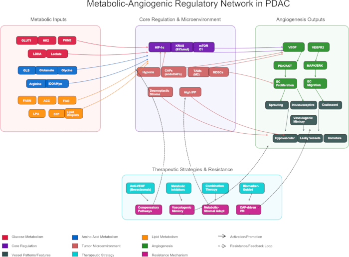
Figure 1: Metabolic-angiogenic regulatory network in PDAC. Metabolic-angiogenic regulatory network in PDAC. Schematic overview of metabolic-angiogenic coupling in pancreatic ductal adenocarcinoma. (Left) Nutrient pathway reprogramming: glucose metabolism (crimson), amino acid metabolism (blue), and lipid metabolism (orange). (Center) Integration of metabolic inputs by central regulators (purple; HIF-1α, mutant KRAS, mTORC1) within the tumor microenvironment (salmon), characterized by hypoxia, desmoplastic stroma with high interstitial fluid pressure (IFP), CAFs (including FAPα⁺CD144⁺ endoCAFs), M2-polarized TAMs, and MDSCs. (Right) VEGF-VEGFR2-driven angiogenic signaling and four distinct vascularization modes (dark slate gray): sprouting, intussusceptive, coalescent angiogenesis, and vasculogenic mimicry (VM). (Bottom) Current therapeutic strategies (teal) and resistance mechanisms (magenta). Solid arrows indicate activation; dashed arrows denote resistance feedback loops. Please click here to view a larger version of this figure.
| Biomarker/Panel | Biological Rationale | Evidence Level | Key Limitations | Translational Stage |
| CA19‑993 | Sialylated Lewis antigen | Clinically established | Low specificity for PDAC vs. biliary obstruction | Clinical standard |
| Serum VEGF (diagnostic)88 | HIF‑1α/VEGF axis activation | Single‑center retrospective | Spectrum bias; resectable‑only cohort | Discovery |
| Serum VEGF (response)89 | Pharmacodynamic marker | Phase I (STARPAC, n=19) | Single‑arm; no control arm | Hypothesis‑generating |
| LDHA/metabolite score90 | Glycolytic flux | Retrospective; single‑center | No benign comparator; small cohort | Discovery |
| Proline‑creatine‑palmitic acid + CA19‑991 | Multi‑omics integration | Discovery + small validation | Single‑institution; modest n | Early validation |
| 7‑gene lipid/macrophage risk model92 | Lipid‑immune crosstalk | Retrospective bioinformatics | No prospective cohort; batch effects | Discovery |
Table 1: Tiered evidence framework for metabolic-angiogenic biomarkers in PDAC. The table stratifies metabolic-angiogenic biomarkers in PDAC by translational maturity across three tiers: (i) the clinically established standard CA19-9; (ii) early-phase candidates, including diagnostic and pharmacodynamic serum VEGF assays and an LDHA/metabolite score; and (iii) discovery-stage signatures, exemplified by a multi-metabolite panel (proline--creatine--palmitic acid plus CA19-9) and a 7-gene lipid/macrophage risk model. A recurrent pattern emerges across emerging candidates: although biological rationale is well-defined, methodological limitations--retrospective or single-center design, absence of benign comparators, small cohorts, and lack of prospective validation--collectively constrain advancement beyond discovery or early hypothesis-generating stages.
Disclosures
The authors declare no competing financial interests.
Acknowledgements
The study was funded by the Digital Microfluidic platform for drug screening based on 3D spheroids of primary tumor cells (FDCT0001/2025/RID).
References
- Siegel RL, Giaquinto AN, Jemal A. Cancer statistics, 2024. CA Cancer J Clin. 2024;74(1):12-49.
- Mackay TM, et al. Implementation of Best Practices in Pancreatic Cancer Care in the Netherlands: A Stepped-Wedge Randomized Clinical Trial. JAMA Surg. 2024;159(4):429-37.
- Latenstein AEJ, et al. Effect of centralization and regionalization of pancreatic surgery on resection rates and survival. Br J Surg. 2021;108(7):826-33.
- Kotecha K, et al. Centralization of Pancreaticoduodenectomy: A Systematic Review and Spline Regression Analysis to Recommend Minimum Volume for a Specialist Pancreas Service. Ann Surg. 2024;279(6):953-60.
- Rahib L, Wehner MR, Matrisian LM, Nead KT. Estimated Projection of US Cancer Incidence and Death to 2040. JAMA Netw Open. 2021;4(4):e214708.
- Han B, Zheng R, Zeng H, et al. Cancer incidence and mortality in China, 2022. J Natl Cancer Cent. 2024;4(1):47-53.
- Sung H, Siegel RL, Rosenberg PS, Jemal A. Emerging cancer trends among young adults in the USA: analysis of a population-based cancer registry. Lancet Public Health. 2019;4(3):e137-47.
- Hughes T, et al. The current and future global burden of cancer among adolescents and young adults: a population-based study. Lancet Oncol. 2024;25(12):1614-24.
- Folkman J. Anti-angiogenesis: new concept for therapy of solid tumors. Annals of Surgery, 1972, 175(3): 409-416.
- Hanahan D, Weinberg RA. Hallmarks of cancer: the next generation. Cell. 2011;144(5):646-74.
- Rhodin JA, Fujita H. Capillary growth in the mesentery of normal young rats. Intravital video and electron microscope analyses. J Submicrosc Cytol Pathol. 1989;21(1):1-34.
- Potente M, Gerhardt H, Carmeliet P. Basic and therapeutic aspects of angiogenesis. Cell. 2011;146(6):873-87.
- De Bock K, et al. Role of PFKFB3-driven glycolysis in vessel sprouting. Cell. 2013;154(3):651-63.
- Jakobsson L, et al. Endothelial cells dynamically compete for the tip cell position during angiogenic sprouting. Nat Cell Biol. 2010;12(10):943-53.
- Lu C, et al. An integrative proteotranscriptomics approach reveals new ADAM9 substrates and downstream pathways 2024;25(4):101538.
- Schoors S, et al. Fatty acid carbon is essential for dNTP synthesis in endothelial cells. Nature. 2015;520(7546):192-7.
- Baum O, et al. VEGF-A promotes intussusceptive angiogenesis in the developing chicken chorioallantoic membrane. Microcirculation. 2010;17(6):447-57.
- Djonov V, Baum O, Burri PH. Vascular remodeling by intussusceptive angiogenesis. Cell Tissue Res. 2003;314(1):107-17.
- Pezzella F, Kerbel RS. On coalescent angiogenesis and the remarkable flexibility of blood vessels. Angiogenesis. 2022;25(1):1-3.
- Nitzsche B, et al. Coalescent angiogenesis—evidence for a novel concept of vascular network maturation. Angiogenesis. 2022;25(1):35-45.
- Sun B, Zhang D, Zhao N, Zhao X. Epithelial-to-endothelial transition and cancer stem cells: two cornerstones of vasculogenic mimicry in malignant tumors. Oncotarget. 2017;8(18):30502-10.
- Maniotis AJ, et al. Vascular channel formation by human melanoma cells in vivo and in vitro: vasculogenic mimicry. Am J Pathol. 1999;155(3):739-52.
- Yang JP, et al. Tumor vasculogenic mimicry predicts poor prognosis in cancer patients: a meta-analysis. Angiogenesis. 2016;19(2):191-200.
- Hexige S, et al. Identification of novel vascular projections with cellular trafficking abilities on the microvasculature of pancreatic ductal adenocarcinoma. J Pathol. 2015;236(2):142-54.
- Olive KP, et al. Inhibition of Hedgehog Signaling Enhances Delivery of Chemotherapy in a Mouse Model of Pancreatic Cancer. Science. 2009;324(5933):1457-61.
- Sofuni A, et al. Differential diagnosis of pancreatic tumors using ultrasound contrast imaging. J Gastroenterol. 2005;40(5):518-25.
- Liu W, Yin B, Liang Z-H, Yu Y, Lu N. Computed tomography perfusion imaging evaluation of angiogenesis in patients with pancreatic adenocarcinoma. World J Clin Cases. 2022;10(8):2393-403.
- Rhim AD, et al. Stromal elements act to restrain, rather than support, pancreatic ductal adenocarcinoma. Cancer Cell. 2014;25(6):735-47.
- Jacobetz MA, et al. Hyaluronan impairs vascular function and drug delivery in a mouse model of pancreatic cancer. Gut. 2013;62(1):112-20.
- Provenzano PP, et al. Enzymatic targeting of the stroma ablates physical barriers to treatment of pancreatic ductal adenocarcinoma. Cancer Cell. 2012;21(3):418-29.
- Nguyen NC, Taalab K, Osman MM. Decreased blood flow with increased metabolic activity: a novel sign of pancreatic tumor aggressiveness. Clin Cancer Res. 2010;16(1):367.
- Büchler P, et al. Hypoxia-inducible factor 1 regulates vascular endothelial growth factor expression in human pancreatic cancer. Pancreas. 2003;26(1):56-64.
- Wei D, et al. Stat3 activation regulates the expression of vascular endothelial growth factor and human pancreatic cancer angiogenesis and metastasis. Oncogene. 2003;22(3):319-29.
- Shibaji T, et al. Prognostic significance of HIF-1 alpha overexpression in human pancreatic cancer. Anticancer Res. 2003;23(6C):4721-7.
- Yang J, et al. HIF-2α promotes the formation of vasculogenic mimicry in pancreatic cancer by regulating the binding of Twist1 to the VE-cadherin promoter. Oncotarget. 2017;8(29):47801-15.
- Neesse A, Algül H, Tuveson DA, Gress TM. Stromal biology and therapy in pancreatic cancer: a changing paradigm. Gut. 2015;64(9):1476-84.
- Wang W-Q, et al. Intratumoral α-SMA enhances the prognostic potency of CD34 associated with maintenance of microvessel integrity in hepatocellular carcinoma and pancreatic cancer. PLoS One. 2013;8(8):e71189.
- Koong AC, et al. Pancreatic tumors show high levels of hypoxia. Int J Radiat Oncol Biol Phys. 2000;48(4):919-22.
- Hu Q, et al. UHRF1 promotes aerobic glycolysis and proliferation via suppression of SIRT4 in pancreatic cancer. Cancer Lett. 2019;452:226-36.
- Raz S, et al. Severe hypoxia induces complete antifolate resistance in carcinoma cells due to cell cycle arrest. Cell Death Dis. 2014;5(2):e1067.
- Parks SK, Mazure NM, Counillon L, Pouysségur J. Hypoxia promotes tumor cell survival in acidic conditions by preserving ATP levels. J Cell Physiol. 2013;228(9):1854-62.
- Jokilehto T, Jaakkola PM. The role of HIF prolyl hydroxylases in tumour growth. J Cell Mol Med. 2010;14(4):758-70.
- Uchida T, et al. Prolonged hypoxia differentially regulates hypoxia-inducible factor (HIF)-1alpha and HIF-2alpha expression in lung epithelial cells: implication of natural antisense HIF-1alpha. J Biol Chem. 2004;279(15):14871-8.
- Bristow RG, Hill RP. Hypoxia and metabolism. Hypoxia, DNA repair and genetic instability. Nat Rev Cancer. 2008;8(3):180-92.
- He G, Jiang Y, Zhang B, Wu G. The effect of HIF-1α on glucose metabolism, growth and apoptosis of pancreatic cancerous cells. Asia Pac J Clin Nutr. 2014;23(1):174-80.
- Tung K-H, et al. CHC promotes tumor growth and angiogenesis through regulation of HIF-1α and VEGF signaling. Cancer Lett. 2013;331(1):58-67.
- Kitamoto S, et al. MUC1 enhances hypoxia-driven angiogenesis through the regulation of multiple proangiogenic factors. Oncogene. 2013;32(39):4614-21.
- Shukla SK, et al. MUC1 and HIF-1alpha Signaling Crosstalk Induces Anabolic Glucose Metabolism to Impart Gemcitabine Resistance to Pancreatic Cancer. Cancer Cell. 2017;32(1):71-87.e7.
- Shi S, et al. VEGF Promotes Glycolysis in Pancreatic Cancer via HIF1α Up-Regulation. Curr Mol Med. 2016;16(4):394-403.
- Logsdon DP, et al. Blocking HIF signaling via novel inhibitors of CA9 and APE1/Ref-1 dramatically affects pancreatic cancer cell survival. Sci Rep. 2018;8:13759.
- Logsdon DP, et al. Regulation of HIF1α under Hypoxia by APE1/Ref-1 Impacts CA9 Expression: Dual Targeting in Patient-Derived 3D Pancreatic Cancer Models. Mol Cancer Ther. 2016;15(11):2722-32.
- Azoitei N, et al. PKM2 promotes tumor angiogenesis by regulating HIF-1α through NF-κB activation. Mol Cancer. 2016;15(1):3.
- Chang LH, Pan SL, Lai CY, Tsai AC, Teng CM. Activated PAR-2 regulates pancreatic cancer progression through ILK/HIF-α-induced TGF-α expression and MEK/VEGF-A-mediated angiogenesis. Am J Pathol. 2013;183(2):566-75.
- McDonald PC, et al. Regulation of pH by Carbonic Anhydrase 9 Mediates Survival of Pancreatic Cancer Cells With Activated KRAS in Response to Hypoxia. Gastroenterology. 2019;157(3):823-37.
- Yang J, et al. β-Pentagalloyl-Glucose Sabotages Pancreatic Cancer Cells and Ameliorates Cachexia in Tumor-Bearing Mice. 2019;47(3):675-689
- Ren H, Zhao T, Sun J. The CX3CL1/CX3CR1 reprograms glucose metabolism through HIF-1 pathway in pancreatic adenocarcinoma[J/OL]. Journal of Cellular Biochemistry, 2013, 114(11): 2603-2611.
- Li X, et al. Extracellular lumican inhibits pancreatic cancer cell growth and is associated with prolonged survival after surgery. 2014 Dec 15;20(24):6529-40.
- Maruggi M, et al. Absence of HIF1A Leads to Glycogen Accumulation and an Inflammatory Response That Enables Pancreatic Tumor Growth. Cancer Res. 2019;79(22):5839-48.
- Kim B, Li J, Jang C, Arany Z. Glutamine fuels proliferation but not migration of endothelial cells. EMBO J. 2017;36(16):2321-33.
- Kay EJ, et al. Cancer-associated fibroblasts require proline synthesis by PYCR1 for the deposition of pro-tumorigenic extracellular matrix. Nat Metab. 2022;4(6):693-710.
- Guo D, et al. Vascular endothelial growth factor signaling requires glycine to promote angiogenesis. Sci Rep. 2017;7(1):14749.
- Rose ML, Madren J, Bunzendahl H, Thurman RG. Dietary glycine inhibits the growth of B16 melanoma tumors in mice. Carcinogenesis. 1999;20(5):793-8.
- Morris SM. Recent advances in arginine metabolism: roles and regulation of the arginases. Br J Pharmacol. 2009;157(6):922-30.
- Vandekeere S, et al. Serine Synthesis via PHGDH Is Essential for Heme Production in Endothelial Cells. Cell Metab. 2018;28(4):573-587.e13.
- Cantelmo AR, et al. Inhibition of the Glycolytic Activator PFKFB3 in Endothelium Induces Tumor Vessel Normalization, Impairs Metastasis, and Improves Chemotherapy. Cancer Cell. 2016;30(6):968-85.
- Mishra RC, et al. L-Serine lowers while glycine increases blood pressure in chronic L-NAME-treated and spontaneously hypertensive rats. J Hypertens. 2008;26(12):2339-48.
- Taylor MW, Feng GS. Relationship between interferon-gamma, indoleamine 2,3-dioxygenase, and tryptophan catabolism. FASEB J. 1991;5(11):2516-22.
- Dey S, et al. IDO1 Signaling through GCN2 in a Subpopulation of Gr-1+ Cells Shifts the IFNγ/IL6 Balance to Promote Neovascularization. Cancer Immunol Res. 2021;9(5):514-28.
- Vander Heiden MG, DeBerardinis RJ. Understanding the Intersections between Metabolism and Cancer Biology. Cell. 2017;168(4):657-69.
- Bruning U, et al. Impairment of Angiogenesis by Fatty Acid Synthase Inhibition Involves mTOR Malonylation. Cell Metab. 2018;28(6):866-880.e15.
- Carracedo A, Cantley LC, Pandolfi PP. Cancer metabolism: fatty acid oxidation in the limelight. Nat Rev Cancer. 2013;13(4):227-32.
- Mills GB, Moolenaar WH. The emerging role of lysophosphatidic acid in cancer. Nat Rev Cancer. 2003;3(8):582-91.
- Chandel NS, et al. Reactive oxygen species generated at mitochondrial complex III stabilize hypoxia-inducible factor-1alpha during hypoxia: a mechanism of O2 sensing. J Biol Chem. 2000;275(33):25130-8.
- Walther TC, Farese RV. Lipid droplets and cellular lipid metabolism. Annu Rev Biochem. 2012;81:687-714.
- Gabrilovich DI, Nagaraj S. Myeloid-derived suppressor cells as regulators of the immune system. Nat Rev Immunol. 2009;9(3):162-74.
- Yang J, et al. The enhancement of glycolysis regulates pancreatic cancer metastasis. Cell Mol Life Sci. 2019;77(2):305-21.
- Sanmarco LM, et al. Lactate limits CNS autoimmunity by stabilizing HIF-1α in dendritic cells. Nature. 2023;620(7975):881-9.
- Brown TP, et al. The lactate receptor GPR81 promotes breast cancer growth via a paracrine mechanism involving antigen-presenting cells in the tumor microenvironment. Oncogene 2020bz;39:3292-304.
- Hoque R, et al. Lactate Reduces Organ Injury in Toll-like Receptor- and Inflammasome-Mediated Inflammation via GPR81-mediated Suppression of Innate Immunity. Gastroenterology 2014ca;146:1763-74.
- Yang X, et al. Lactate-Modulated Immunosuppression of Myeloid-Derived Suppressor Cells Contributes to the Radioresistance of Pancreatic Cancer. Cancer Immunol Res 2020cb;8:1440-51.
- Yang L, et al. Lactate Metabolism: The String-Puller for the Development of Pancreatic Cancer. Biology (Basel). 2025;14(9):1213.
- Gao F, et al. Lactate metabolism reprogramming in PDAC: Potential for tumor therapy. Biochim Biophys Acta Rev Cancer. 2025;1880:189373.
- Chen Q, et al. Targeting lactylation and the STAT3/CCL2 axis to overcome immunotherapy resistance in pancreatic ductal adenocarcinoma. J Clin Invest. 2025;135(7):e191422.
- Spencer L, et al. The effect of omega-3 FAs on tumour angiogenesis and their therapeutic potential. Eur J Cancer. 2009;45(14):2077-86.
- Eibl G, et al. PGE2 is generated by specific COX-2 activity and increases VEGF production in COX-2-expressing human pancreatic cancer cells. Biochem Biophys Res Commun. 2003;306(4):887-97.
- Wang M, et al. FABP4 in lipid metabolism and the tumor microenvironment: mechanisms and therapeutic potential. Lipids Health Dis. 2025;25(1):29.
- Gardner GL, Stuart JA. Tumor microenvironment-like conditions alter pancreatic cancer cell metabolism and behavior. Am J Physiol Cell Physiol. 2024;327(11):C959-78.
- Wiewiora M, et al. Evaluations of the combined use of blood- and tissue-based protein biomarkers for pancreatic cancer. Clin Hemorheol Microcirc. 2024;86(4):383-93.
- Hughes CS, Blyuss O, Kocher HM. Plasma vascular endothelial growth factor levels are a potential therapy-response biomarker for pancreatic cancer. Front Oncol. 2025;15:1672385.
- Jiang W, et al. Aberrant lactate dehydrogenase A signaling contributes metabolic signatures in pancreatic cancer. Ann Transl Med. 2021;9(4):358.
- Zhao R, et al. Biomarkers for pancreatic cancer based on tissue and serum metabolomics analysis in a multicenter study. Cancer Med. 2023;12(5):5158-71.
- Wu L, et al. Identification of prognostic and therapeutic biomarkers associated with macrophage and lipid metabolism in pancreatic cancer. Sci Rep. 2025;15:14584.
- Ballehaninna UK, Chamberlain RS. The clinical utility of serum CA 19-9 in the diagnosis, prognosis and management of pancreatic adenocarcinoma: An evidence based appraisal. J Gastrointest Oncol 2012da;3:105-19.
- Dutta P, et al. Combining Hyperpolarized Real-Time Metabolic Imaging and NMR Spectroscopy to Identify Metabolic Biomarkers in Pancreatic Cancer. J Proteome Res. 2019;18(7):2826-34.
- Shakerian N, et al. Current Understanding of Therapeutic and Diagnostic Applications of Exosomes in Pancreatic Cancer. Pancreas. 2025;54(3):e255-67.
- Grobbelaar C, Steenkamp V, Mabeta P. Vascular Endothelial Growth Factor Receptors in the Vascularization of Pancreatic Tumors: Implications for Prognosis and Therapy. Curr Issues Mol Biol. 2025;47(3):179.
- Kindler HL, et al. Gemcitabine Plus Bevacizumab Compared With Gemcitabine Plus Placebo in Patients With Advanced Pancreatic Cancer: Phase III Trial of the Cancer and Leukemia Group B (CALGB 80303). J Clin Oncol. 2010;28(22):3617-22.
- Van Cutsem E, et al. Phase III trial of bevacizumab in combination with gemcitabine and erlotinib in patients with metastatic pancreatic cancer. J Clin Oncol. 2009;27(15):2231-7.
- Rougier P, et al. Randomised, placebo-controlled, double-blind, parallel-group phase III study evaluating aflibercept in patients receiving first-line treatment with gemcitabine for metastatic pancreatic cancer. Eur J Cancer. 2013;49(12):2633-42.
- Spano J-P, et al. Efficacy of gemcitabine plus axitinib compared with gemcitabine alone in patients with advanced pancreatic cancer: an open-label randomised phase II study. Lancet. 2008;371(9630):2101-8.
- Bergers G, Hanahan D. Modes of resistance to anti-angiogenic therapy. Nat Rev Cancer. 2008;8(8):592-603.
- Sun X, et al. Endothelial-like cancer-associated fibroblasts facilitate pancreatic cancer metastasis via vasculogenic mimicry and paracrine signalling. Gut. 2025;74(9):1437-51.
- Liu C, et al. Lactate inhibits lipolysis in fat cells through activation of an orphan G-protein-coupled receptor, GPR81. J Biol Chem 2009cx;284:2811-22.
- Roland CL, et al. Cell surface lactate receptor GPR81 is crucial for cancer cell survival. Cancer Res. 2014;74(18):5301-10.
- Swierczynski J, Hebanowska A, Sledzinski T. Role of abnormal lipid metabolism in development, progression, diagnosis and therapy of pancreatic cancer. World J Gastroenterol. 2014;20(9):2279-303.