This study found that the expression of lncRNA H19 was downregulated in the hearts of mice with myocardial ischemia/reperfusion injury and diabetes. H19 overexpression can improve cardiac function, reduce infarct size, and regulate autophagy/apoptosis through the miR-4735-3p/HIF-1α axis. H19 is a potential therapeutic target of MIRI for diabetes.
Research Article
Long Non-Coding RNA H19 Protection Against Myocardial Ischemia/Reperfusion Injury in Diabetic Mice Through Hypoxia-Inducible Factor 1 Alpha Signaling
* These authors contributed equally
In This Article
Summary
Abstract
Diabetes can aggravate myocardial ischemia/reperfusion injury (MIRI), but the role of long non-coding RNA H19 (lncRNA H19) is still unclear. The purpose of this study was to investigate the effect of lncRNA H19 on MIRI in diabetic mice. C57BL/KsJ db/db diabetes mice and db/+ normal mice (n = 40/group) were divided into sham operation, ischemia/reperfusion (I/R), and I/R+Lv-H19 (lentivirus-mediated H19 overexpression) groups. Echocardiography evaluation of cardiac function and Evans Blue/triphenyltetrazolium chloride staining were used to determine the infarct area; terminal deoxynucleotidyl transferase dUTP nick end labeling (TUNEL) and caspase-3 to detect cell apoptosis; western blot to detect autophagy markers (LC3-II/I, p62) and HIF-1α protein expression; and dual luciferase reporter genes to validate the competitive endogenous RNA (ceRNA) mechanism. H19 expression in the hearts of diabetic mice after I/R was significantly lower than that of normal mice (P < 0.05). H19 overexpression significantly improved cardiac function (left ventricular ejection fraction and fractional shortening restored to near-normal levels), reduced infarct size, and lowered serum CK, CK-MB, and LDH levels (P < 0.05). H19 overexpression regulated autophagic flow (LC3-II/I ratio and p62 expression reversal) and inhibited caspase-3 activity (P < 0.05). Mechanistically, H19 acts as a ceRNA for miR-4735-3p, releasing its inhibition of hypoxia-inducible factor 1 alpha (HIF-1α) and activating the HIF-1α signaling pathway. The results of this study provide mechanistic insight into the molecular processes underlying myocardial I/R injury in diabetic mice and identify LNCRNA H19 as a candidate target for further preclinical research.
Introduction
Cardiovascular disease is a prominent public health issue worldwide1. In China, the rise in cardiovascular diseases is driven by an aging population, inadequate preventive measures, and an increase in young people affected, making it the leading cause of health hazards2,3,4,5. According to the Chinese Cardiovascular Report, approximately 290 million individuals are at high risk for cardiovascular and cerebrovascular diseases. Diabetes, with a prevalence rate of 11.6%, is a major risk factor for these diseases and is on the rise6. Approximately 50% of diabetic patients may require surgical treatment at some stage, with perioperative complications nearly five times more likely than in non-diabetic individuals due to surgical trauma and stress7,8. Hyperglycemia-induced disturbances in myocardial energy metabolism, primarily due to ischemia/reperfusion (I/R) injury, significantly contribute to the morbidity and mortality from perioperative complications in diabetes9. Preventing myocardial ischemia/reperfusion injury (MIRI) in diabetic patients and reducing perioperative complications and mortality is an urgent clinical challenge.
During cardiac I/R injury, selective mitochondrial autophagy is crucial10,11,12. Cardiac I/R injury, a pathology triggered by re-oxygenation of the heart after hypoxic conditions, exacerbates damage to cardiac cells13. Basal autophagy in the myocardium is vital for maintaining tissue homeostasis14,15. Defects in autophagosome production or clearance can lead to heart failure and other cardiac dysfunctions16. The HIF-1α-BCL2/adenovirus E1B 19 kDa-interacting protein 3 (BNIP3) pathway plays a critical role in mitochondrial quality control through selective autophagy or mitophagy17,18,19. HIF-1α, stabilized under hypoxic conditions, translocates to the nucleus to activate gene expression20. Activating this pathway through HIF-1α-mediated mitophagy could mitigate damage by selectively removing damaged mitochondria, thus preserving cardiac cell function and viability21,22,23. Enhancing the HIF-1α pathway could be a viable therapeutic strategy to protect the heart from I/R injury, reduce cell death, and improve recovery after ischemic events24,25.
The interaction between autophagy and HIF-1α signaling is particularly important in the context of diabetes-related MIRI. Under normoxic conditions, HIF-1α is synthesized in a constitutive manner, but rapidly degraded through hydroxylation mediated by the prolyl hydroxylase domain (PHD) and subsequent von Hippel-Lindau (VHL)-dependent ubiquitination26. Under hypoxic conditions, such as during myocardial ischemia, PHD activity is inhibited, leading to HIF-1α stabilization and nuclear translocation. Once inside the nucleus, HIF-1α and HIF-1β form heterodimers and bind to hypoxia response elements (HRE) in the promoter of target genes, including genes encoding BNIP3, BNIP3L/NIX, and FUNDC1, which are key regulators of mitophagy27,28. In addition to hypoxia, HIF-1α can also be activated by reactive oxygen species (ROS), inflammatory cytokines, and growth factors through non-hypoxic mechanisms involving the PI3K/AKT/mTOR and MAPK pathways29,30. In diabetes, hyperglycemia-induced oxidative stress and damage to HIF-1α stability are related to defects in mitochondrial quality control, leading to increased I/R damage. Therefore, the HIF-1α-mediated mitophagy pathway represents the key link between cellular stress response and mitochondrial homeostasis in MIRI of diabetes.
Non-coding RNAs (ncRNAs), including long non-coding RNAs (lncRNAs) and microRNAs (miRNAs), have become important research topics31,32,33. These molecules are known to regulate various biological processes and diseases. Previous studies have shown strong associations between certain lncRNAs and MIRI, which is characterized by tissue damage resulting from blood flow restoration after ischemia. For example, studies by Li et al. and Zhao et al have shown that knocking down lncRNA KCNQ1OT1 and downregulating MALAT1 can protect against myocardial ischemia-reperfusion injury and reduce cardiomyocyte apoptosis, respectively31,34. Despite its recognized significance in retinal injury35, the specific mechanisms and roles of lncRNA H19 in myocardial ischemia-reperfusion injury have not been fully elucidated in diabetic mice. Additional investigation into the lncRNA H19 may shed light on its regulatory processes and aid in the development of a novel MIRI therapy.
In this study, a myocardial I/R model was established in normal and diabetic mice, the expression of lncRNA H19 in cardiomyocytes and tissues was evaluated, and the impact of lncRNA H19 upregulation on apoptosis and autophagy via the HIF-1α signaling pathway was assessed. Staining methods were used to measure infarct size and tissue damage. These findings could lead to new therapeutic approaches for MIRI.
Protocol
Experimental animals and treatments
This animal study was approved by the Institutional Animal Care and Use Committee of The Second Affiliated Hospital of Nanchang University (NCUFII-2023523). Experimental procedures followed the Guidelines of Animal Use and Care of The Second Affiliated Hospital of Nanchang University. Adult db/db mice (male, C57BL/KsJ) and healthy adult male C57BL/KsJ db/+mice (aged 6–8 weeks, weighing 20–25 g) were housed in a controlled environment at a constant temperature of 26°C, with light/dark cycling for 12 h and relative humidity maintained between 60% and 80%. Prior to the experiments, the mice underwent a 24 h fasting period. To prevent disease transmission, they were housed under specific pathogen-free (SPF) conditions. C57BL/KsJ db/+ mice served as the normal control group, while C57BL/KsJ db/db mice represented the diabetic group, with their biological characteristics detailed in Supplemental Table S1. Randomization was done using computer-generated random number sequences.
All researchers who performed echocardiography, histological analysis, and biochemical measurements were blinded to the grouping. The exclusion criteria are as follows: (1) mice that die during or immediately after surgery (within 6 h); (2) mice with failed LAD ligation were confirmed to have ST segment elevation or no significant myocardial whitening; (3) mice weighing outside the range of 20–25 g. C57BL/KsJ db/+ mice were divided into two subgroups: (1) sham group (NM Sham, n = 10 for echocardiography, n = 30 for histology/biochemistry) and (2) normal Ischemia/Reperfusion group (NM I/R, n = 10 for echocardiography, n = 30 for histology/biochemistry). C57BL/KsJ db/db mice were divided into three subgroups: (3) diabetic sham group (DM Sham, n=10 for echocardiography, n = 30 for histology/biochemistry), (4) diabetic ischemia/reperfusion group (DM I/R, n = 10 for echocardiography, n = 30 for histology/biochemistry), and (5) diabetic Ischemia/Reperfusion + Lv-lncRNA H19 treatment group (DM I/R + Lv-H19, n = 10 for echocardiography, n = 30 for histology/biochemistry). Total n = 40 per group. An additional lentivirus control group (DM I/R + Lv-NC, n = 10 for echocardiography, n = 30 for histology/biochemistry) was included.
To investigate the protective effect of lncRNA H19 overexpression on cardiac ischemia-reperfusion (I/R) injury, lentiviral (LV) was employed for delivery via tail vein injection. Specifically, 7 days prior to I/R surgery, mice were injected intravenously with a lentiviral vector encoding lncRNA H19 (1 × 10⁹ transducing units [TU] per mouse), diluted in 200 μL of PBS. To control for lentivirus, an empty vector lentivirus (Lv NC) was constructed using the same third-generation HIV-1-based lentiviral system, without the H19 insert. The NC grade (1 × 10⁹ DM I/R+Lv NC control group (n = 10 on echocardiography, n = 30 on histology/biochemistry) received TU in 200μL of PBS via tail vein injection 7 days before I/R surgery. This empty vector control was used to exclude non-specific effects of lentiviral transduction.
MIRI surgery
According to the MIRI model construction scheme described by Yu et al.36, the mice were anesthetized with 3%–5% isoflurane in 100% oxygen for 3 min, and anesthesia was maintained with 1.5%–3% isoflurane via a nasal cone connected to the small-animal anesthesia system. The depth of anesthesia was confirmed by the absence of the pedal retraction reflex and regular breathing. A No. 22 venous catheter was used for a midline neck incision for tracheal intubation. The starting parameters for mechanical ventilation were as follows: tidal volume of 0.2–0.3 mL, respiratory rate of 110–130 breaths per min, positive end-expiratory pressure (PEEP) of 2–3 cm H2O. Throughout the surgical process, a heating pad with a rectal temperature-probe feedback system was used to maintain body temperature at 37.0 ± 0.5 °C. The heart was exposed by performing a left thoracotomy through the fourth intercostal space and opening the pericardium.
The left anterior descending coronary artery (LAD) was visually recognized at its origin from the left main coronary artery. The occlusion of the left anterior descending branch was confirmed through the following methods: (1) immediate elevation of the ST segment on the electrocardiogram (ECG) (lead II), (2) myocardial whitening (pale discoloration) visible on the left ventricular anterior wall, and (3) reduced local wall motion. A conical needle was used to thread the 7–0 Prolene suture around the distal end of the left atrial appendage, approximately 2–3 mm around the LAD. The continuous changes in the electrocardiogram and myocardial whitening confirm that the occlusion has lasted for 30 min. Reperfusion was achieved by loosening the suture and confirming the recovery of myocardial color and regression of ST-segment elevation, followed by 120 min of reperfusion. For the sham surgery, the mice underwent the same surgical preparation, including anesthesia, intubation, mechanical ventilation, thoracotomy, and pericardiotomy, but the left anterior descending artery suture was passed without ligation. The chest was sutured layer by layer with 5-0 silk thread. After surgery, the mice were injected with buprenorphine (0.05 mg/kg, subcutaneous injection) for pain relief and placed in a warm recovery room (30 °C) with supplemental oxygen until fully awake. Monitor every 4 h for the first 24 h after surgery, and then every day thereafter.
Euthanasia
The humane endpoint was predefined as: (1) a sustained weight loss of >20% within 24 h; (2) inability to walk or obtain food/water; (3) difficulty breathing or cyanosis; (4) signs of severe pain (vocalization, aggression, or self-harm) that are not relieved by analgesic treatment. Animals that reached these endpoints were immediately euthanized. At the end of the experimental protocol, mice were euthanized by cervical dislocation under deep isoflurane anesthesia (5% isoflurane for 5 min), and death was confirmed by respiratory arrest, absence of heartbeat, and bilateral pupil dilation. Heart tissue was quickly removed for downstream analysis.
Real-time qRT-PCR
Total cellular RNA was extracted, followed by reverse transcription of 2 μg of the RNA using the referenced cDNA synthesis kit (see the Table of Materials). The real-time qPCR kit was used to determine relative transcript abundance; primer information is shown in Supplemental Table S2. For normalization, U6 was used for miRNA and Gapdh for lncRNA. Relative gene expression levels were calculated using the 2-ΔΔCT method11.
Creation of lentiviral vectors for lncRNA H19
A lentivirus specifically designed to overexpress the mouse lncRNA H19, incorporating 20 μg of plasmid DNA containing the full-length H19 coding sequence into the vector, was used in this work. To achieve safe and efficient gene delivery, we employed a third-generation HIV-1-based lentiviral system. This system comprised essential packaging plasmids, specifically psPAX2, which provided the viral structural (Gag), enzymatic (Pol), and regulatory (Rev) proteins necessary for lentiviral particle assembly and maturation, alongside the envelope plasmid pMD2.G, which expressed the vesicular stomatitis virus G glycoprotein (VSV-G) to confer broad cellular tropism and enhance viral infectivity. After co-transfection with the packaging vector, the supernatant was collected at 48 h and 72 h post transfection; the titers at these time points were comparable.
The supernatants from both time points were combined to maximize the yield. The referenced lentivirus concentration reagent was used to achieve a virus titer of 1 × 10⁹ PFU/mL, followed by resuspension in phosphate-buffered saline (PBS) (pH 7.4). The lentiviral titer was determined by qPCR titration. 293T cells were seeded in a 96-well plate (1 × 10⁴ cells/well) and concentrated lentivirus (100 to 10-8) was continuously diluted 10-fold 72 h after transduction. Genomic DNA was extracted, and the integration of lentiviral vector copy number was quantified by qPCR using WPRE element-specific primers. A standard curve was generated using continuously diluted plasmid DNA with known copy numbers. The titer was calculated in units of transduction per milliliter (TU/mL). The final titers of Lv-H19 and Lv-NC were both 1 × 10⁹ TU/mL. The empty vector lentiviral control (Lv NC) was generated using the same packaging system but without the H19 insert, containing only the skeletal plasmid pGFP-V-RS. Lv-H19 and Lv NC were both produced and concentrated using the same protocol to ensure equivalent titers (1 × 10⁹ PFU/mL, confirmed by qPCR-based titer determination). For in vivo delivery, all lentiviral constructs (Lv-H19, Lv-NC) were administered via tail vein injection (200 μL total volume per mouse, containing 1 × 10⁹ TU) 7 days prior to I/R surgery.
Echocardiographic assessment
Echocardiography measurement was performed 120 min after reperfusion following I/R surgery. For the sham surgery group, measurements were taken at the same time point after thoracotomy. The same isoflurane regimen described above was used (induction: 3%–5%; maintenance: 1.5% –3%) to anesthetize mice again for echocardiography evaluation. The mice were fixed, and echocardiography was performed using a 15 MHz linear probe. During ultrasound imaging, a heating pad with temperature-feedback control was used to maintain body temperature at 37.0 ± 0.5°C. Heart rate was maintained between 400 beats/min and 500 beats/min; mice with heart rates exceeding this range were excluded from analysis. All ultrasound measurements were performed by a senior operator who was blinded to the groups. Image acquisition and analysis are performed by the same operator to ensure consistency. For each mouse, three consecutive cardiac cycles tracked in the same M-mode were measured, and the average was calculated. In addition, each mouse underwent three independent image acquisitions and repeated measurements, and the average of these acquisitions was used for statistical analysis. The left ventricular dimensions (left ventricular end-diastolic diameter [LVEDD], left ventricular end-systolic diameter [LVESD], left ventricular dysfunction [LVDV], left ventricular stroke volume [LVSV]) were averaged over three heartbeats. The left ventricular fractional shortening (LVFS) and left ventricular ejection fraction (LVEF) were calculated using the Simpson method.


These measurements were repeated thrice, and the mean values were recorded. Echocardiographic evaluation was performed in 10 randomly selected mice per group. All ultrasound measurements were performed by a senior operator who blinded the groups, and the average of three consecutive cardiac cycles was taken for each mouse.
Haematoxylin-eosin (HE) and Masson staining
To conduct histopathological examination, left ventricular tissue samples were collected from the middle of the cardiac papilla to ensure consistent sampling across all animals. Following collection, the tissue was fixed in 10% neutral-buffered formalin for 24 h at room temperature. Decalcification was subsequently carried out in 10% EDTA (pH 7.4) for 14 days at room temperature, with fresh solution provided every 48 h. After decalcification, the samples were processed for paraffin embedding by sequential dehydration in 70%, 80%, 95%, and 100% ethanol (1 h per step), followed by two xylene washes (30 min each). Paraffin-embedded tissues were sectioned at 4 μm and prepared for H&E staining by deparaffinization in xylene (2 × 5 min), rehydration through descending concentrations of ethanol (100%, 95%, 80%, and 70%; 2 min each), and rinsing with distilled water. The sections were stained with hematoxylin for 5 min, washed in tap water, differentiated in 1% acidic alcohol (1% HCl in 70% ethanol) for 3 s, and blued in Scott's tap water for 30 s. Following counterstaining with 1% eosin in 95% ethanol for 2 min, the sections were dehydrated, cleared, and mounted.
For Masson's trichrome staining, sections were deparaffinized and rehydrated as described above. The cell nuclei were stained with Weigert's hematoxylin solution for 5 min, rinsed in distilled water, and stained with 0.7% Mason Lichun Red acidic magenta solution for 10 min. Then, the slices were differentiated in 2% phosphomolybdic acid for 4 min, rinsed in 2% glacial acetic acid, and stained with 2% aniline blue for 5 min. All staining steps were performed at room temperature. After graded ethanol dehydration, xylene clarification, and neutral resin installation, the slices were imaged using an optical microscope. All staining was completed in a single batch, with 10 mice per group (n = 10 mice/group).
Infarct size determination
After hemodynamic evaluation, left anterior descending artery ligation was performed to assess infarct size, and an I/R injury A carotid artery catheter was used to administer 1 mL of 2.0% Evans Blue dye, delineating the at-risk myocardial region in vivo. The heart was removed quickly, frozen (-20 °C, 30 min), and sliced (1 mm). Each heart yielded 5–6 slices. Staining with 2,3,5-triphenyltetrazolium chloride (TTC) (1.0% PBS, pH 7.4, 37 °C, 15 min) was used to observe surviving myocardium. Image analysis software was employed to differentiate and quantify the ischemic (area at risk, AAR), infarct (INF), and non-ischemic (area not at risk, ANAR) regions. The infarct area of each slice was calculated, and the average of all slices of each heart was calculated to obtain the final percentage of infarct area (). Ten mice were analyzed per group (n = 10 mice/group).
Myocardial enzyme determination
Following a 24 h reperfusion period, the mice received ether inhalation for anesthetic induction (Induction: 3%–5%; Maintenance: 1.5%–3%). Blood samples were collected from the posterior orbital sinus using a heparinized glass capillary (inner diameter 0.5 mm). After collection, the sample was immediately transferred to a 1.5 mL microcentrifuge tube and allowed to solidify at room temperature for 30 min. Centrifugation was performed at 3,000 × g for 15 min at 4 °C to separate the serum; we collected approximately 50̲–70 μL of blood per mouse. Immediately after blood collection, direct digital pressure was applied to the closed eyelid for 60 s to ensure complete hemostasis and prevent hematoma formation or bleeding37. Serum lactate dehydrogenase (LDH), creatine kinase (CK), and CK-myocardial band (CK-MB) levels were measured using a semi-automatic biochemical analyzer and a commercially available reagent kit. All tests were conducted according to the manufacturer's instructions. The interbatch and intrabatch coefficients of variation were <5% and <3%, respectively. Eight mice per group (n = 8 per group) were analyzed.
Western blotting assay
Heart tissue and cultured cells were lysed in RIPA buffer supplemented with protease and phosphatase inhibitors to extract total protein. Protein concentrations were subsequently quantified using the bicinchoninic acid (BCA) assay with bovine serum albumin (BSA) as the standard, and the sample was normalized to an equal concentration (1 μg/μL). A 10% or 12% polyacrylamide gel was used (10% for HIF-1α, ATG5, ATG7, and p62; LC3A/B and α-SMA are 12%). Proteins (20 μg per lane) were separated by SDS-PAGE and transferred onto a PVDF membrane under wet-transfer conditions (100 V, 90 min, 4 °C). The membrane was blocked with 5% skim milk in TBST (Tris-buffered saline containing 0.1% Tween-20) at room temperature for 1 h, and then incubated overnight with primary antibody at 4 °C. Each antibody type required a separate membrane to prevent peeling and replication. The primary antibodies used are anti-APG5L/ATG5 (1:1,000), anti-Apg7 (1:1,000), anti-α-Smooth Muscle Actin (1:500), anti-SQSTM1/p62 (1:1,000), anti-LC3A/B (1:1,000), anti-Tubulin (1:2,000), and HIF-1α (1:300). After washing with TBST for 3 x 5 min, the membrane was incubated with HRP conjugated secondary antibody (anti rabbit/mouse IgG, 1:5,000) at room temperature for 1 h. Protein bands were visualized by enhanced chemiluminescence, imaged for intensity quantification with ImageJ, and the intensity was normalized to microtubule protein as a loading control. Each experiment was conducted with three biological replicates and three technical replicates.
Caspase-3 activity assay
To evaluate caspase-3 activity, cells were treated with 90 μL of lysis reagent and 10 μL of Ac-DEVD-pNA substrate. The reaction was carried out at room temperature for 2 h, and absorbance was measured at 405 nm using a colorimetric reader. The degree of caspase-3 activation is determined by the absorbance ratio of apoptotic cells to control cells. Standard curves were generated using different concentrations of p-nitroaniline (pNA) (0–100 μM) to quantify the amount of pNA released. Caspase-3 activity is expressed as μmol pNA released per minute per milligram of protein. The control group (untreated cells) served as a baseline reference, and all samples were tested three times (technical replicates). The entire experiment was independently repeated three times (three biological replicates).
Dual-luciferase reporter gene assay
To validate the predicted interactions between miR-4735-3p and its potential targets (lncRNA H19 and HIF-1α), we constructed four dual-luciferase reporter vectors using the pmirGLO vector. The wild-type (WT) fragment of mouse lncRNA H19 containing the putative miR-4735-3p binding site, as well as the WT 3‘-untranslated region (UTR) of mouse Hif-1α mRNA containing the predicted miR-4735-3p target sequence, were amplified from mouse genomic DNA by PCR and cloned downstream of the firefly luciferase gene.
The corresponding mutant (MUT) constructs were generated via site-directed mutagenesis by introducing a four-base substitution (changing the seed-matching sequence AAAGGU to UCGACA) within the complementary region of the miR-4735-3p seed sequence. These mutations were designed to disrupt the base-pairing between miR-4735-3p and the target transcripts while leaving the flanking sequences intact. The specific binding sites of miR-4735-3p on lncRNA H19 and the Hif-1α 3’UTR were predicted using bioinformatics databases (TargetScan and StarBase v2.0) and confirmed by the subsequent mutagenesis studies.
For the luciferase reporter assays, 293T cells were seeded at a density of 2 × 10⁵ cells per well in 24-well plates 24 h prior to transfection, achieving 70%–80% confluence at the time of transfection. The cells were co-transfected with either the WT or MUT reporter plasmid (100 ng/well), along with either miR-4735-3p mimics (50 nM), miR-4735-3p inhibitor (100 nM), or their respective negative controls. At 48 h post transfection, cells were lysed using passive lysis buffer, and firefly and Renilla luciferase activities were measured. Firefly luciferase activity was normalized to Renilla luciferase activity to determine relative luciferase activity (for normalization) and then normalized to the control group. Three technical replicates were performed for each transfection condition, and the entire experiment was independently repeated three times (biological replicates, n = 3).
Isolation and culture of primary cells
Primary cardiomyocytes were isolated from mature C57BL/KsJ db/+ mice. For isolation of myocardial cells, the mice were anesthetized with 2% isoflurane; the heart was removed and perfused with free buffer (37°C, 3 min) using Ca2+, then digested with collagenase B/D and supplemented with 50 mmol/L Ca2+. The left ventricle was digested (37°C, 15 min), centrifuged (900 × g, 2 min), and the myocardial cells were collected. The cells were resuspended sequentially in 125 mmol/L and 250 mmol/L Ca2+, with a gravity-settling buffer used between steps (10 min). After 60 min of cultivation, non-adherent cells were washed with PBS, and adherent cells were divided into high-glucose (33 mmol/L) and control groups (5 mmol/L).
Hypoxia/reoxygenation (H/R) injury cell model
Using the H/R scheme to simulate myocardial cell hypoxia-reoxygenation injury, the experimental protocol includes 3 h of hypoxia (5% CO₂/95% N₂) followed by 3 h of reoxygenation (5% CO₂/95 % O₂). Control cells were incubated under normoxic conditions (37 °C, 5% CO2) for equal periods.
Construction of the shRNA plasmid and cell transfection
AC16 human cardiac myocytes were cultivated in DMEM (10% FBS) at 37 °C in a 5% CO₂ incubator. AC16 cells were used for mechanistic studies to verify whether the observed H19/miR-4735-3p/HIF-1α regulatory axis is conserved across species. To knock down lncRNA-H19 expression, interfering RNA sequences were designed utilizing the BLOCK-iT RNAi Designer. Oligonucleotide strands incorporating HindIII and BamHI restriction sites were engineered into the 3′- and 5′UTRs of the shRNAs. These shRNAs were subsequently ligated into the pGFP-V-RS plasmid vector. The amplified plasmid was extracted and subsequently transfected into cells. Cells were then harvested for subsequent molecular analyses. The miR-4735-3p mimic (50 nM; 5’-AAAGGUGCUCAAAUUAGACAU-3’), mimic control (50 nM; 5’-UAGUCUCGGGAGACUCACUACC-3’), miR-4735-3p inhibitor(5’-AUGUCUAAUUUGAGCACCU-3’, full-length nucleotide 2'-methoxy modification), and inhibitor control (5'-CAGUACUUUUGUGUAGUACAA-3') were obtained commercially. The shRNA sequences were as follows: lncRNA H19 shRNA: 5’-GCAAGAAGCGGGTCTGTTTCT-3’; lncRNA H19 shNC: 5’-TTGCACGCTAACTTAGGTTCGA-3’; Negative control shRNA (NC shRNA): 5’-TTCTCCGAACGTGTCACGTTTC-3’; HIF-1α shRNA: 5’-GAAACTCTTCCAAGCAATTTT-3’. HIF-1α KD in AC16 cells was performed per manufacturer's instructions using GenBank-derived sequences (http://blast.ncbi.nlm.nih.gov/Blast). Subsequently, Ambion's siRNA online design tool was used to identify targeted sequences against the HIF-1α gene (https://www.thermofisher.com/uk/en/home/brands/invitrogen/ambion.html). General Biosystems was commissioned to design the HIF-1α target sequences, which were then screened for target specificity using Blast (http://blast.ncbi.nlm.nih.gov/Blast).
Lactate dehydrogenase, superoxide dismutase, and malondialdehyde determination
Following the establishment of the cardiomyocyte hypoxia/reoxygenation (H/R) model, the expression levels of malondialdehyde (MDA), superoxide dismutase (SOD), and lactate dehydrogenase (LDH) were quantified using specific assay kits36,38,39.
MTT assay
Cardiomyocytes were seeded into 96-well plates at a density of 1,000–10,000 cells per well in 200μL of culture medium. After incubation at 37°C in a humidified atmosphere containing 5% CO₂ to allow cell attachment, the cells were treated for 24h in accordance with the experimental protocol. At the end of the treatment period, 20μL of MTT solution was added to each well to achieve a final concentration of 0.5 mg/mL, and the plates were incubated at 37°C in the dark for 4 h to allow the formation of formazan crystals. After incubation, the culture medium was removed, and 150μL of dimethyl sulfoxide (DMSO) was added to each well to solubilize the formazan crystals. The plate was gently agitated at room temperature for 10 min to facilitate complete dissolution. Absorbance was recorded at 570 nm with background correction at 630 nm using a microplate reader.
Cell viability = absorbance in treated cells/absorbance in control cells × 100%.
Each experimental condition included five technical replicates, and all experiments were independently performed three times.
Statistical analysis
Results are presented as the mean ± standard error of the mean (SEM) from at least three independent experiments. Statistical differences between the two groups were evaluated using an unpaired two-tailed t-test. For comparisons involving multiple groups, one-way or two-way analysis of variance (ANOVA) followed by Bonferroni's post hoc test was used. Homogeneity of variance was assessed before performing ANOVA. When the assumption of equal variances was not met, Welch's one-way ANOVA was performed. Logarithmic transformation was applied before two-way ANOVA when appropriate. Correlations were evaluated using Pearson's correlation coefficient (R2). Differences were considered statistically significant at P < 0.05, P < 0.01, and P < 0.001.
Results
Effects of lncRNA H19 upregulation on the expression of LDH, CK-MB, and CK in diabetic mice
In this study, C57BL/KsJ db/db mice exhibited significantly higher serum insulin and blood glucose levels than C57BL/KsJ db/+ mice, which served as models of diabetic (DM) and non-diabetic (NM) conditions, respectively. qRT-PCR revealed that lncRNA H19 expression was significantly reduced following I/R injury in diabetic mice, indicating a potentially critical role in MIRI pathophysiology (Figure 1A). Post ischemia/reperfusion (I/R), creatine kinase (CK) levels were higher in DM than NM; however, overexpression of lncRNA H19 significantly reduced CK levels (Figure 1B,C). CK-MB levels followed a similar pattern, with substantial reduction upon lncRNA H19 overexpression (Figure 1D). Likewise, lactate dehydrogenase (LDH) levels were initially higher in DM following I/R, but markedly decreased with lncRNA H19 overexpression (Figure 1E). These findings indicate that upregulation of lncRNA H19 mitigates myocardial ischemia in diabetic mice.
Effects of lncRNA H19 upregulation on cardiac function in diabetic mice
To evaluate the effect of MIRI on mouse cardiac function, echocardiography measurements were performed. In DM and NM models, LVEDD and LVESD significantly increased following I/R injury. However, upregulation of lncRNA H19 markedly reduced LVEDD and LVESD compared to the I/R group alone (Figure 2A,B). Additionally, LVEF and shortening fraction (FS) decreased post I/R in both DM and NM. Overexpression of lncRNA H19 restored FS and LVEF to near-normal levels (Figure 2C,D), demonstrating its protective effects in diabetic models of myocardial infarction/injury.
Effects on MIRI in diabetic mice
H&E staining demonstrated extensive myocardial damage in both DM and NM mice post-I/R, evidenced by the increased blue-purple staining areas (Figure 3A). The damage was notably more severe in DM. In this experimental group, elevated expression of lncRNA H19 protects against I/R-induced myocardial tissue injury. Masson's trichrome staining showed that myocardial fibers were dyed red and collagen fibers blue. Compared to the sham group, the I/R groups showed dysorganized myocardial fibers and increased collagen deposition, which was moderated by lncRNA H19 overexpression (Figure 3B). Evans Blue/TTC staining indicated myocardial infarctions in both groups post-I/R, with a significantly larger infarct size in DM, which was substantially reduced by lncRNA H19 overexpression (Figure 3C). After I/R, the levels of α-SMA protein in the myocardial tissue of NM and DM groups were significantly reduced, reflecting a decrease in myofibroblast activation. This reduction may indicate impaired wound healing or reduced pathological fibrosis. Upregulation of lncRNA H19 restored α-SMA levels, indicating that H19 promotes appropriate myofibroblast-mediated tissue repair without inducing excessive fibrosis deposition (Figure 3D,E). Thus, in the diabetic model, lncRNA H19 overexpression significantly enhances recovery following I/R.
Effects of lncRNA H19 on apoptosis in DM cardiomyocytes
TUNEL labeling revealed that I/R induced apoptosis in both DM and NM cardiomyocytes, with a significantly higher rate in DM compared to NM. Overexpression of lncRNA H19 substantially decreased the apoptosis in cardiomyocytes of diabetic mice (Figure 4A,B). The results from the caspase-3 activity test corroborated these findings, showing similar protective effects against apoptosis through lncRNA H19 overexpression (Figure 4C). These results suggest that lncRNA H19 overexpression provides significant protection to cardiac cells from apoptosis. Overexpression of lncRNA H19 regulates autophagy activity in DM cardiomyocytes. I/R enhanced the expression of autophagy-related proteins Atg7 and Atg5, as well as the LC3-II/LC3-I ratio, while inhibiting the expression of p62, indicating an increase in autophagy flux. However, upregulation of lncRNA H19 reversed these effects, indicating that H19 can prevent excessive autophagy (Figure 4D).
Alleviation of MIRI in DM by activation of HIF-1α signaling
In the shH19 group, lncRNA H19 expression was downregulated, as shown in Figure 5A, accompanied by decreased protein and mRNA levels of HIF-1α (Figure 5B-D). Conversely, lncRNA H19 expression was elevated in the Lv-H19 group in AC16 cells (Figure 5E). These changes confirmed effective transfection and subgroup differentiation, illustrating lncRNA H19's regulatory effect on HIF-1α expression. To elucidate the contribution of autophagy to H/R injury, cells were pretreated with the autophagy inhibitor 3-MA. H/R exposure significantly increased LDH levels, with a greater increase observed under high-glucose conditions than in the control group. It is worth noting that overexpression of Lv-H19 or HA-HIF-1α, or administration of 3-MA, effectively reduced LDH release in a high-glucose environment, indicating reduced H/R-induced cell damage. And Lv-H19 and 3-MA attenuated the effect of shHIF-1α on LDH concentration (Figure 5I). MTT analysis showed that H/R exposure significantly reduced cell survival, with the H/R+shHIF-1α group showing the greatest decrease. It is worth noting that the loss of survival rate was effectively restored by co-transfection with Lv-H19 or 3-MA, indicating that both interventions offset the adverse effects of HIF-1α knockout (Figure 5J). SOD levels were suppressed post-H/R, more so under high-glucose conditions, but were restored in groups treated with HA-HIF-1α, 3-MA, and Lv-H19 (Figure 5K). MDA levels increased under high-glucose conditions and were regulated downward by 3-MA, Lv-H19, and HA-HIF-1α, whereas shHIF-1α had the opposite effect (Figure 5L). Thus, lncRNA H19 ameliorates H/R injury by inhibiting cardiomyocyte autophagy and activating the HIF-1α signaling pathway
LncRNA H19 as a ceRNA for miR-4735-3p and its effect on HIF-1α expression
Targeted scan and StarBase v2.0 were two mature microRNA bioinformatics databases used to explore the potential mechanisms by which lncRNA H19 regulates HIF-1α. Both lncRNA H19 and HIF-1α were identified as binding partners of miR-4735-3p (Figure 6A,B). Overexpression of miR-4735-3p, achieved through transfection with mimics, led to reduced HIF-1α levels in AC16 cells (Figure 6C-F). Luciferase assays showed that miR-4735-3p overexpression significantly reduced the luciferase activity of the HIF1A-WT 3’UTR reporter but did not affect the HIF1A-MUT reporter (Figure 6G). Conversely, miR-4735-3p inhibitors increased HIF-1α levels and its WT 3'UTR activity, without affecting the Mut 3'UTR (Figure 6H-L) in AC16 cells. Similarly, the H19-WT reporter activity was suppressed by miR-4735-3p mimics, whereas the H19-MUT reporter remained unaffected (Figure 6M,N). Manipulating miR-4735-3p levels modulated the effects of altered lncRNA H19 expression on HIF-1α: downregulation mitigated the impact of lncRNA H19 knockdown (Figure 6O-Q), whereas upregulation reduced the effects of lncRNA H19 overexpression in AC16 cells (Figure 6R-T). These findings support lncRNA H19's role as a ceRNA for miR-4735-3p in regulating HIF-1α expression.
Data Availability
The data generated in the present study are available at the Figshare DOI: https://doi.org/10.6084/m9.figshare.33511567.
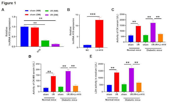
Figure 1: Effects of lncRNA H19 upregulation on CK, CK-MB, and LDH levels. (A) Relative lncRNA H19 expression determined by qRT-PCR in normal mice (NM) and diabetic mice (DM) following myocardial ischemia/reperfusion (I/R). (B) Relative lncRNA H19 expression following lentiviral H19 overexpression (Lv-H19). (C–E) Serum levels of creatine kinase (CK), creatine kinase-MB (CK-MB), and lactate dehydrogenase (LDH) in the indicated experimental groups. Data are presented as the mean ± SEM. P < 0.05, P < 0.01, P < 0.001. Abbreviations: CK = creatine kinase; CK-MB = creatine kinase-MB; DM = diabetic mice; I/R = ischemia/reperfusion; LDH = lactate dehydrogenase; lncRNA H19 = long non-coding RNA H19; Lv-H19 = lentiviral H19 overexpression; NC = negative control; NM = normal mice; qRT-PCR = quantitative reverse transcription polymerase chain reaction. Please click here to view a larger version of this figure.
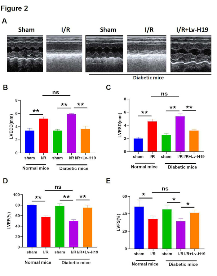
Figure 2: Cardiac function in diabetic mice following lncRNA H19 overexpression. (A) Representative M-mode echocardiographic images of the left ventricle from normal sham, normal ischemia/reperfusion, diabetic sham, diabetic ischemia/reperfusion, and diabetic ischemia/reperfusion mice treated with lentiviral H19. (B) Left ventricular end-diastolic diameter. (C) Left ventricular end-systolic diameter. (D) Left ventricular ejection fraction. (E) Left ventricular fractional shortening. Data are presented as the mean ± SEM. ns, not significant; P < 0.05; P < 0.01. Abbreviations: DM = diabetic mice; I/R = ischemia/reperfusion; LVEF = left ventricular ejection fraction; LVEDD = left ventricular end-diastolic diameter; LVESD = left ventricular end-systolic diameter; LVFS = left ventricular fractional shortening; Lv-H19 = lentiviral H19 overexpression; NM = normal mice. Please click here to view a larger version of this figure.
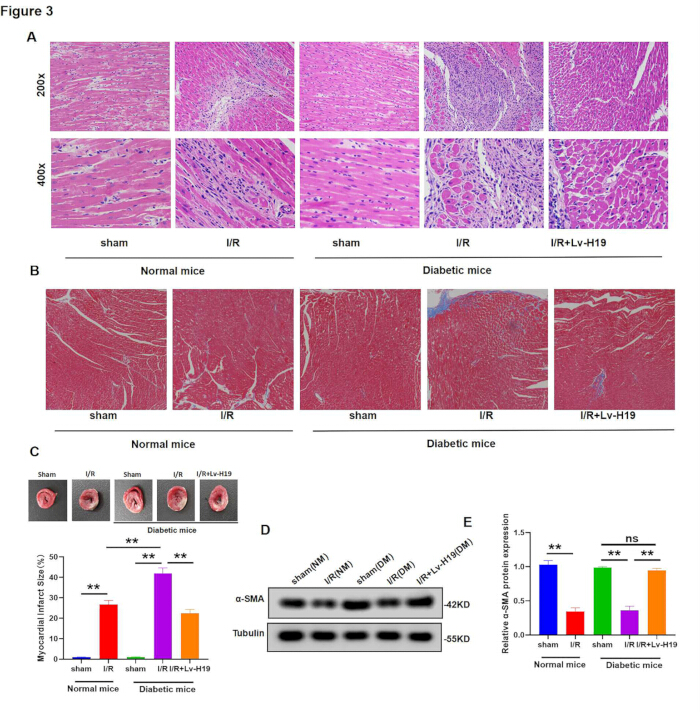
Figure 3: Histopathological changes and myocardial infarct size following lncRNA H19 overexpression. (A) Representative H&E-stained myocardial sections showing histopathological changes following myocardial ischemia/reperfusion. (B) Representative Masson's trichrome-stained sections showing myocardial fibrosis and collagen deposition (blue). (C) Representative Evans blue/TTC-stained heart sections and quantification of infarct size. (D) Representative western blot analysis of α-smooth muscle actin (α-SMA). (E) Quantification of α-SMA protein expression. Data are presented as the mean ± SEM. ns, not significant; P < 0.01. Abbreviations: α-SMA = alpha-smooth muscle actin; DM = diabetic mice; H&E = hematoxylin and eosin; I/R = ischemia/reperfusion; NM = normal mice; TTC = triphenyl tetrazolium chloride. Please click here to view a larger version of this figure.
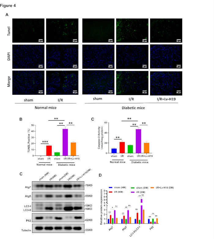
Figure 4: Cardiomyocyte apoptosis and autophagy following lncRNA H19 overexpression. (A) Representative TUNEL-stained myocardial sections. (B) Quantification of apoptotic cardiomyocytes. (C) Caspase-3 activity in the indicated experimental groups. (D) Representative western blots of autophagy-related proteins. (E–H) Quantification of Atg7, Atg5, p62, and the LC3-II/LC3-I ratio. Data are presented as the mean ± SEM. ns, not significant; P < 0.05; P < 0.01; P < 0.001. Abbreviations: Atg = autophagy-related protein; DM = diabetic mice; I/R = ischemia/reperfusion; LC3 = microtubule-associated protein 1 light chain 3; NM = normal mice; p62 = sequestosome 1; TUNEL = terminal deoxynucleotidyl transferase dUTP nick-end labeling. Please click here to view a larger version of this figure.
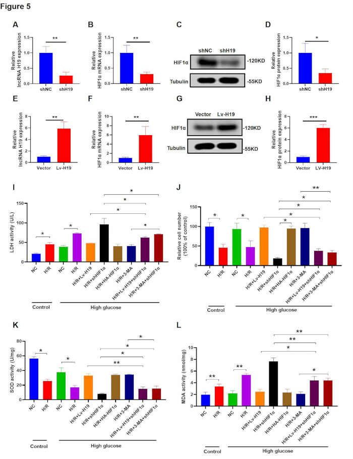
Figure 5: Regulation of HIF-1α signaling by lncRNA H19 in AC16 cells. (A,B) Relative lncRNA H19 and hypoxia-inducible factor 1 alpha (HIF-1α) mRNA expression following lncRNA H19 knockdown in AC16 cells. (C,D) Representative western blots and quantification of HIF-1α protein expression after lncRNA H19 knockdown. (E,F) Relative lncRNA H19 and HIF-1α mRNA expression following lncRNA H19 overexpression. (G,H) Representative western blots and quantification of HIF-1α protein expression after lncRNA H19 overexpression. (I) Lactate dehydrogenase (LDH) activity. (J) Cell viability determined by MTT assay. (K) Superoxide dismutase (SOD) activity. (L) Malondialdehyde (MDA) levels. Data are presented as the mean ± SEM. ns, not significant; P < 0.05; P < 0.01. Abbreviations: 3-MA = 3-methyladenine; H/R = hypoxia/reoxygenation; HA-HIF-1α = HIF-1α overexpression plasmid; HIF-1α = hypoxia-inducible factor 1 alpha; LDH = lactate dehydrogenase; Lv-H19 = lentiviral H19 overexpression; MDA = malondialdehyde; MTT = 3-(4,5-dimethylthiazol-2-yl)-2,5-diphenyltetrazolium bromide; qRT-PCR = quantitative reverse transcription polymerase chain reaction; shHIF-1α = HIF-1α short hairpin RNA; SOD = superoxide dismutase. Please click here to view a larger version of this figure.
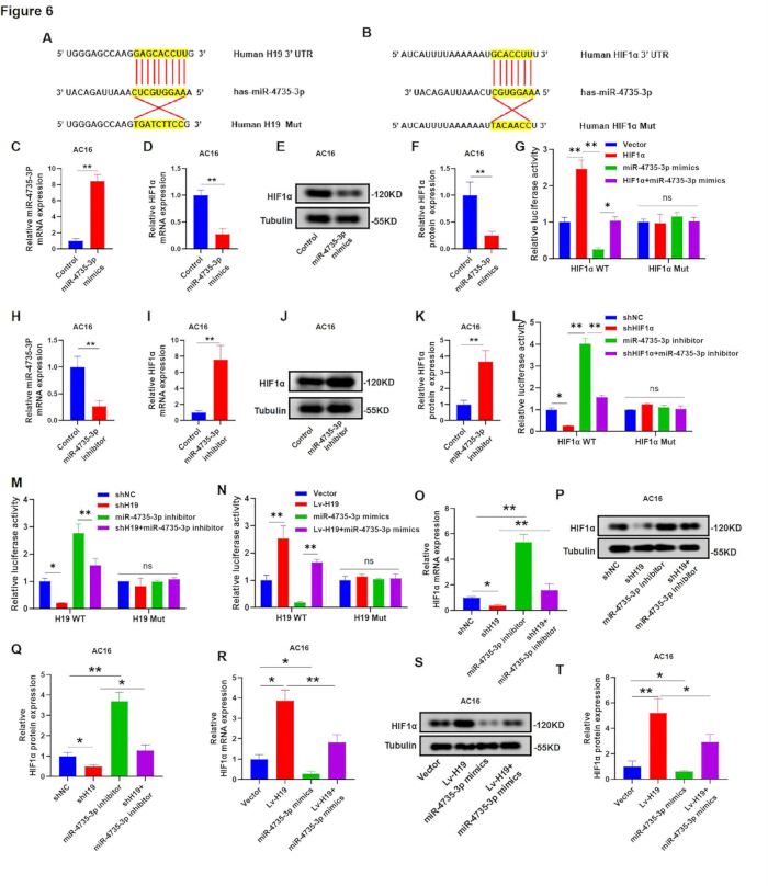
Figure 6: Competing endogenous RNA network involving lncRNA H19, miR-4735-3p, and HIF-1α. (A,B) Predicted binding sites between miR-4735-3p and H19 or HIF-1α were generated using bioinformatics databases. (C–F) Relative miR-4735-3p and HIF-1α expression and HIF-1α protein levels following transfection with miR-4735-3p mimics. (G) Dual-luciferase reporter assay showing the interaction between miR-4735-3p and the HIF-1α 3′UTR. (H–L) Relative miR-4735-3p and HIF-1α expression, HIF-1α protein levels, and luciferase activity following transfection with a miR-4735-3p inhibitor. (M,N) Dual-luciferase reporter assays demonstrating direct binding between miR-4735-3p and lncRNA H19. (O–T) Relative HIF-1α mRNA and protein expression following co-transfection with the indicated plasmids and miR-4735-3p inhibitor or mimics. Data are presented as the mean ± SEM. ns, not significant; P < 0.05; P < 0.01. Abbreviations: ceRNA = competing endogenous RNA; HIF-1α = hypoxia-inducible factor 1 alpha; 3′UTR = 3′ untranslated region; HIF1A-MUT = mutant HIF-1α 3′UTR; HIF1A-WT = wild-type HIF-1α 3′UTR; H19-MUT = mutant H19; H19-WT = wild-type H19; MUT = mutant; qRT-PCR = quantitative reverse transcription polymerase chain reaction; UTR = untranslated region; WT = wild type. Please click here to view a larger version of this figure.
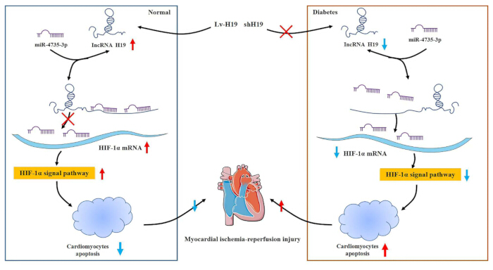
Figure 7: Proposed mechanism by which lncRNA H19 attenuates myocardial ischemia/reperfusion injury in diabetic mice. Schematic illustration of the proposed mechanism in which lncRNA H19 functions as a competing endogenous RNA by sponging miR-4735-3p, thereby promoting HIF-1α signaling and attenuating myocardial ischemia/reperfusion injury in diabetic mice. Abbreviations: ceRNA = competing endogenous RNA; HIF-1α = hypoxia-inducible factor 1 alpha; lncRNA H19 = long non-coding RNA H19; miR-4735-3p = microRNA-4735-3p. Please click here to view a larger version of this figure.
Supplemental Table S1: Biological characteristics of C57BL/KsJ db/+ and C57BL/KsJ db/db mice. Please click here to download this file.
Supplemental Table S2: Primer sequences for qRT-PCR Please click here to download this file.
Discussion
Myocardial ischemia-reperfusion (I/R) injury is a key issue in cardiovascular disease, particularly exacerbated in patients with diabetes mellitus (DM), where hyperglycemia and metabolic dysregulation contribute to poor outcomes40,41,42. Researchers have found that diabetes mellitus aggravates myocardial ischemia-reperfusion injury (MIRI) and affects the effectiveness of ischemic preconditioning and certain cardioprotective drugs43,44. To further understand the functions of lncRNA H19 in cardiomyocytes, we performed a comprehensive screening of lncRNA expression in I/R models of both diabetic (DM) and non-diabetic (NM) mice. Chip analysis and qRT-PCR revealed that several lncRNAs, including Inpp5d, Adapt33, AK082072, and Rmst, were similarly downregulated following I/R injury. However, among these, the lncRNA H19 showed the most significant reduction, suggesting a potentially critical role in MIRI pathophysiology. Additionally, lncRNA H19 has been reported to play a crucial role in cardiovascular biology, including embryonic heart development and cellular stress responses, particularly in ischemia/reperfusion injury and vascular remodeling, suggesting its involvement in cardioprotective mechanisms. Given its pronounced downregulation, prior evidence of its cardioprotective role, and its known interactions with key signaling pathways involved in myocardial injury and repair, we selected lncRNA H19 for further investigation. The study demonstrated that the lncRNA H19 attenuates MIRI in diabetic mice, indicating a protective mechanism that may enhance cell survival and functional recovery post-ischemia. Our findings showed that H19 expression levels in the DM and NM models differed significantly. In DM, lncRNA H19 expression was markedly downregulated following I/R injury, suggesting a potential protective role. This finding aligns with previous studies indicating that lncRNA H19 can act as a cardioprotective agent by modulating various molecular pathways involved in stress responses and cellular survival. In contrast, DM models exhibited a blunted upregulation of lncRNA H19, which may contribute to the exacerbated I/R injury observed in diabetic conditions.
Apoptosis and autophagy are critical cellular processes implicated in responding to I/R injury45. Our study revealed that upregulating lncRNA H19 significantly reduced apoptosis rates and modulated autophagy levels in cardiomyocytes. Notably, the pro-apoptotic activity of Caspase-3 was significantly suppressed by lncRNA H19 upregulation. This shift in the balance of apoptotic regulators likely contributes to the observed cardioprotective effects. In terms of autophagy, overexpression of lncRNA H19 regulates the expression of autophagy-related genes, including LC3-II and p62. Autophagy acts as a dual player in I/R injury, where excessive autophagy can lead to cell death, while moderate autophagy can promote cellular survival by removing damaged organelles and proteins46. The current research results indicate that lncRNA H19 can prevent excessive autophagy and restore autophagy flux to a protective level, thereby reducing I/R damage, rather than just enhancing or inhibiting autophagy.
As a pivotal regulator of cellular responses to hypoxia, HIF-1α showed a similar trend. In NM models, HIF-1α expression was significantly elevated post-I/R injury, potentially facilitating adaptive responses to hypoxic stress. However, in DM models, the increase in HIF-1α expression was less pronounced, suggesting impaired hypoxic response mechanisms in diabetic cardiomyocytes. These findings underscore the complex interplay between diabetes, hypoxia, and cardiac injury, emphasizing the necessity of targeted therapeutic strategies. In our results, lncRNA H19 alleviates H/R injury by activating the HIF-1α signaling pathway. The activation of this pathway by lncRNA H19 signifies a crucial adaptive response that not only protects cardiomyocytes from ischemic damage but also enhances the myocardium's overall resilience during I/R episodes. This indicates that lncRNA H19 may be essential for the metabolic response of cardiac cells to stress, thereby enhancing blood flow and oxygen supply to the affected areas.
Next, we investigated the mechanism whereby lncRNA H19 regulated HIF-1α. Research is still ongoing, but it appears that miRNAs and lncRNAs are involved in nearly all cellular processes, including myocardial I/R injury. The complex interaction between miRNAs and lncRNAs in the control of gene expression is highlighted by this competing endogenous RNA (ceRNA) activity. This mechanism of action underscores the intricate interplay between lncRNAs and miRNAs in gene regulation. In this study, TargetScan and StarBase v2.0 predicted miR-4735-3p as a target of lncRNA H19 via a putative binding site. While direct interactions have not been widely reported, miR-4735-3p has been implicated in vascular remodeling and cellular stress responses, processes also influenced by lncRNA H1940. Collectively, these findings support a model in which lncRNA H19 regulates miR-4735-3p during I/R injury, and its function as a ceRNA for this miRNA adds a critical regulatory layer governing HIF-1α expression. By functioning as a molecular sponge for miR-4735-3p, lncRNA H19 prevents this miRNA from binding to its target, thereby alleviating the repressive effects on HIF-1α. The following observations form the basis of this conclusion. First, miR-4735-3p overexpression reduced HIF-1α expression. MiR-4735-3p reduced the luciferase activity of the wild-type (WT) 3'UTR of HIF-1α but not that of the mutant (MUT) 3'UTR. Second, luciferase reporter assays confirmed that miR-4735-3p and lncRNA H19 were bound together. Moreover, these observations demonstrated that downregulated miR-4735-3p attenuated the impact of lncRNA H19 knockdown on HIF-1α, and upregulated miR-4735-3p attenuated the influence of lncRNA H19 overexpression on HIF-1α. Overall, these findings demonstrated that lncRNA H19 acts as a ceRNA for miR-4735-3p, thereby contributing to HIF-1α expression.
At present, a consistent mechanistic model of the heart-protective effect of lncRNA H19 in MIRI of diabetes has been identified. Under the condition of diabetes, miR-4735-3p was upregulated and combined with 3'UTR of lncRNA H19 and HIF-1α mRNA to inhibit the expression of HIF-1α. This inhibition can impair the HIF-1α-mediated autophagic response, leading to the accumulation of damaged mitochondria, excessive ROS production, and exacerbation of I/R damage. When lncRNA H19 is overexpressed, it acts as a competitive endogenous RNA (ceRNA), isolating miR-4735-3p and thereby releasing HIF-1α translation. The increase in HIF-1α protein levels promotes autophagy through BNIP3 and FUNDC1, facilitates the clearance of damaged mitochondria, reduces oxidative stress, and maintains the vitality of myocardial cells. This comprehensive model links the non-coding RNA regulatory network (lncRNA H19/miR-4735-3p) with the functional protection pathway (HIF-1α/autophagy), and provides a framework for understanding the molecular basis of H19-mediated MIRI cardioprotection in diabetes.
This study used both primary mouse cardiomyocytes and human AC16 cells. Although the core H19/miR-4735-3p/HIF-1α regulatory axis appears conserved across species, several considerations warrant discussion. Firstly, species-specific differences in miRNA seed sequences and target-site affinity may affect the quantitative aspects of regulatory interactions. Secondly, the AC16 cell line, derived from human ventricular cardiomyocytes fused with SV40-transformed fibroblasts, may not fully reproduce the complex metabolic and contractile phenotypes of primary adult cardiomyocytes. Third, the diabetic environment in mice involves systemic metabolic disorders and cannot be fully recapitulated in cell culture. Despite these limitations, the consistent identification of the two cell types supports the effectiveness of the proposed mechanism and indicates that the H19/miR-4735-3p/HIF-1α pathway is a conserved regulatory module that warrants further research in human diabetes MIRI.
It is acknowledged that the manuscript does not directly compare lncRNA H19 levels and its impact between normal and diabetic mice following ischemia/reperfusion (I/R). Our primary focus was on the diabetic mouse model, as diabetes exacerbates MIRI, making it particularly relevant for investigating the role of lncRNA H19. While a comparison with normal mice could offer additional insights, our study aimed to assess H19's therapeutic potential specifically in diabetic MIRI. We recognize this limitation and suggest future studies could further explore its effects in both normal and diabetic models. In addition, Xie et al. explored the relationship between lncRNA H19 and HIF-1α, but their study differs from ours in experimental models and mechanisms47. While our research focuses on diabetic mice with myocardial ischemia/reperfusion injury (MIRI), emphasizing H19's role as a ceRNA for miR-4735-3p that activates the HIF-1α signaling pathway, Xie’s work investigates hypoxia-induced injury in cardiomyocytes and myocardial infarction (MI) rats, where H19 stabilizes HIF-1α through direct transcriptional regulation. These differences highlight the complex and context-dependent roles of lncRNA H19 in myocardial injury, underscoring the need for further research to fully understand its multifaceted functions in different pathological settings.
In summary, the intricate control of gene expression in the heart is underscored by the protective effects of lncRNA H19 in MI/RI via activation of the HIF-1α signaling pathway and its role as a ceRNA for miR-4735-3p (Figure 7). The results of this study indicate that strategies aimed at enhancing the expression of lncRNA H19 or regulating its interaction with the HIF-1α pathway deserve further study in the preclinical model of diabetes MIRI. However, it is important to emphasize that the current data come from individual mouse models and cultured cell systems; therefore, these observational results need to be studied in large-animal models and final human tissues in the future to evaluate the translational relevance of the H19/miR-4735-3p/HIF-1α axis. This study lays the foundation for future research of this kind.
Disclosures
The authors have no conflicts of interest to declare.
Authors' contributions
Conceptualization, X.D. and K.Q.; methodology, L.G.; software, Y.L.; validation, Y.C., J.S., and X.H.; formal analysis, L.G.; investigation, L.G.; resources, L.G.; data curation, Y.L.; writing-original draft preparation, L.G.; writing-review and editing, X.D. and K.Q.; visualization, Y.L.; supervision, X.D. and K.Q.; project administration, L.G.; funding acquisition, X.D. and K.Q. All authors have read and agreed to the published version of the manuscript.
Acknowledgements
This research was funded by the National Natural Science Foundation of China (No. 82160364 and No.82460228) and the Science and Technology Foundation of Jiangxi Province (No. 20212BAB216056 and 20232BCJ22016).
Materials
| Name | Company | Catalog Number | Comments |
|---|---|---|---|
| 15-MHz linear transducer | Sequoia Acuson, Siemens, Erlangen, Germany | ||
| 293T cells | BeNa Culture Collection in Shanghai,china | ||
| 293T cells | BeNa Culture Collection in Shanghai, China | BNCC340228 | |
| anti-APG5L/ATG5 | ab108327, Abcam | ||
| anti-Apg7 | 2631, Cell Signaling Technology, | ||
| anti-LC3A/B | ab62721, Abcam, | ||
| anti-rabbit/mouse IgG H&L (HRP) secondary antibody | ZXGB-Bio, | ||
| anti-SQSTM1/p62 | 5114, Cell Signaling Technology, | ||
| anti-Tubulin | 11224-1-AP, Proteintech, | ||
| anti-α-Smooth Muscle Actin | 19245, Cell Signaling Technology, | ||
| BLOCK-iT RNAi Designer | Invitrogen | ||
| caspase-3 activity assay kit | BestBio, China | C1115 | |
| CK assay kit | Nanjing Jiancheng Bioengineering Institute, Nanjing, China | ||
| CK-MB assay kit | Nanjing Jiancheng Bioengineering Institute, Nanjing, China | ||
| Collagenase type B/D | R&D Systems, Minneapolis, MN, USA | 11088858001 | |
| ECL Plus | Life Technology, USA | ||
| HIF-1α | 36169, Cell Signaling Technology, | ||
| Human cardiomyocyte AC16 cells | Shanghai Hengya Biotechnology Co., Ltd. | AC335797 | |
| Knockdown (KD) of HIF-1α expression in AC16 | Genepharma Corporation Ltd.,Shanghai, China | ||
| LDH Cytotoxicity Test Kit | Beyotime, Shanghai, China | ||
| light microscope | Leica Microsystems, Wetzlar, Germany | LS2 | |
| Lipofectamine 3000 | Life Technologies, USA | L3000001 | |
| Malondialdehyde Assay Kit | Solarbio | ||
| Mason-Ponceau acidic magenta solution | Sigma-Aldrich, St. Louis, MO, USA | 210-043-9 | |
| miRNA target expression vector | Promega, Madison, WI, USA | E1330 | |
| Pmir-GLO dual-luciferase miRNA target expression vector | Promega, Madison, WI, USA | ||
| PrimeScript 1st Strand cDNA Synthesis Kit | TaKaRa, Japan | ||
| PVDF membrane | Millipore, USA | ||
| QuantiTect SYBR Green RT-PCR Kit | QIAGEN, Germany | ||
| RIPA lysis buffer | Beyotime, China | L13578 | |
| siRNA-HIF- 1α | Thermo Fisher Scientific,Inc. | ||
| The Total Superoxide Dismutase Assay Kit | Solarbio, Beijing, China | ||
| TRIzol reagent | Invitrogen, USA | ||
| Wiegert's iron hematoxylin solution | Sigma-Aldrich, St. Louis, MO, USA | MFCD00132590 | |
| ImageJ | National Institutes of Health (NIH) | N/A | Image analysis |
| TargetScan | TargetScan | N/A | miRNA target prediction database |
| StarBase v2.0 | Sun Yat-sen University | N/A | RNA interaction database |
| GenBank BLAST | National Center for Biotechnology Information (NCBI) | N/A | Sequence alignment |
| Ambion siRNA Designer | Thermo Fisher Scientific | N/A | siRNA design tool |
| psPAX2 plasmid | Addgene | 12260 | Lentiviral packaging plasmid |
| pMD2.G plasmid | Addgene | 12259 | Lentiviral envelope plasmid |
| pGFP-V-RS plasmid | OriGene Technologies | N/A | Empty/shRNA vector |
References
- Goldsborough E 3rd, Osuji N, Blaha MJ. Assessment of cardiovascular disease risk: a 2022 update. Endocrinol Metab Clin North Am. 2022;51(3):483-509.
- Cunha PS, Laranjo S, Heijman J, Oliveira MM. The atrium in atrial fibrillation—a clinical review on how to manage atrial fibrotic substrates. Front Cardiovasc Med. 2022;9:879984.
- Lv Y, Zhao S. Diagnosis of cardiac rehabilitation after percutaneous coronary intervention in acute myocardial infarction patients by emission computed tomography image features under filtered back projection reconstruction algorithm. J Healthc Eng. 2021;2021:6844549.
- Vieta E, et al. Bipolar disorders. Nat Rev Dis Primers. 2018;4:18008.
- Khanji MY, et al. Personalized E-coaching in cardiovascular risk reduction: a randomized controlled trial. Ann Glob Health. 2019;85(1):107.
- Shen S, et al. Waist-to-height ratio is an effective indicator for comprehensive cardiovascular health. Sci Rep. 2017;7:43046.
- Atef Abdelsattar Ibrahim H, et al. Glucose indices as inflammatory markers in children with acute surgical abdomen: a cross-sectional study. Ann Med. 2023;55(2):2248454.
- Han H, et al. Development and validation of a prediction model of perioperative hypoglycemia risk in patients with type 2 diabetes undergoing elective surgery. BMC Surg. 2022;22(1):167.
- Boťanská B, et al. Matrix metalloproteinases and their role in mechanisms underlying effects of quercetin on heart function in aged Zucker diabetic fatty rats. Int J Mol Sci. 2021;22(9):4457.
- Le LTT, Nhu CXT. The role of long non-coding RNAs in cardiovascular diseases. Int J Mol Sci. 2023;24(18):13805.
- Jin Z, et al. Notoginsenoside R1 protects hypoxia-reoxygenation deprivation-induced injury by upregulation of miR-132 in H9c2 cells. Hum Exp Toxicol. 2021;40(12 Suppl):S29-38.
- Nong SX, et al. Differential expression and significance of peripheral blood genes in coronary artery heart disease. J Thorac Dis. 2022;14(9):3415-28.
- Ferenczyová K, et al. Pharmacology of catechins in ischemia-reperfusion injury of the heart. Antioxidants (Basel). 2021;10(9):1390.
- Ma B, et al. Dapper1 promotes autophagy by enhancing the Beclin1-Vps34-Atg14L complex formation. Cell Res. 2014;24(8):912-24.
- Glantzounis GK, et al. Recent advances in the surgical management of hepatocellular carcinoma. Ann Gastroenterol. 2021;34(4):453-65.
- Andreescu M. Recent advances in serum biomarkers for cardiological risk stratification and insight into the cardiac management of the patients with hematological malignancies treated with targeted therapy. Cureus. 2023;15(11):e49696.
- Zhao C, et al. PINK1/Parkin-mediated mitophagy regulation by reactive oxygen species alleviates rocaglamide A-induced apoptosis in pancreatic cancer cells. Front Pharmacol. 2019;10:968.
- Chu CT, Bayır H, Kagan VE. LC3 binds externalized cardiolipin on injured mitochondria to signal mitophagy in neurons: implications for Parkinson disease. Autophagy. 2014;10(2):376-8.
- Miyamoto Y, et al. Possible existence of lysosome-like organella within mitochondria and its role in mitochondrial quality control. PLoS One. 2011;6(1):e16054.
- Liu Y, Shah KM, Luo J. Strategies for articular cartilage repair and regeneration. Front Bioeng Biotechnol. 2021;9:770655.
- Wang S, et al. The mitophagy pathway and its implications in human diseases. Signal Transduct Target Ther. 2023;8(1):304.
- Mu J, et al. BRD4 inhibition by JQ1 prevents high-fat diet-induced diabetic cardiomyopathy by activating PINK1/Parkin-mediated mitophagy in vivo. J Mol Cell Cardiol. 2020;149:1-14.
- Edmunds LR, et al. Liver-specific Prkn knockout mice are more susceptible to diet-induced hepatic steatosis and insulin resistance. Mol Metab. 2020;41:101051.
- He Q, et al. Natural products: protective effects against ischemia-induced retinal injury. Front Pharmacol. 2023;14:1149708.
- Wang M, et al. Mesenchymal stem cell secretions improve donor heart function following ex vivo cold storage. J Thorac Cardiovasc Surg. 2022;163(4):e277-92.
- Bao Q, et al. CNP ameliorates macrophage inflammatory response and atherosclerosis. Circ Res. 2024;134(8):e72-91.
- Zhang W, et al. Mitophagy mediated by HIF-1α/FUNDC1 signaling in tubular cells protects against renal ischemia/reperfusion injury. Ren Fail. 2024;46(1):2332492.
- Chen Z, et al. Bioinformatics integration reveals key genes associated with mitophagy in myocardial ischemia-reperfusion injury. BMC Cardiovasc Disord. 2024;24(1):183.
- Akhter N, et al. Endoplasmic reticulum stress promotes the expression of TNF-α in THP-1 cells by mechanisms involving ROS/CHOP/HIF-1α and MAPK/NF-κB pathways. Int J Mol Sci. 2023;24(20):15186.
- Ba X, et al. WTD attenuating rheumatoid arthritis via suppressing angiogenesis and modulating the PI3K/AKT/mTOR/HIF-1α pathway. Front Pharmacol. 2021;12:696802.
- Li P, Liu C, Yu Z, Wu M. New insights into regulatory T cells: exosome- and non-coding RNA-mediated regulation of homeostasis and resident Treg cells. Front Immunol. 2016;7:574.
- Greene J, et al. Differential CircRNA expression signatures may serve as potential novel biomarkers in prostate cancer. Front Cell Dev Biol. 2021;9:605686.
- Rutkowska-Zapała M, et al. Transcriptome profiling of regulatory T cells from children with transient hypogammaglobulinemia of infancy. Clin Exp Immunol. 2023;214(3):275-88.
- Zhao L, et al. LncRNA KCNQ1OT1 promotes the development of diabetic nephropathy by regulating miR-93-5p/ROCK2 axis. Diabetol Metab Syndr. 2021;13(1):108.
- Luo H, et al. The lncRNA H19/miR-675 axis regulates myocardial ischemic and reperfusion injury by targeting PPARα. Mol Immunol. 2019;105:46-54.
- Yu SY, et al. Knockdown of lncRNA AK139328 alleviates myocardial ischaemia/reperfusion injury in diabetic mice via modulating miR-204-3p and inhibiting autophagy. J Cell Mol Med. 2018;22(10):4886-98.
- Cao HX, et al. Effects of rutin on myocardial enzyme and cardiac morphology in diabetic mice. Sichuan Da Xue Xue Bao Yi Xue Ban. 2018;49(4):570-4.
- Liao H, et al. Silencing hsa_circ_0049271 attenuates hypoxia-reoxygenation (H/R)-induced myocardial cell injury via the miR-17-3p/FZD4 signaling axis. Ann Transl Med. 2023;11(2):99.
- Zhou Y, et al. The anti-photoaging effect of C-phycocyanin on ultraviolet B-irradiated BALB/c-nu mouse skin. Front Bioeng Biotechnol. 2023;11:1229387.
- Wu Y, et al. (-)-Epigallocatechin-3-gallate attenuates myocardial injury induced by ischemia/reperfusion in diabetic rats and in H9c2 cells under hyperglycemic conditions. Int J Mol Med. 2017;40(2):389-99.
- Klapper-Goldstein H, et al. VDAC1 in the diseased myocardium and the effect of VDAC1-interacting compound on atrial fibrosis induced by hyperaldosteronism. Sci Rep. 2020;10(1):22101.
- Hu M, et al. Metformin protects H9C2 cardiomyocytes from high-glucose and hypoxia/reoxygenation injury via inhibition of reactive oxygen species generation and inflammatory responses: role of AMPK and JNK. J Diabetes Res. 2016;2016:2961954.
- Yan T, et al. Effects of myocardial fibrosis on ischemic preconditioning against myocardial ischemia-reperfusion injury in type 2 diabetic rats. Zhonghua Yi Xue Za Zhi. 2021;101(47):3857-63.
- Liu X, et al. Remote liver ischemic preconditioning attenuates myocardial ischemia/reperfusion injury in streptozotocin-induced diabetic rats. Sci Rep. 2021;11(1):1903.
- Wang H, et al. Propofol attenuates hypoxia/reoxygenation-induced apoptosis and autophagy in HK-2 cells by inhibiting JNK activation. Yonsei Med J. 2019;60(12):1195-202.
- Wang X, Lu W, Liu B, Xu Y. Thrombin aggravates hypoxia/reoxygenation injury of astrocytes by activating the autophagy pathway mediated by SPRED2. Exp Ther Med. 2021;22(4):1107.
- Xie L, et al. The interplay between H19 and HIF-1α in mitochondrial dysfunction in myocardial infarction. Cell Signal. 2023;112:110919.