This retrospective cohort study examines adjunctive Huoxue Huayu therapy in cataract patients with allergic conjunctivitis. Compared with standard therapy alone, adjunctive treatment was associated with better visual outcomes, fewer ocular-surface symptoms, lower serum inflammatory-marker levels, better quality of life, and fewer 30-day macular-edema events, although causality cannot be established.
Research Article
Adjunctive Huoxue Huayu Therapy and Postoperative Outcomes in Cataract Patients with Allergic Conjunctivitis: A Retrospective Cohort Study
* These authors contributed equally
In This Article
Summary
Abstract
This retrospective cohort study evaluated the association of adjunctive Huoxue Huayu therapy with postoperative outcomes in cataract patients with concomitant allergic conjunctivitis. We analyzed 158 patients who underwent phacoemulsification and intraocular lens implantation between June 2024 and June 2025, with one predefined study eye included per patient. All patients received standard postoperative pharmacologic therapy, and 79 additionally received Huoxue Huayu therapy through routine physician–patient decision-making. Patient-level propensity scores were estimated using multivariable logistic regression, followed by 1:1 nearest-neighbor matching without replacement; repeated outcomes were analyzed using generalized estimating equations. After matching, 78 patients were retained in each group with balanced baseline characteristics. At postoperative day 7, near visual acuity was 0.30 ± 0.10 in the adjunctive-treatment group and 0.20 ± 0.08 in the control group, while distance visual acuity was 0.70 ± 0.20 and 0.60 ± 0.15, respectively. Ocular Surface Disease Index scores were consistently lower in the adjunctive-treatment group at 7 days, 1 month, and 3 months. Day-7 serum interleukin-6 levels were 18.43 ± 4.27 versus 26.71 ± 5.93 pg/mL, and tumor necrosis factor-α levels were 23.56 ± 5.18 versus 34.82 ± 7.41 pg/mL. The adjunctive-treatment group also had lower aqueous flare grades, higher visual-function and cataract-specific quality-of-life scores, and fewer 30-day macular-edema events (2.56% vs. 14.10%). No serious treatment-related adverse events were documented. Adjunctive Huoxue Huayu therapy was associated with more favorable postoperative clinical, patient-reported, and serum inflammatory-marker outcomes; however, the retrospective nonrandomized design precludes conclusions regarding efficacy or causality.
Introduction
Cataract is the leading cause of blindness globally. According to the World Health Organization, over half of all blindness is attributable to cataract1,2. Phacoemulsification, with its small incision (2–3 mm), rapid postoperative recovery, and reliable visual improvement, has become the gold standard for cataract surgery worldwide. However, the procedure may disrupt the corneal epithelium, tear film, and blood–aqueous barrier, thereby promoting local ocular inflammation and increasing the risk of postoperative complications3,4,5. Among these, the incidence of dry eye can be as high as 10% to 85%, often manifesting as shortened tear film break-up time, positive corneal fluorescein staining, foreign body sensation, photophobia, and visual fatigue, markedly compromising patients’ postoperative quality of life and visual satisfaction.
A further clinical challenge is that a substantial proportion of patients with cataract also have allergic conjunctivitis. This condition is triggered by allergens such as pollen and dust mites, which provoke IgE-mediated type I hypersensitivity, leading to conjunctival hyperemia, edema, and increased mucus secretion, leaving the ocular surface in a chronic inflammatory “primed” state. When these patients undergo phacoemulsification, surgical trauma may compound the pre-existing allergic inflammation, creating a possible “two-hit” effect. Contact with surgical instruments, ultrasound energy, and irrigating solutions may further activate mast cells and eosinophils and promote the release of local inflammatory mediators. Meanwhile, pre-existing ocular-surface inflammation may increase corneal nerve sensitivity and disrupt tear-secretion regulation, thereby aggravating postoperative ocular discomfort and inflammatory responses6,7,8. Studies have shown that cataract patients with coexisting allergic conjunctivitis exhibit significantly greater postoperative inflammatory reactions than non-allergic patients, and their risk of serious complications such as macular edema and persistent anterior chamber inflammation is markedly elevated.
Beyond conventional lubricating and anti-inflammatory treatments, biologically derived products have increasingly been investigated as adjunctive approaches for severe ocular-surface inflammation and impaired epithelial repair. Human umbilical cord blood-derived preparations contain growth factors, cytokines, and anti-inflammatory proteins that may support epithelial regeneration and modulate the ocular-surface inflammatory environment. A prospective pilot study reported improvements in symptoms, tear-film parameters, visual acuity, and epithelial healing after treatment with umbilical cord blood serum in patients with severe ocular-surface disorders associated with systemic autoimmune diseases and other refractory conditions9. Similarly, umbilical cord blood platelet lysate eyedrops were associated with improvements in patient-reported symptoms and clinical ocular-surface findings in patients with severe ocular graft-versus-host disease10. Although these topical perinatal tissue-derived therapies differ substantially from adjunctive Huoxue Huayu therapy and were evaluated in more severe disease populations, they illustrate the broader development of adjunctive ocular-surface therapies that combine immunomodulatory and regenerative objectives.
From the perspective of traditional Chinese medicine (TCM), the core pathogenesis of post-cataract surgery with allergic conjunctivitis can be summarized as “blood stasis obstructing the collaterals, yin deficiency with blood dryness, and external invasion of wind pathogen.” The surgical wound by the metal blade causes extravasated blood to stagnate in the eye collaterals, impeding the normal flow of qi and blood; aging and physical decline lead to liver and kidney yin deficiency, leaving insufficient yin-blood to nourish the ocular orifices; on top of this, wind pathogen, carrying the allergens, invades from the exterior—wind predominance gives rise to itching, while heat predominance leads to swelling. As emphasized in Ten Lectures by Eminent Ophthalmologists of Traditional Chinese Medicine, the postoperative use of formulas that activate blood and resolve stasis while nourishing yin, such as modified Fu Yuan Huo Xue Decoction, can promote the absorption of residual cortical material, accelerate wound healing and restoration of transparency of the refractive media, reduce inflammatory responses, and also protect the optic nerve and maintain intraocular pressure stability11,12. The Huoxue Huayu principle of activating blood and resolving stasis has long been a core strategy in TCM ophthalmology for treating ocular blood stasis syndromes.
However, evidence regarding adjunctive Huoxue Huayu therapy in cataract patients with coexisting allergic conjunctivitis remains limited. Previous studies have generally focused on postoperative dry eye or nonspecific inflammation after uncomplicated cataract surgery and have not specifically examined patients with pre-existing allergic conjunctivitis, a clinically distinct subgroup that may experience a heightened postoperative inflammatory burden. The novelty of the present study lies in its focused evaluation of this population and its integrated assessment of visual acuity, ocular-surface symptoms, aqueous flare, serum inflammatory markers, patient-reported visual function and quality of life, and OCT-confirmed macular edema within a propensity-score-matched retrospective cohort. Accordingly, this study evaluated the association between adjunctive Huoxue Huayu therapy and postoperative outcomes in cataract patients with allergic conjunctivitis.
Protocol
Study design and ethics statement
The study protocol was approved by the Ethics Committee of Jingxing County Hospital (Approval No. YKZYYWLC2404). Because this was a retrospective study using existing clinical records, the Ethics Committee waived the requirement for informed consent. All data were de-identified before analysis, and the study was conducted in accordance with the Declaration of Helsinki. This single-center retrospective cohort study included patients who underwent phacoemulsification and intraocular lens implantation at Jingxing County Hospital between June 2024 and June 2025. The study was reported in accordance with the STROBE statement.
Patient screening and eligibility assessment
Hospital electronic medical records and cataract surgery records were screened for patients aged 50–80 years who underwent phacoemulsification and intraocular lens implantation during the study period. Demographic characteristics, ophthalmic diagnoses, operative records, postoperative treatment regimens, laboratory findings, and follow-up records were reviewed before treatment-group classification and outcome extraction.
Patients were included when all of the following criteria were met: diagnosis of age-related cortical, nuclear, or posterior subcapsular cataract13,14; completion of phacoemulsification and intraocular lens implantation; preoperative diagnosis of allergic conjunctivitis; regular use of olopatadine eye drops twice daily for at least 2 weeks before surgery, with allergic symptoms clinically controlled; uneventful surgery without posterior capsule rupture, vitreous loss, or another serious intraoperative complication; and complete clinical records covering at least 3 months of follow-up.
Patients were excluded when any of the following conditions were present: glaucoma, uveitis, retinal detachment, diabetic retinopathy, Sjögren’s syndrome, rheumatoid arthritis, or another ocular or systemic condition likely to affect the assessment of postoperative inflammation; systemic glucocorticoid or immunosuppressive treatment within 3 months before surgery; a documented allergy to any component of the Huoxue Huayu formula; severe hepatic or renal dysfunction, defined as alanine aminotransferase or aspartate aminotransferase greater than twice the upper limit of normal or serum creatinine greater than 177 µmol/L; incomplete clinical data; follow-up of less than 3 months; or loss to follow-up.
For patients who underwent bilateral surgery during the study period, only the first-operated eye was selected as the study eye. The fellow eye was excluded from propensity score matching and outcome analyses. Throughout the manuscript, “patient” refers to an enrolled participant, whereas “study eye” refers to the single predefined eye included in eye-specific analyses.
Diagnosis and grading of allergic conjunctivitis
Allergic conjunctivitis was diagnosed according to the Chinese Expert Consensus on the Diagnosis and Treatment of Allergic Conjunctivitis. The diagnosis was based on compatible ocular symptoms and signs, including ocular itching, conjunctival hyperemia, conjunctival papillary reaction, or chemosis, together with a positive skin-prick test or serum allergen-specific immunoglobulin E result.
Disease severity was recorded as mild or moderate according to the preoperative clinical grading documented by the attending ophthalmologist. Only patients whose allergic conjunctivitis had been clinically stabilized with olopatadine eye drops and were graded as mild or moderate before surgery were included. Patients with uncontrolled severe allergic conjunctivitis, acute exacerbation, or corneal involvement were not included in the analytical cohort.
Preoperative preparation
Before surgery, all patients used olopatadine eye drops twice daily for at least 2 weeks to control allergic conjunctivitis. Surgery was performed only after ocular itching, conjunctival hyperemia, and other allergic symptoms had become clinically stable and were graded as mild or moderate.
The preoperative assessment included distance and near visual acuity, tear film break-up time, Ocular Surface Disease Index score, slit-lamp examination, baseline serum IL-6 and TNF-α measurement, and macular optical coherence tomography. These assessments were obtained as part of routine clinical care and were retrospectively extracted from the medical records.
Phacoemulsification procedure
All surgeries were performed by the same surgical team using the Infiniti phacoemulsification system. A standardized 2.8 mm temporal clear corneal incision was created, followed by continuous curvilinear capsulorhexis with a target diameter of approximately 5.5 mm. Phacoemulsification was performed using a low-energy ultrasound strategy, with a mean ultrasound time of less than 1.5 min. A foldable intraocular lens was then implanted. The surgical procedure and intraoperative management were performed according to the same institutional protocol in both treatment groups.
Treatment allocation and postoperative conventional drug regimen
Treatment allocation was nonrandom and occurred at the patient level during routine postoperative care. All patients received the same standard postoperative pharmacologic regimen. Adjunctive Huoxue Huayu therapy was discussed with eligible patients as an additional treatment option. The final decision was made through discussion between the treating physician and the patient and primarily reflected the patient’s willingness and preference to receive oral herbal treatment. No randomization or prespecified treatment-allocation algorithm was used. Patients were classified retrospectively according to the postoperative treatment actually received and documented in the medical records.
The control group received tobramycin–dexamethasone eye drops four times daily, with the administration frequency tapered weekly until discontinuation; sodium hyaluronate eye drops four times daily; and oral loratadine 10 mg once daily. The conventional postoperative regimen continued for 4 weeks.
The observation group received the same standard postoperative pharmacologic regimen plus adjunctive Huoxue Huayu therapy, administered as a decoction beginning on postoperative day 1. Patients who underwent bilateral surgery received the same patient-level treatment regimen for both eyes.
Standardization, preparation, and administration of the Huoxue Huayu formula
The fixed base formula consisted of Salviae Miltiorrhizae Radix 15 g, Angelicae Sinensis Radix 12 g, Carthami Flos 6 g, Chuanxiong Rhizoma 10 g, Paeoniae Radix Rubra 10 g, and Rehmanniae Radix 15 g. All crude herbal materials were supplied and dispensed by the hospital's traditional Chinese medicine pharmacy. Before dispensing, the identity, appearance, and quality of each herbal material were checked by qualified pharmacy personnel in accordance with routine hospital procedures and the applicable standards of the Chinese Pharmacopoeia. The same pharmacopeial names, medicinal parts, dose specifications, and dispensing procedures were used throughout the study period.
Individualized additions were selected by the treating traditional Chinese medicine physician before the first dose according to the predominant clinical presentation documented in the medical record. When ocular itching was the predominant allergic symptom, Saposhnikoviae Radix 10 g and Tribuli Fructus 10 g were added. When marked conjunctival hyperemia was the predominant slit-lamp finding, Scutellariae Radix 10 g and Lonicerae Japonicae Flos 15 g were added. When persistent ocular dryness was the predominant complaint and a yin-deficiency pattern was documented during the traditional Chinese medicine assessment, Scrophulariae Radix 15 g and Ophiopogonis Radix 10 g were added.
When more than one clinical feature was present, the treating physician selected one modification according to the predominant presentation. Each patient, therefore, received either the fixed base formula alone or one predefined modification. The selected prescription was maintained throughout the 4-week treatment period unless treatment was discontinued because of intolerance or another safety concern.
All prescriptions were prepared centrally by the hospital's traditional Chinese medicine pharmacy using the same routine decoction procedure. One daily dose of the prescribed herbal materials was soaked in water for 30 min and decocted twice. The first decoction was performed for approximately 30 min, and the second for approximately 20 min. The two filtrates were combined and concentrated to approximately 250 mL. The final decoction was divided into two 125-mL portions and labeled with the patient identification and preparation date. Patients were instructed to take one 125-mL portion orally in the morning and the other in the evening for 4 consecutive weeks.
Treatment adherence was assessed using pharmacy dispensing records and postoperative follow-up documentation. The prescribed course comprised 56 administrations. Adherence was calculated as the number of documented administrations taken divided by 56 and multiplied by 100%. Adequate adherence was defined as completion of at least 80% of the prescribed administrations. Early discontinuation was defined as permanent cessation of the decoction before reaching the 80% adherence threshold. Temporary missed doses followed by continued treatment were not classified as discontinuation. The timing and documented reason for discontinuation were recorded.
Safety and tolerability were assessed retrospectively from medical records, pharmacy dispensing records, and follow-up documentation during the 3-month follow-up period. The review focused on events potentially related to the herbal decoction, including allergic reactions, gastrointestinal symptoms, treatment interruption or discontinuation, and serious treatment-related adverse events. When documented, the type of event, timing, management, and clinical outcome were extracted. Available alanine aminotransferase, aspartate aminotransferase, and serum creatinine results before treatment and at week 4 were also reviewed.
Blood sample collection and serum processing
Fasting venous blood samples were collected in the early morning before surgery and on postoperative day 7. For each assessment, 3 mL of venous blood was collected into a serum-separation tube. The samples were allowed to clot at room temperature for 30 min and were then centrifuged at approximately 1,000 × g for 15 min. The serum was separated into aliquots and stored at -80 °C until analysis. The preoperative and postoperative samples from the same patient were processed using the same procedure.
ELISA measurement of IL-6 and TNF-α
Serum IL-6 and TNF-α concentrations were measured using commercial enzyme-linked immunosorbent assay kits according to the manufacturer’s instructions. All serum samples and reagents were brought to the required assay temperature before testing. Standards and serum samples were added to the designated wells according to the kit protocol. Following the required incubation and washing steps, the detection reagents and substrate solution were added sequentially. The reaction was terminated using the stop solution supplied with the kit, and absorbance was measured using a microplate reader. Cytokine concentrations were calculated from the corresponding standard curves and expressed in pg/mL. Each serum sample was tested in duplicate, and the mean of the two measurements was used for statistical analysis.
Slit-lamp aqueous flare grading
Anterior chamber inflammation was evaluated on postoperative day 7 by the same trained ophthalmologist using slit-lamp biomicroscopy. Aqueous flare was graded according to the Standardization of Uveitis Nomenclature Working Group criteria: grade 0, no flare; grade 1+, faint flare; grade 2+, moderate flare with clear iris and lens details; grade 3+, marked flare with hazy iris and lens details; and grade 4+, intense flare with fibrin or plastic aqueous. The aqueous flare grade of each study eye was recorded before treatment-group data were used for statistical comparison.
OCT diagnosis of postoperative macular edema
Postoperative macular edema was evaluated using spectral-domain optical coherence tomography. Macular OCT examinations were performed preoperatively and on postoperative day 30. An additional examination was performed before day 30 when unexplained visual deterioration or metamorphopsia occurred.
A 6 × 6 mm macular cube scan centered on the fovea was obtained by the same trained technician using the same OCT system throughout the study period. The central 1-mm subfield thickness was calculated automatically. Automated segmentation boundaries and the presence of intraretinal cystoid spaces were reviewed by an ophthalmologist and manually corrected when necessary. Scans with obvious motion artifacts, poor centration, or signal strength below 7/10 were repeated. Macular-edema status was recorded before group-level statistical comparison.
Postoperative macular edema was defined as the new appearance of intraretinal cystoid hyporeflective spaces in the foveal or parafoveal region, accompanied by an increase of at least 10% in central macular thickness compared with the preoperative measurement. Events meeting these criteria within 30 days after surgery were recorded as OCT-confirmed postoperative macular edema. All 156 matched study eyes had evaluable preoperative and postoperative day 30 OCT records and were included in this analysis.
Visual acuity and questionnaire assessments
Distance and near visual acuity were evaluated separately under standardized illumination. Distance visual acuity was measured at 5 m using the International Standard E Chart, whereas near visual acuity was measured at 33 cm using a standard near-vision chart. All visual acuity values presented in this study were recorded and reported in decimal notation, with higher decimal values indicating better visual acuity. Assessments were performed preoperatively, on postoperative days 1 and 7, and at 1 month postoperatively.
Ocular-surface symptoms were assessed using the validated Chinese version of the 12-item Ocular Surface Disease Index questionnaire. The total score ranges from 0 to 100, with higher scores indicating more severe symptoms. OSDI assessments were performed on postoperative day 7 and at 1 and 3 months postoperatively.
Visual function was evaluated using the Chinese version of the 14-item Visual Function Index. The total score ranges from 0 to 100, with higher scores indicating better visual function. VF-14 assessments were performed in 1 and 3 months postoperatively.
Quality of life was assessed using the Chinese-language version of the 12-item cataract-specific Quality of Life instrument15. The questionnaire comprises four domains: self-care, mobility, social interaction, and mental well-being. Each item is rated on a 4-point scale according to the degree of difficulty experienced. Domain scores are linearly transformed to a scale of 0–100, and the overall score is calculated by equally weighting the four domain scores. Higher scores indicate a better quality of life. Quality-of-life assessments were performed at 1 and 3 months postoperatively.
Because this was a retrospective study based on routine clinical care, patients, treating clinicians, and clinical outcome assessors were not prospectively masked to postoperative treatment. Visual acuity, questionnaire scores, slit-lamp findings, OCT findings, and laboratory results were extracted from the medical records using the predefined assessment time points and outcome criteria described above. Aqueous-flare grades and OCT-defined macular-edema status had been documented before group-level statistical comparison; however, no independent masked re-adjudication of the clinical outcomes was performed.
Propensity score matching and statistical workflow
Statistical analyses were performed using IBM SPSS Statistics and R. Before propensity score matching, the analytical dataset was restricted to one predefined study eye per patient. Patient-level propensity scores were estimated using multivariable logistic regression. Treatment group was entered as the dependent variable, and age, sex, cataract type, preoperative distance visual acuity, preoperative near visual acuity, preoperative tear film break-up time, preoperative OSDI score, allergic conjunctivitis severity, operative time, and ultrasound energy were entered as covariates. Adjunctive Huoxue Huayu therapy was coded as 1 and standard postoperative pharmacologic therapy alone as 0, and all prespecified covariates were entered simultaneously as main-effect terms in the logistic regression model.
Patients were matched using 1:1 nearest-neighbor matching without replacement, with a caliper of 0.02 on the propensity-score scale. Covariate balance was assessed using absolute standardized mean differences. An absolute standardized mean difference below 0.10 was considered indicative of adequate post-matching balance.
Continuous variables are presented as mean ± standard deviation, and categorical variables are presented as number and percentage. Between-group comparisons were performed using the independent-samples t-test, Mann–Whitney U test, chi-squared test, or Fisher’s exact test, as appropriate. Repeated continuous outcomes were analyzed using generalized estimating equations, with treatment group, assessment time, and the group-by-time interaction included in the model. The group-by-time interaction was used to determine whether longitudinal changes differed between the treatment groups. Because each patient contributed only one predefined study eye, additional adjustment for bilateral-eye clustering was not required. All statistical tests were two-sided, and P < 0.05 was considered statistically significant.
Results
Baseline characteristics and PSM results
During the study period, 213 patients with coexisting allergic conjunctivitis who underwent phacoemulsification were screened. Fifty-five patients were excluded, including 28 who were lost to follow-up, 14 with diabetic retinopathy, 8 with incomplete data, and 5 with hepatic or renal dysfunction, leaving 158 eligible patients. Among them, 130 underwent unilateral surgery, and 28 underwent bilateral surgery. For patients undergoing bilateral surgery, only the first-operated eye was included according to the predefined study-eye selection criterion. Consequently, the analytical cohort consisted of 158 patients and 158 study eyes, with each patient contributing one eye. The overall patient-screening, treatment-classification, propensity-score-matching, and outcome-assessment workflow is summarized in Figure 1. Before propensity score matching, 79 patients were included in each group.
Before propensity score matching, 79 patients, each contributing one study eye, were included in each group. Several baseline covariates showed absolute SMDs above 0.10 before matching. After 1:1 nearest-neighbor matching, 78 matched pairs were retained; one patient from each group was excluded because no suitable match was available within the prespecified caliper. Before matching, the absolute SMDs ranged from 0.101 to 0.215, whereas all post-matching SMDs ranged from 0.029 to 0.074, indicating adequate covariate balance after matching. The baseline characteristics before and after matching are presented in Supplementary Table 1 and Table 1, respectively.
Among the 78 patients in the matched observation group, 18 patients (23.1%) received the fixed base formula without additional herbs. The ocular itching-predominant modification was prescribed to 26 patients (33.3%), the conjunctival hyperemia-predominant modification to 19 patients (24.4%), and the ocular dryness/yin-deficiency modification to 15 patients (19.2%). Each patient received only one modification according to the predominant clinical presentation.
Visual acuity
In the matched cohort (n = 78 study eyes per group), preoperative distance and near visual acuity, both reported in decimal notation, were comparable between the two groups (Table 1). On postoperative day 1, near visual acuity was 0.08 ± 0.06 in the observation group and 0.07 ± 0.07 in the control group (t = 0.835, P = 0.405), whereas distance visual acuity was 0.10 ± 0.08 and 0.09 ± 0.07, respectively (t = 0.812, P = 0.419). On postoperative day 7, near visual acuity was 0.30 ± 0.10 versus 0.20 ± 0.08, and distance visual acuity was 0.70 ± 0.20 versus 0.60 ± 0.15 (both P < 0.001). At 1 month postoperatively, near visual acuity was 0.40 ± 0.10 versus 0.20 ± 0.08, and distance visual acuity was 0.80 ± 0.10 versus 0.70 ± 0.12 (both P < 0.001). Generalized estimating equation analysis showed significant group-by-time interactions for near visual acuity (Wald χ2 = 207.08, df = 2, P < 0.001) and distance visual acuity (Wald χ2 = 49.23, df = 2, P < 0.001), indicating that the longitudinal changes differed between the groups (Table 2).
Ocular surface disease index
Among the matched patients (n = 78 per group), the OSDI score on postoperative day 7 was 42.3 ± 12.1 in the observation group and 55.6 ± 14.3 in the control group (t = 6.218, P < 0.001); at 1 month postoperatively, it decreased to 22.5 ± 8.6 in the observation group and 31.8 ± 10.4 in the control group (t = 5.876, P < 0.001); at 3 months postoperatively, it further decreased to 10.2±5.3 in the observation group and 15.7 ± 7.8 in the control group (t = 5.134, P < 0.001). Generalized estimating equation analysis showed a significant group-by-time interaction for OSDI scores (Wald χ2 = 24.90, df = 2, P < 0.001), indicating that the longitudinal changes differed between the groups (Table 3).
Slit-lamp examination
Among the matched study eyes (n = 78 per group), slit-lamp examination on postoperative day 7 showed that aqueous flare in the observation group was predominantly grade 0 or I. Mild-to-moderate flare (grade I + II) was observed in 22 study eyes (28.2%), and severe flare (grade III) was not observed. In the control group, mild-to-moderate flare was observed in 49 study eyes (62.8%), and severe flare was observed in 4 study eyes (5.1%). The distributions of aqueous flare grades differed significantly between the two groups (Table 4).
Serum inflammatory markers
Among the matched patients (n = 78 per group), preoperative serum levels of IL-6 and TNF-α were comparable between the two groups, with no statistically significant difference. On postoperative day 7, serum concentrations of both inflammatory markers increased relative to baseline in both groups, whereas the magnitude of increase was smaller in the observation group than in the control group. Specifically, the IL-6 level was 18.43 ± 4.27 pg/mL in the observation group versus 26.71 ± 5.93 pg/mL in the control group, a significant between-group difference (t = 9.862, P < 0.001); the TNF-α level was 23.56 ± 5.18 pg/mL in the observation group versus 34.82 ± 7.41 pg/mL in the control group, also a significant difference (t = 10.734, P < 0.001, Table 5). Generalized estimating equation analysis showed significant group-by-time interactions for serum IL-6 (Wald χ2 = 145.64, df = 1, P < 0.001) and TNF-α (Wald χ2 = 170.99, df = 1, P < 0.001), indicating that the preoperative-to-postoperative changes differed between the groups.
Visual function index
Among the matched patients (n = 78 per group), the VF-14 score at 1 month postoperatively was 92.81 ± 1.52 in the observation group and 81.21 ± 2.32 in the control group (t = 28.564, P < 0.001); at 3 months postoperatively, the corresponding scores were 96.23 ± 1.23 and 88.51 ± 2.14, respectively (t = 25.873, P < 0.001). Generalized estimating equation analysis showed a significant group-by-time interaction for VF-14 scores (Wald χ2 = 206.98, df = 1, P < 0.001), indicating different longitudinal changes between the groups (Table 6).
Cataract-specific quality of life
Among the matched patients (n = 78 per group), the 12-item cataract-specific QOL score at 1 month postoperatively was 93.7 ± 2.1 in the observation group and 85.6 ± 2.2 in the control group (t = 22.341, P < 0.001); at 3 months postoperatively, the corresponding scores were 97.1 ± 1.4 and 91.3 ± 2.0, respectively (t = 21.456, P < 0.001). Generalized estimating equation analysis showed a significant group-by-time interaction for cataract-specific QOL scores (Wald χ2 = 86.43, df = 1, P < 0.001), indicating different longitudinal changes between the groups (Table 7).
Postoperative macular edema, treatment adherence, and safety
Among the matched study eyes (n = 78 per group), postoperative macular edema within 30 days occurred in 2 study eyes (2.56%) in the observation group and 11 study eyes (14.10%) in the control group according to the prespecified OCT diagnostic criteria, with a statistically significant between-group difference (χ2 = 6.842, P = 0.009; Table 8). Among the 78 patients receiving adjunctive Huoxue Huayu therapy, 75 patients (96.2%) completed at least 80% of the prescribed 4-week treatment course. Three patients (3.8%) discontinued the decoction early: two patients (2.6%) because of mild gastrointestinal discomfort and one patient (1.3%) because of poor palatability. The gastrointestinal symptoms resolved after discontinuation without additional treatment. All three patients completed scheduled follow-up and were retained in the outcome analyses. No suspected herbal-related allergic reactions or serious treatment-related adverse events were documented. Paired hepatic and renal function results before treatment and at week 4 were available for 72 patients (92.3%), and no patient met the predefined criteria for clinically meaningful hepatic or renal abnormalities.
Overall, the predefined retrospective workflow was completed for 158 eligible patients and 158 study eyes, and propensity score matching yielded 78 patients with one study eye per group (Figure 1; Supplementary Table 1; Table 1). Across the matched analyses, the adjunctive-treatment group had more favorable postoperative visual acuity, OSDI, aqueous flare, serum inflammatory-marker, VF-14, and cataract-specific quality-of-life results, together with fewer OCT-confirmed 30-day macular-edema events (Table 2, Table 3, Table 4, Table 5, Table 6, Table 7, and Table 8). Treatment adherence was high, and no serious treatment-related adverse events or clinically meaningful hepatic or renal abnormalities were documented in the available records.
DATA AVAILABILITY:
All aggregate data generated or analyzed during this study are included in this article and its accompanying tables. The de-identified participant-level data underlying the results presented in Table 1, Table 2, Table 3, Table 4, Table 5, Table 6, Table 7, and Table 8 are provided in Supplementary File 1.
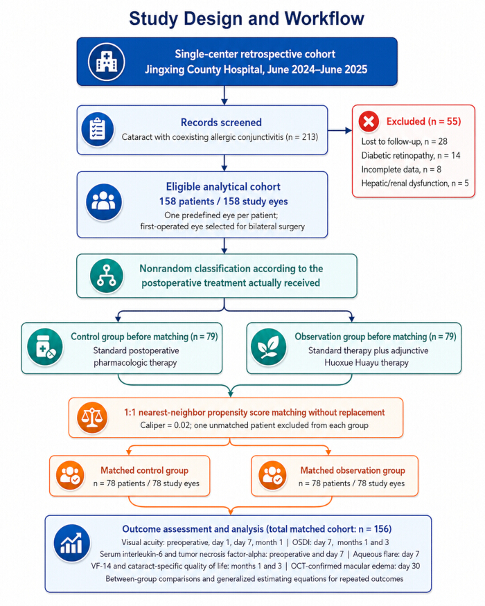
Figure 1: Schematic of the study design and workflow. Electronic medical records of 213 cataract patients with coexisting allergic conjunctivitis were screened. After the exclusion of 55 patients, 158 patients, each contributing one predefined study eye, were classified according to the postoperative treatment received. Before propensity score matching, 79 patients were included in each group. One-to-one nearest-neighbor propensity score matching without replacement, using a caliper of 0.02, retained 78 patients in each group. Clinical, laboratory, patient-reported, and optical coherence tomography outcomes were assessed at prespecified postoperative time points. PSM, propensity score matching; OSDI, Ocular Surface Disease Index; VF-14, Visual Function 14-Item Index; OCT, optical coherence tomography. Please click here to view a larger version of this figure.
| Observation group | Control group | t/χ² | P | SMD | |
| Number of patients | 78 | 78 | - | - | - |
| Age (years) | 65.3 ± 7.8 | 66.1 ± 8.2 | 0.621 | 0.536 | 0.062 |
| Gender (Male/Female) | 38/40 | 41/37 | 0.347 | 0.556 | 0.051 |
| Cataract classification (cortical/nuclear/subcapsular) | 32/28/18 | 30/30/18 | 0.172 | 0.918 | 0.029 |
| Preoperative distance visual acuity (decimal) | 0.150 ± 0.108 | 0.143 ± 0.094 | 0.432 | 0.666 | 0.069 |
| Preoperative near visual acuity (decimal) | 0.112 ± 0.072 | 0.107 ± 0.073 | 0.431 | 0.667 | 0.069 |
| Preoperative BUT (s) | 6.2 ± 1.8 | 6.0 ± 2.0 | 0.674 | 0.502 | 0.053 |
| Preoperative OSDI score | 38.5 ± 10.2 | 39.8 ± 11.5 | 0.726 | 0.469 | 0.074 |
| Allergy grading (mild/moderate) | 52/26 | 48/30 | 0.512 | 0.474 | 0.054 |
| Surgical duration (min) | 12.3 ± 3.1 | 12.8 ± 3.4 | 0.947 | 0.345 | 0.061 |
| Ultrasonic energy (%) | 38.5 ± 8.2 | 39.1 ± 7.9 | 0.468 | 0.641 | 0.05 |
Table 1: Comparison of baseline characteristics between the two groups after propensity score matching (n = 78 patients with one study eye per group). Data are presented as mean ± standard deviation or number. PSM, propensity score matching; SMD, standardized mean difference; OSDI, Ocular Surface Disease Index. An absolute SMD < 0.10 indicated adequate covariate balance. Each patient contributed one predefined study eye. Patient-level outcomes are reported by number of patients, whereas eye-specific outcomes are reported by number of study eyes.
| Observation group | Control group | t | P | ||
| Number of eyes | - | 78 | 78 | - | - |
| 1 day after surgery | Near vision | 0.08±0.06 | 0.07±0.07 | 0.835 | 0.405 |
| - | Far vision | 0.10±0.08 | 0.09±0.07 | 0.812 | 0.419 |
| 7 days after surgery | Near vision | 0.30±0.10 | 0.20±0.08 | 6.723 | <0.001 |
| - | Far vision | 0.70±0.20 | 0.60±0.15 | 3.648 | <0.001 |
| 1 month post-operation | Near vision | 0.40±0.10 | 0.20±0.08 | 13.042 | <0.001 |
| - | Far vision | 0.80±0.10 | 0.70±0.12 | 5.491 | <0.001 |
Table 2: Comparison of decimal near and distance visual acuity at different postoperative time points between the two groups. Repeated measurements were analyzed using generalized estimating equations; the corresponding group-by-time interaction results are reported in the text. Visual acuity was recorded in decimal notation, with higher values indicating better visual acuity. Near and distance visual acuity were analyzed separately.
| Observation group | Control group | t | P | |
| Number of patients | 78 | 78 | - | - |
| 7 days after surgery | 42.3 ± 12.1 | 55.6 ± 14.3 | 6.218 | <0.001 |
| 1 month postoperatively | 22.5 ± 8.6 | 31.8 ± 10.4 | 5.876 | <0.001 |
| 3 months postoperatively | 10.2 ± 5.3 | 15.7 ± 7.8 | 5.134 | <0.001 |
Table 3: Comparison of OSDI scores at different postoperative time points between the two groups (score). Repeated measurements were analyzed using generalized estimating equations; the corresponding group-by-time interaction results are reported in the text.
| Observation group | Control group | χ² | P | |
| Number of study eyes | 78 | 78 | - | - |
| Level 0 (no flicker) | 56 (71.8) | 25 (32.1) | - | - |
| Grade I (mild) | 22 (28.2) | 49 (62.8) | - | - |
| Grade II (moderate) | 0 (0) | 0 (0) | - | - |
| Mild to moderate total (Grade I+II) | 22 (28.2) | 49 (62.8) | 14.862 | <0.001 |
Table 4: Comparison of slit-lamp aqueous flare grades between the two groups on postoperative day 7. Aqueous flare was graded according to the Standardization of Uveitis Nomenclature Working Group criteria: grade 0, no flare; grade 1+, faint flare; grade 2+, moderate flare with clear iris and lens details; grade 3+, marked flare with hazy iris and lens details; and grade 4+, intense flare with fibrin or plastic aqueous. Data is presented as number (percentage). Each patient contributed one predefined study eye.
| IL-6 | TNF-α | |||
| Observation group | Control group | Observation group | Control group | |
| Number of patients | 78 | 78 | 78 | 78 |
| Preoperative | 8.72 ± 2.15 | 8.95 ± 2.31 | 10.34 ± 2.86 | 10.61 ± 3.02 |
| 7 days after surgery | 18.43 ± 4.27 | 26.71 ± 5.93 | 23.56 ± 5.18 | 34.82 ± 7.41 |
| Compare t-values within the group | 19.843 | 26.572 | 20.912 | 29.156 |
| P | <0.001 | <0.001 | <0.001 | <0.001 |
| Inter group comparison t-value (postoperative 7 days) | 9.862 | - | 10.734 | - |
| P | <0.001 | - | <0.001 | - |
Table 5: Comparison of serum IL-6 and TNF-α levels between the two groups before surgery and on postoperative day 7 (pg/mL). Repeated measurements were analyzed using generalized estimating equations; the corresponding group-by-time interaction results are reported in the text.
| Observation group | Control group | t | P | |
| Number of patients | 78 | 78 | - | - |
| 1 month postoperatively | 92.81 ± 1.52 | 81.21 ± 2.32 | 28.564 | <0.001 |
| 3 months postoperatively | 96.23 ± 1.23 | 88.51 ± 2.14 | 25.873 | <0.001 |
Table 6: Comparison of postoperative VF-14 scores between the two groups. Repeated measurements were analyzed using generalized estimating equations; the corresponding group-by-time interaction results are reported in the text.
| Observation group | Control group | t | P | |
| Number of patients | 78 | 78 | - | - |
| 1 month postoperatively | 93.7 ± 2.1 | 85.6 ± 2.2 | 22.341 | <0.001 |
| 3 months postoperatively | 97.1 ± 1.4 | 91.3 ± 2.0 | 21.456 | <0.001 |
Table 7: Comparison of postoperative 12-item cataract-specific Quality of Life scores between the two groups. Repeated measurements were analyzed using generalized estimating equations; the corresponding group-by-time interaction results are reported in the text. QOL, quality of life. The total score ranges from 0 to 100, with higher scores indicating better quality of life.
| Observation group | Control group | χ² | P | |
| Number of study eyes | 78 | 78 | - | - |
| Macular edema | 2(2.56) | 11(14.10) | 6.842 | 0.009 |
Table 8: Comparison of OCT-confirmed postoperative macular edema within 30 days between the two groups. Postoperative macular edema was defined as newly developed intraretinal cystoid spaces accompanied by an increase of at least 10% in central macular thickness relative to the preoperative OCT measurement. Data is presented as number (percentage).
Supplementary Table 1: Baseline characteristics of the two groups before propensity score matching: Continuous variables are presented as mean ± standard deviation, and categorical variables are presented as number. Standardized mean differences were used to assess baseline covariate balance between the groups. An absolute standardized mean difference of less than 0.10 was considered indicative of adequate balance. Each patient contributed one predefined study eye. OSDI, Ocular Surface Disease Index; SMD, standardized mean difference.Please click here to download this file.
Supplementary File 1: De-identified participant-level data underlying Tables 1–8. The Excel workbook contains separate worksheets for the matched observation and control groups, with 78 patients and one predefined study eye per group. The dataset includes study identification codes, matched-pair identifiers, baseline demographic and clinical characteristics, postoperative distance and near visual acuity, Ocular Surface Disease Index scores, aqueous flare grades, serum interleukin-6 and tumor necrosis factor-α levels, Visual Function 14-Item Index scores, cataract-specific quality-of-life scores, and 30-day macular-edema outcomes. All direct patient identifiers were removed before the dataset was prepared for publication.Please click here to download this file.
Discussion
Interpretation of findings and potential mechanisms
In this retrospective cohort, receipt of adjunctive Huoxue Huayu therapy was associated with more favorable postoperative visual acuity, OSDI, aqueous flare, serum IL-6 and TNF-α, visual function, and quality-of-life outcomes than standard postoperative pharmacologic therapy alone. Fewer OCT-confirmed macular-edema events were observed in the observation group than in the control group within 30 days (2.56% vs. 14.10%). Given the retrospective design, nonrandom treatment allocation, and limited number of events, this finding should not be interpreted as evidence that Huoxue Huayu therapy reduced the risk of postoperative macular edema. From a traditional Chinese medicine perspective, the formula was selected according to the theoretical concepts of blood stasis, wind, heat, and yin deficiency. Within this framework, postoperative ocular discomfort and visual disturbance may be interpreted as manifestations related to impaired circulation of qi and blood and disturbance of the ocular collaterals. Dan Shen and Dang Gui were included as principal herbs traditionally used to activate and nourish the blood, while Hong Hua, Chuan Xiong, and Chi Shao were included to support the resolution of blood stasis and the regulation of the collaterals. Sheng Di Huang was included to nourish yin and cool the blood. These traditional indications provided the clinical rationale for formula selection; however, the present retrospective study did not evaluate or validate these theoretical pathogenetic concepts or herb-specific effects16,17,18.
Preclinical and pharmacological studies have reported anti-inflammatory, antioxidant, microcirculatory, and vascular-permeability-related effects for several constituents of the herbs included in the formula. For example, previous experimental studies have linked tanshinone IIA, Angelica polysaccharides, safflor yellow, tetramethylpyrazine, and total paeony glycosides to inflammatory signaling, oxidative stress, platelet activity, or vascular function19,20,21. These observations provide possible hypotheses for future investigation but should not be interpreted as mechanisms demonstrated in the present cohort. In this study, serum IL-6 and TNF-α levels on postoperative day 7 were 30.9% and 32.3% lower, respectively, in the observation group than in the control group, indicating an association between adjunctive treatment and a lower systemic inflammatory-marker profile. Because tear fluid, conjunctival tissue, aqueous humor, NF-κB signaling, platelet activity, and ocular microcirculation were not measured, neither the responsible biological pathway nor a direct local ocular effect can be inferred from these data.
Anterior chamber inflammation after phacoemulsification is one of the key factors affecting visual prognosis22,23. In this study, mild-to-moderate aqueous flare was observed in 22 study eyes (28.2%) in the observation group and 49 study eyes (62.8%) in the control group. The lower aqueous flare grades observed in the adjunctive-treatment group represent a clinical association with less postoperative anterior chamber inflammation. Previous experimental studies have reported vascular, microcirculatory, and NF-κB-related effects for several constituents of the formula, and these findings offer potential explanations that could be examined in future mechanistic studies24,25,26,27,28. However, ocular vascular permeability, blood–aqueous barrier integrity, local inflammatory-cell infiltration, NF-κB signaling, and cytokine concentrations in ocular samples were not evaluated in the present study. Therefore, the between-group difference in aqueous flare cannot be attributed to any specific pharmacological pathway or to direct preservation of the blood–aqueous barrier. Of note, among the 4 study eyes with severe flare in the control group, 2 developed macular edema (50%), whereas no study eye in the observation group had severe flare and macular edema occurred in only 2 study eyes (2.56%). These concurrent findings describe an association among treatment exposure, aqueous flare, and macular-edema events, but they do not demonstrate preservation of the blood–aqueous barrier or any direct protective mechanism.
Allergic conjunctivitis and surgical trauma may jointly contribute to a heightened postoperative inflammatory response; however, the present study was not designed to establish a local synergistic biological mechanism. In the control group, mean serum TNF-α increased from 10.61 pg/mL preoperatively to 34.82 pg/mL on postoperative day 7. This change reflects an alteration in a systemic inflammatory marker after surgery. Because tear fluid, conjunctival tissue, aqueous humor, and other ocular samples were not analyzed, the ocular source and local biological significance of the serum cytokine changes cannot be determined. The lower serum IL-6 and TNF-α levels observed in the adjunctive-treatment group may be compatible with differences in systemic inflammatory status, but the responsible pathways cannot be determined from the present data. Previous experimental and pharmacological studies have described anti-inflammatory, microcirculatory, and NF-κB-related effects of several herbal components included in the formula29,30,31,32,33. These mechanisms remain hypothetical in the context of the present cohort because platelet activity, microcirculation, NF-κB signaling, and local ocular cytokines were not directly measured.
Beyond objective inflammatory markers, this study also incorporated the VF-14 and the 12-item cataract-specific QOL instrument to evaluate visual function and quality of life from the patients’ perspective. Postoperatively, the observation group had higher scores on both patient-reported measures. The parallel between-group differences in OSDI, VF-14, quality-of-life scores, serum inflammatory markers, aqueous flare, and macular-edema events indicate a consistent pattern across several patient-reported and clinical domains. Nevertheless, postoperative tear-film stability, local ocular cytokines, and potential mediation pathways were not directly assessed. Therefore, the present data cannot establish a causal pathway linking lower serum IL-6 and TNF-α levels to improved ocular-surface comfort, visual function, or macular outcomes.
The significance of the present study lies primarily in its focused evaluation of cataract patients with coexisting allergic conjunctivitis, a clinically relevant subgroup that may have a greater postoperative inflammatory and ocular-surface burden than patients undergoing otherwise uncomplicated cataract surgery. By integrating visual acuity, ocular-surface symptoms, slit-lamp findings, serum inflammatory markers, patient-reported visual function and quality of life, and OCT-confirmed macular-edema events within a propensity-score-matched cohort, the study identified a consistent, hypothesis-generating pattern across multiple outcome domains. These findings do not establish clinical efficacy, but they provide preliminary estimates and outcome-selection information that may assist the design of adequately powered prospective studies in this specific population.
This study has several limitations. First, it was a single-center retrospective cohort study with nonrandom treatment allocation influenced by physician–patient decision-making and patient preference. The single-center setting may limit the generalizability of the findings, while the retrospective and nonrandomized design may introduce selection bias. Although propensity score matching improved the balance of measured baseline characteristics, it could not eliminate residual confounding related to unmeasured factors, including treatment preference, health beliefs, adherence, socioeconomic considerations, and other elements of clinical judgment. Therefore, the observed between-group differences should be interpreted as associations rather than causal treatment effects. In addition, no formal masking of patients, treating clinicians, or clinical outcome assessors was implemented during routine care. Consequently, patient-reported outcomes and clinician-graded findings may have been influenced by expectation or observer bias, although standardized laboratory procedures and prespecified OCT criteria reduced subjectivity for some outcomes. Moreover, only one predefined study eye was included for patients undergoing bilateral surgery to maintain statistical independence; therefore, potential differences between the two eyes of the same patient were not evaluated. Second, although a fixed base prescription, predefined modification criteria, and centralized preparation and dispensing procedures were used, individualized additions introduced some treatment heterogeneity. Because this retrospective study was based on routine clinical treatment, batch-specific chemical fingerprinting and quantitative analysis of marker compounds were unavailable. Furthermore, the study was neither designed nor adequately powered to compare outcomes among the different prescription patterns. Therefore, these patterns were reported descriptively without modification-specific efficacy analyses. Third, the follow-up period was limited to 3 months, preventing evaluation of the long-term effectiveness and safety of adjunctive Huoxue Huayu therapy. Safety outcomes were retrospectively extracted from routine medical records, and week-4 hepatic and renal function results were unavailable for six patients. The sample size and follow-up duration were also insufficient to detect uncommon or delayed adverse events. Therefore, the absence of documented serious treatment-related adverse events should not be interpreted as definitive evidence of safety. Fourth, IL-6 and TNF-α were measured only in serum, whereas tear fluid, conjunctival tissue, aqueous humor, and other local ocular samples were not analyzed. These biomarkers should therefore be interpreted as systemic inflammatory markers, and the present findings cannot establish direct modulation of local ocular-surface cytokines or a mechanistic relationship between serum cytokine changes and ocular outcomes. Other potentially relevant systemic and local inflammatory mediators, including IL-1β, IL-8, vascular endothelial growth factor, and matrix metalloproteinase-9, were also not assessed. Finally, because no herbal-medicine-only group was included, the independent contribution of Huoxue Huayu therapy could not be separated from that of standard postoperative pharmacologic therapy. Future research should proceed through adequately powered, multicenter prospective studies with prespecified primary outcomes, concealed random allocation where feasible, masked outcome assessment, and longer follow-up. Such studies should use standardized formula composition, batch-level quality control, predefined adherence criteria, and systematic monitoring of hepatic, renal, gastrointestinal, allergic, and ocular adverse events. Parallel measurement of tear-fluid or aqueous-humor cytokines, tear-film parameters, anterior chamber inflammation, retinal thickness, and relevant signaling or microcirculatory markers would be required to test—not assume—the proposed biological mechanisms. Larger samples are also needed to evaluate relatively uncommon outcomes such as postoperative macular edema and serious adverse events, while prespecified analyses may determine whether allergic-conjunctivitis severity or formula modification influences the observed associations.
The present findings are exploratory and do not justify routine recommendation of adjunctive Huoxue Huayu therapy. Standard postoperative pharmacologic therapy should remain the primary management strategy for cataract patients with coexisting allergic conjunctivitis. Nevertheless, the consistency of the observed associations across visual, ocular-surface, inflammatory-marker, patient-reported, and OCT-defined outcomes suggests that this adjunctive strategy merits formal prospective evaluation in this clinically distinct subgroup. The current results may help identify candidate outcomes and approximate event rates for future trial design, particularly for OSDI, aqueous flare, visual-function measures, serum inflammatory markers, and postoperative macular edema. Until prospective efficacy and safety data are available, serum IL-6 and TNF-α should be regarded only as exploratory systemic biomarkers and should not be used independently to guide treatment selection or postoperative management.
In this single-center retrospective cohort, adjunctive Huoxue Huayu therapy was associated with more favorable postoperative clinical and patient-reported outcomes, lower serum IL-6 and TNF-α levels, and fewer 30-day macular-edema events than standard postoperative pharmacologic therapy alone. Because treatment allocation was nonrandom and residual confounding cannot be excluded, these findings do not establish efficacy, causality, or a reduction in macular-edema risk. The serum cytokine results do not demonstrate local ocular modulation or a mechanism involving IL-6, TNF-α, microcirculation, or the blood–aqueous barrier. Although no serious treatment-related adverse events were documented, the available retrospective safety data are insufficient to establish safety or support routine clinical recommendation. These associations require confirmation in adequately powered prospective multicenter randomized controlled trials incorporating standardized herbal quality control, longer follow-up, systematic safety monitoring, and prespecified assessment of local ocular biomarkers and mechanistic endpoints.
Disclosures
The authors declare that they have no competing interests.
Acknowledgements
This work was supported by the Scientific Research Plan Project of Hebei Provincial Administration of Traditional Chinese Medicine (Grant No. 2023176).
Materials
| Name | Company | Catalog Number | Comments |
|---|---|---|---|
| AcrySof IQ aspheric foldable intraocular lens | Alcon Laboratories, Inc. | SN60WF.215; https://accessgudid. nlm.nih.gov/devices/00380655093221 | Model SN60WF (diopter-specific catalog suffix); Foldable posterior-chamber intraocular lens implanted after phacoemulsification; lens power was selected individually. |
| Angelicae Sinensis Radix | Jingxing County Hospital Traditional Chinese Medicine Pharmacy | https://ydz.chp.org.cn/ | Pharmacopeial-grade crude herb; 12 g in the fixed base formula. |
| BD Vacutainer SST serum-separation tube, 5 mL | Becton, Dickinson and Company | 367986 | Serum-separation tube with clot activator and polymer gel; 3 mL venous blood was collected at each assessment. |
| Carthami Flos | Jingxing County Hospital Traditional Chinese Medicine Pharmacy | https://ydz.chp.org.cn/ | Pharmacopeial-grade crude herb; 6 g in the fixed base formula. |
| Centrifuge 5702 | Eppendorf SE | 5702000010 | Benchtop low-speed centrifuge used for serum separation for 15 min. |
| Chuanxiong Rhizoma | Jingxing County Hospital Traditional Chinese Medicine Pharmacy | https://ydz.chp.org.cn/ | Pharmacopeial-grade crude herb; 10 g in the fixed base formula. |
| Claritin loratadine tablets, 10 mg | Bayer HealthCare LLC | NDC 11523-0800-2 | Administered orally once daily for 4 weeks as part of the standard postoperative regimen. |
| Tumbling E ETDRS roll-up distance chart, 4-5 m | Good-Lite Co. | SKU 500090 | Used for distance visual-acuity assessment at 5 m under standardized illumination. |
| Human interleukin-6 ELISA kit | Invitrogen, Thermo Fisher Scientific | BMS213-2 | 96-test sandwich ELISA kit used to quantify serum interleukin-6; each sample was tested in duplicate. |
| Human tumor necrosis factor-alpha ELISA kit | Invitrogen, Thermo Fisher Scientific | BMS223-4 | 96-test sandwich ELISA kit used to quantify serum tumor necrosis factor-alpha; each sample was tested in duplicate. |
| HYLO COMOD sodium hyaluronate eye drops, 0.1%, 10 mL | URSAPHARM Arzneimittel GmbH | PZN 00495970 | Preservative-free sodium hyaluronate ophthalmic solution administered four times daily for 4 weeks. |
| IBM SPSS Statistics, version 26.0 | IBM Corp. | https://www.ibm.com/support/pages/ibm -spss-statistics-26-documentation | Used for data management and statistical analysis; RRID:SCR_016479. |
| INFINITI Vision System | Alcon Manufacturing, Ltd. | https://www.accessdata.fda.gov/scripts/ cdrh/cfdocs/cfpmn/pmn.cfm?id=K120912 | Phacoemulsification platform used for cataract surgery. |
| Lonicerae Japonicae Flos | Jingxing County Hospital Traditional Chinese Medicine Pharmacy | https://ydz.chp.org.cn/ | Pharmacopeial-grade crude herb; 15 g added when marked conjunctival hyperemia was predominant. |
| Multiskan FC microplate photometer | Thermo Fisher Scientific | 51119000 | Absorbance microplate reader used for ELISA endpoint measurements. |
| Olopatadine hydrochloride ophthalmic solution, 0.1% | Gland Pharma Limited | NDC 68083-477-01 | Administered twice daily for at least 2 weeks before surgery to stabilize allergic conjunctivitis. |
| Ophiopogonis Radix | Jingxing County Hospital Traditional Chinese Medicine Pharmacy | https://ydz.chp.org.cn/ | Pharmacopeial-grade crude herb; 10 g added for persistent ocular dryness with a yin-deficiency pattern. |
| Paeoniae Radix Rubra | Jingxing County Hospital Traditional Chinese Medicine Pharmacy | https://ydz.chp.org.cn/ | Pharmacopeial-grade crude herb; 10 g in the fixed base formula. |
| Near Vision Card | Precision Vision | SKU 2841 | Near-vision chart used at 33 cm under standardized illumination. |
| R statistical software, version 4.3.1 | R Foundation for Statistical Computing | https://stat.ethz.ch/pipermail/ r-announce/2023/000694.html | Used for propensity-score matching and statistical analysis; RRID:SCR_001905. |
| Rehmanniae Radix | Jingxing County Hospital Traditional Chinese Medicine Pharmacy | https://ydz.chp.org.cn/ | Pharmacopeial-grade crude herb; 15 g in the fixed base formula. |
| Salviae Miltiorrhizae Radix | Jingxing County Hospital Traditional Chinese Medicine Pharmacy | https://ydz.chp.org.cn/ | Pharmacopeial-grade crude herb; 15 g in the fixed base formula. |
| Saposhnikoviae Radix | Jingxing County Hospital Traditional Chinese Medicine Pharmacy | https://ydz.chp.org.cn/ | Pharmacopeial-grade crude herb; 10 g added when ocular itching was predominant. |
| Scrophulariae Radix | Jingxing County Hospital Traditional Chinese Medicine Pharmacy | https://ydz.chp.org.cn/ | Pharmacopeial-grade crude herb; 15 g added for persistent ocular dryness with a yin-deficiency pattern. |
| Scutellariae Radix | Jingxing County Hospital Traditional Chinese Medicine Pharmacy | https://ydz.chp.org.cn/ | Pharmacopeial-grade crude herb; 10 g added when marked conjunctival hyperemia was predominant. |
| BQ 900 slit-lamp biomicroscope | Haag-Streit AG | https://haag-streit.com/en/products/categories /general-diagnostics/slit-lamps/bq-900 | Used for postoperative slit-lamp examination and aqueous-flare grading. |
| CIRRUS 5000 spectral-domain optical coherence tomography system | Carl Zeiss Meditec AG | https://www.zeiss.com/meditec/en/ products/optical-coherence-tomography -devices/cirrus-500-5000.html | Used for 6 × 6 mm macular cube scans and postoperative macular-edema assessment. |
| Tobramycin 0.3% and dexamethasone 0.1% ophthalmic suspension | Bausch + Lomb Incorporated | NDC 24208-295-05 | Administered four times daily with weekly tapering as part of the 4-week postoperative regimen. |
| Tribuli Fructus | Jingxing County Hospital Traditional Chinese Medicine Pharmacy | https://ydz.chp.org.cn/ | Pharmacopeial-grade crude herb; 10 g added when ocular itching was predominant. |
| Forma 900 Series ultra-low-temperature freezer, model 905 | Thermo Fisher Scientific | https://knowledge1.thermofisher.com/ | Model 905; Used to store serum aliquots at −80 °C until analysis. |
References
- Mishra D, et al. Enzymatic and biochemical properties of lens in age-related cataract versus diabetic cataract: a narrative review. Indian J Ophthalmol. 2023;71(6):2379–84.
- Liu YC, et al. Cataracts. Lancet. 2017;390(10094):600–12.
- Dick HB. [Cataract surgery]. Klin Monbl Augenheilkd. 2026;243(3):239-53. German.
- Price FW Jr, Price MO. Combined cataract/DSEK/DMEK: changing expectations. Asia Pac J Ophthalmol (Phila). 2017;6(4):388–92.
- Chen SP, Woreta F, Chang DF. Cataracts: a review. JAMA. 2025;333(23):2093-2103.
- Colin J. The role of NSAIDs in the management of postoperative ophthalmic inflammation. Drugs. 2007;67(9):1291–308.
- Lucena NP, et al. Intracameral moxifloxacin after cataract surgery: a prospective study. Arq Bras Oftalmol. 2018;81(2):92–4.
- Qian L, Wei W. Identified risk factors for dry eye syndrome: a systematic review and meta-analysis. PLoS One. 2022;17(8):e0271267.
- Foti R, et al. Umbilical cord blood-derived products in autoimmune systemic syndromes with severe dryness: a pilot study. Medicina (Kaunas). 2024;60(11):1764.
- Gagliano C, et al. Umbilical cord blood platelet lysate eyedrops for the treatment of severe ocular surface disorders in graft-versus-host disease patients: clinical study. Life (Basel). 2024;14(10):1268.
- Wong KY, et al. A review of using traditional Chinese medicine in the management of glaucoma and cataract. Clin Exp Optom. 2024;107(2):156–70.
- Liu XM, Shi H, Li W. Review on the potential roles of traditional Chinese medicines in the prevention, treatment, and postoperative recovery of age-related cataract. J Ethnopharmacol. 2024;324:117786.
- Zhang K, Zhu X, Lu Y. The proteome of cataract markers: focus on crystallins. Adv Clin Chem. 2018;86:179–210.
- Lee CM, Afshari NA. The global state of cataract blindness. Curr Opin Ophthalmol. 2017;28(1):98–103.
- Lau J, Michon JJ, Chan WS, Ellwein LB. Visual acuity and quality of life outcomes in cataract surgery patients in Hong Kong. Br J Ophthalmol. 2002;86(1):12–7.
- Ma H, et al. Baicalein in ophthalmology: recent progress and emerging therapeutic applications. J Ocul Pharmacol Ther. 2025;41(9):490–502.
- Radomska-Leśniewska DM, et al. Therapeutic potential of curcumin in eye diseases. Cent Eur J Immunol. 2019;44(2):181–9.
- Lei L, et al. [Chemical functiomics: a new direction for modern research on traditional Chinese medicine—a case study of the core component group of hemostatic and blood-activating/stasis-resolving Chinese herbal drugs]. Zhongguo Zhong Yao Za Zhi. 2022;47(24):6803–9. Chinese.
- Lu A, et al. Recent progress and research trend of anti-cataract pharmacology therapy: a bibliometric analysis and literature review. Eur J Pharmacol. 2022;934:175299.
- Tao L, et al. Pleiotropic effects of herbs characterized with blood-activating and stasis-resolving functions on angiogenesis. Chin J Integr Med. 2016;22(10):795–800.
- Zhang JS, Zhang BX, Hui XS, Wang J. [Traditional Chinese medicine theoretical mechanism of blood-activating and stasis-resolving therapy combined with thrombolysis/thrombectomy in improving clinical efficacy]. Zhongguo Zhong Yao Za Zhi. 2024;49(17):4812–7. Chinese.
- Qureshi MH, Steel DHW. Retinal detachment following cataract phacoemulsification—a review of the literature. Eye (Lond). 2020;34(4):616–31.
- Foster GJL, et al. Phacoemulsification of the rock-hard dense nuclear cataract: options and recommendations. J Cataract Refract Surg. 2018;44(7):905–16.
- Yu L, et al. Rehmannia glutinosa polysaccharides enhance intestinal immunity of mice through regulating the microbiota. Int J Biol Macromol. 2024;283(Pt 3):137878.
- Chen Z, Cao Y, He S, Qiao Y. Development of models for classification of action between heat-clearing herbs and blood-activating stasis-resolving herbs based on the theory of traditional Chinese medicine. Chin Med. 2018;13:12.
- Liu H, et al. Effectiveness and safety of traditional Chinese medicine in treatment of primary Sjögren's syndrome patients: a meta-analysis. Comb Chem High Throughput Screen. 2023;26(14):2554–71.
- Shi YH, et al. Monoterpene derivatives with anti-allergic activity from red peony root, the root of Paeonia lactiflora. Fitoterapia. 2016;108:55–61.
- Tan H, Ma X, Wang M. Effect of Targeted Nursing Combined with Psychological Intervention on Perioperative Anxiety and Postoperative Complications in Elderly Cataract Patients. J Mod Nurs Pract Res, 2021; 1(2): 7. DOI: 10.53964/jmnpr.2021007.
- Li X, et al. Traditional Chinese medicine, Ziyin-Mingmu decoction, regulates cholesterol metabolism, oxidative stress, inflammation and gut microbiota in age-related macular degeneration models. Pharm Res. 2025;42(7):1101–18.
- Zhou YM, Cao YH, Guo J, Cen LS. Potential prospects of Chinese medicine application in diabetic retinopathy. World J Diabetes. 2024;15(10):2010-4.
- Yu J, et al. Investigation of traditional Chinese medicine constitution in individuals afflicted by dry eye disease: a retrospective study. Medicine (Baltimore). 2024;103(38):e39675.
- Zhang RX, Li MX, Jia ZP. Rehmannia glutinosa: review of botany, chemistry and pharmacology. J Ethnopharmacol. 2008;117(2):199–214.
- Cao F, Wang Y, Chu B, Liu X, Wu C, Chen X, Zeng S, Feng H, Tian M, Wang H, Ni J, Hou S. Appetitive Lifestyles and Obesity with Risk of Senile Cataract: An Univariable and Multivariable Mendelian Randomization Study. Clin Mol Epidemiol, 2024; 1: 6. DOI: 10.53964/cme.2024006