The video demonstrates the separation of EVs from virions and other contaminants by density gradient centrifugation, followed by capture and concentration with hydrogel nanoparticles. Washed fractions are then ready for downstream identification of those enriched in EVs.
Bu içeriği görüntülemek için JoVE aboneliğiniz gereklidir. Giriş yapın veya ücretsiz denemenizi bugün başlatın.
Yöntem makalesi
Separation and Enrichment of Extracellular Vesicles in the Presence of Virions
175 görüntülenme
⸱
31 Temmuz 2026
Bu makalede
Özet
Protokol
1. Filtration and Precipitation of Extracellular Vesicles (EVs)
- To prepare the culture supernatant from infected or transfected cells (i.e., cell lines and/or primary cells), culture approximately 10 mL of late-log cells for 5 days at 37 °C and 5% CO2 in appropriate culture medium (i.e., Roswell Park Memorial Institute medium [RPMI] or Dulbecco’s modified Eagle’s medium [DMEM] with 10% fetal bovine serum [FBS]).
NOTE: All culture medium reagents should be free of EVs, and can be either purchased (see Table of Materials) or prepared in-house by pre-ultracentrifugation of serum at 100,000 × g for 90 min. This protocol has been successful for several commonly-used cell lines including: CEM, Jurkat, 293T, U937 (uninfected lines), U1, J1.1, ACH2, HUT102, MT-2 (HIV-1 and HTLV-1 infected lines), multiple transfected cells, and primary myeloid and T-cells (both infected and uninfected); however, this protocol can be used for any cell type, including those that require specialized media or culture conditions. Density of cells may need to be optimized for different cell types. It is recommended that the highest density be used with minimal cell death after 5 days. - Centrifuge the culture at 3,000 × g for 5 min to pellet cells and discard the pellet.
- Filter the culture supernatant using a sterile 0.22 µm filter and collect filtrate in a clean tube.
- Add equal volume of PEG precipitation reagent (1:1 ratio) to filtered supernatant. Invert tube several times to ensure a homogenous mixture.
NOTE: Do not vortex. - Incubate mixture at 4 °C overnight (O/N).
- Centrifuge mixture at 1,500 × g for 30 min at room temperature (RT) to yield a heterogeneous EV pellet.
NOTE: EV pellets should appear white or off-white in color. - Discard the EV-depleted culture supernatant.
- Resuspend the EV pellet in 150–300 µL of 1× phosphate-buffered saline (PBS) without calcium and magnesium and keep on ice.
2. Construction of a Density Gradient
- Mix iodixanol density gradient medium with 1× PBS to create 11 different 1 mL density fractions from 6 to 18% iodixanol in 1.2% increments in separate microcentrifuge tubes, as shown in Figure 1A.
- Vortex each tube to mix.
- Layer density fractions into a pre-cleaned and dry swinging bucket ultracentrifuge tube, starting with fraction #18 and ending with fraction #6 as indicated in Figure 1B.
NOTE: All tubes should be sanitized using a 10% bleach spray followed by rinsing 3× with deionized water and a final wash of sterile deionized water prior to loading of the gradient fractions. - Add resuspended EV pellets (300 µL) to the top of the layered gradient in the ultracentrifuge tube.
- Ultracentrifuge at 100,00 × g at 4 °C for 90 min.
- Carefully remove the 1 mL fractions from the ultracentrifuge tube and transfer each fraction into new microcentrifuge tubes.
3. Enrichment of EV Fractions Using Nanoparticles
- Create a 30% slurry of nanoparticles using equal volumes of NT80, NT82, and 1× PBS.
NOTE: The mixture should be vortexed prior to use to ensure homogeneity. - Add 30 µL of the slurry to each microcentrifuge tube containing the density fractions and pipette/invert them several times to mix.
- Rotate EV-enriching nanoparticle-containing density fraction microcentrifuge tubes overnight (O/N) at 4 °C at approximately 20 rpm.
- Centrifuge density fraction microcentrifuge tubes at 20,000 × g for 5 min at RT.
- Discard the liquid and wash the EV pellet twice with 1× PBS.
NOTE: Nanoparticle pellets can be frozen at -20 °C or immediately used for various downstream assays (i.e., polymerase chain reaction (PCR), Western blot, mass spectrometry, and other assays).
Erişim kısıtlı. Bu içeriği görüntülemek için lütfen giriş yapın veya deneme sürümünü başlatın.
Sonuçlar
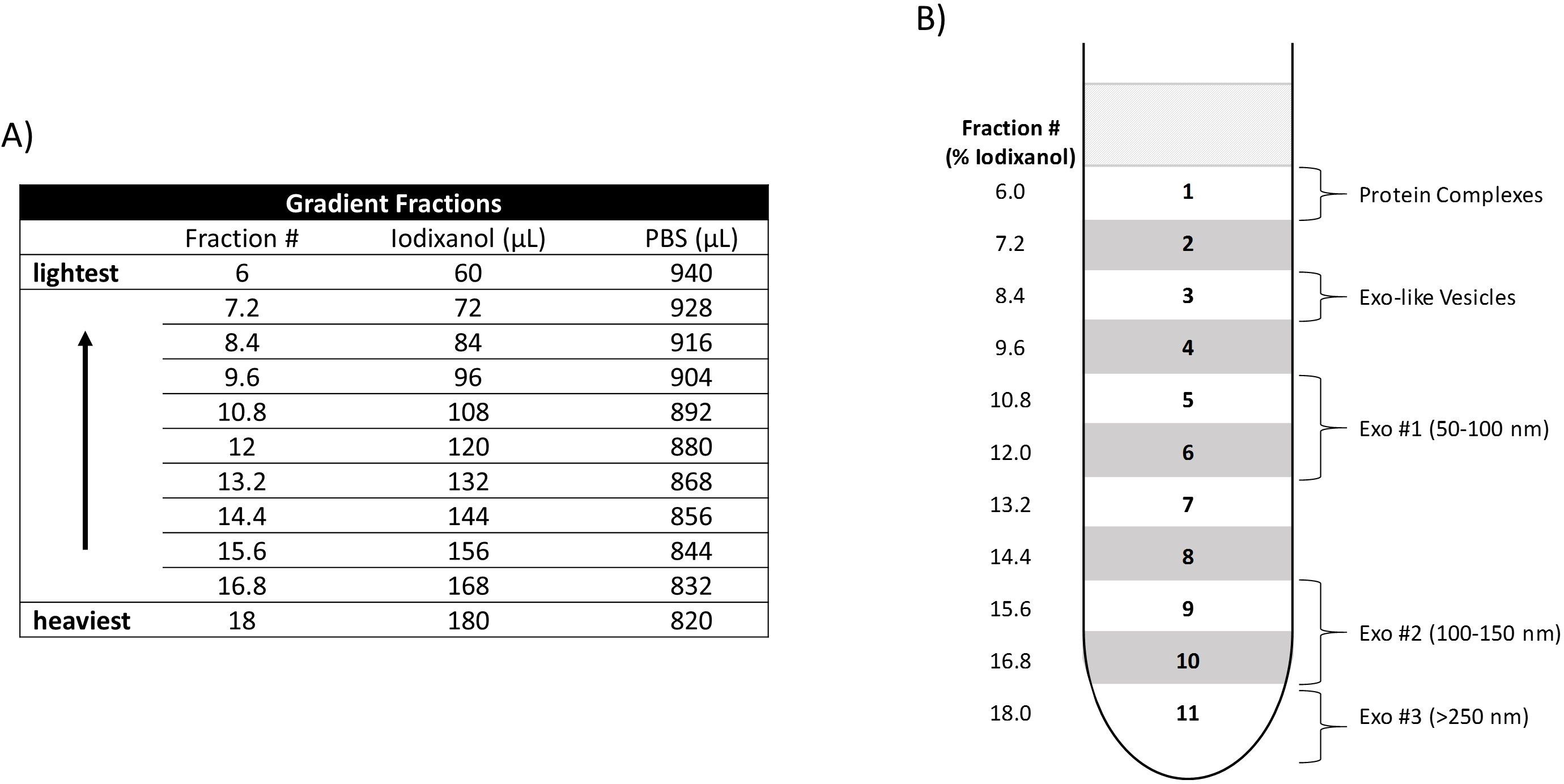
Figure 1: Construction of an iodixanol density gradient. (A) Relative amounts of iodixanol and PBS utilized to create each density fraction (Fraction #). The Fraction # denotes the percentage of iodixanol ...
Erişim kısıtlı. Bu içeriği görüntülemek için lütfen giriş yapın veya deneme sürümünü başlatın.
Malzemeler
| Ad | Şirket | Katalog numarası | Yorumlar |
|---|---|---|---|
| CEM CD4+ Cells | NIH AIDS Reagent Program | 117 | CEM |
| DPBS without Ca and Mg (1x) | Quality Biological | 114-057-101 | |
| Exosome-Depleted FBS | Thermo Fisher Scientific | A2720801 | |
| Fetal Bovine Serum | Peak Serum | PS-FB3 | Serum |
| HIV-1 infected U937 Cells | NIH AIDS Reagent Program | 165 | U1 |
| Nalgene Syringe Filter 0.2 µm SFCA | Thermo Scientific | 723-2520 | |
| Nanotrap (NT80) | Ceres Nanosciences | CN1030 | Reactive Red 120 core |
| Nanotrap (NT82) | Ceres Nanosciences | CN2010 | Cibacron Blue F3GA core |
| Optima XE-980 Ultracentrifuge | Beckman Coulter | A94471 | |
| OptiPrep Density Gradient Medium | Sigma-Aldrich | D1556-250mL | Iodixanol |
| SW 41 Ti Swinging-Bucket Rotor | Beckman Coulter | 331362 | |
| Ultra-Clear Tube, 14 mm x 89 mm | Beckman Coulter | 344059 |
