Bu protokol, miyokard enfarktüsü sonrası kardiyak yeniden şekillenmede CRACD'nin rolünü incelemek amacıyla CRISPR/Cas9 teknolojisi kullanılarak bir C57BL/6N-Cracdem1(c.538-83 to c.3352+255 del) fare modelinin oluşturulmasını ve fenotipik karakterizasyonunu açıklamaktadır.
Yöntem makalesi
Miyokard Enfarktüsü Sonrası Yeniden Şekillenme Çalışmaları İçin CRISPR/Cas9 ile Düzenlenmiş Cracd-Eksik Fare Modelinin Üretimi ve Fenotipik Karakterizasyonu
Bu makalede
Özet
Özet
Myokard infarktüsü (MI) dünya çapında morbidite ve mortalitenin önemli bir nedeni olmaya devam etmektedir. Bu protokol, bir MI modelinin oluşturulmasını ve karakterizasyonunu sağlayan bir yöntem tanımlamaktadır. CracdC57BL/6N arka planında CRISPR/Cas9 teknolojisi kullanılarak CRACD eksik fare hattı oluşturuldu. Zigotlara, 3153-bp'lik genomik bir silinme oluşturacak şekilde tasarlanmış Cas9 mRNA'sı, bir gRNA yapısı ve bir donör şablonu birlikte enjekte edildi. Düzenlenmiş alel, PCR genotipleme ve Sanger dizileme yöntemiyle doğrulandı. CRACD'nin post-MI remodelleme sürecindeki fonksiyonel rolünü değerlendirmek amacıyla Cracd-eksik ve vahşi tip (WT, C57BL/6N) farelerde sol ön inen koroner arterin kalıcı ligasyonu ile miyokard infarktüsü (MI) oluşturuldu. Postoperatif 7. günde transtorasik ekokardiyografi ve nokta izleme birim görüntüleme yöntemiyle kardiyak fonksiyonlar ve sol ventrikül duvar hareketleri değerlendirildi, ardından H&&E ve Masson trikrom boyama. Temsili sonuçlar, bunu gösterdi: Cracdeksik farelerde ventriküler dilatasyon azalmış ve sistolik fonksiyon WT kontrol gruplarına kıyasla korunmuştur. Bu protokol, kardiyak patofizyolojide CRACD'nin mekanistik çalışmalarına güvenilir bir deneysel platform sunar.
Giriş
Miyokard enfarktüsü (MI), dünya çapında morbidite ve mortalitenin önde gelen nedenlerinden biri olmaya devam etmekte ve küresel kardiyovasküler sağlık üzerinde önemli bir yüke katkıda bulunmaktadır1. MI sonrası uzun vadeli prognoz, öncelikle ventriküler dilatasyon, duvar incelmesi ve interstisiyel fibrozisi içeren3,4 ve kalp yetmezliğine (HF) ilerlemenin temel belirleyicileri olan sol ventrikül (LV) yeniden şekillenme süreciyle belirlenir2. Post-MI kardiyak yeniden şekillenme sonucu ortaya çıkan HF'nin küresel insidansı, kardiyovasküler hastalık yükünü daha da artırmaktadır5. Bu yerleşik klinik gözlemlere rağmen, post-MI yeniden şekillenmede spesifik genlerin rolünü değerlendirmek için hassas ve tekrarlanabilir in vivo modeller kullanan sistematik çalışmalar eksiktir. Mevcut hiçbir protokol, CRACD gibi bir hücre iskeleti regülatörünü değerlendirmek için CRISPR/Cas9 ile mühendisliği yapılmış bir nakavt modelini gelişmiş strain görüntüleme ile birleştirmemektedir. Bu nedenle, hassas genetik manipülasyon ve yüksek çözünürlüklü kardiyak fenotipleme sağlayan bir yönteme acilen ihtiyaç duyulmaktadır.
KIAA1211 olarak da bilinen aktin dinamiğinin inhibe edici regülatörü kaplama proteini (CRACD), ağırlıklı olarak sitozolde yerleşmiş olan ve hücre iskeleti dinamiklerinde kritik bir rol oynayan bir aktin polimerizasyon regülatörüdür6. Aktin kaplama proteinlerine (CAPZA1, CAPZA2, CAPZB) bağlanarak ve onların aktin filamentlerinin dikenli uçlarını kaplamasını önleyerek aktin polimerizasyonunu pozitif yönde regüle eder7. Epitel dokularda CRACD, aktin hücre iskeletinin bütünlüğünü koruyarak hücre morfolojisinin ve sinyal iletiminin sürdürülmesinde kritik bir rol oynar8,9. Tümör hücrelerinde, regülasyon bozukluğu artan hücre metastazı ve proliferasyonu ile ilişkilidir6,10. CRACD'nin kalpteki rolü henüz tam olarak anlaşılamamıştır. Kardiyak transkriptom ve deneysel müdahale çalışmaları, Cracd gen ekspresyonunun patolojik strese yanıt verdiğini ve ventriküler yeniden yapılanma sırasında hücre iskeleti dinamikleriyle ilgili genleri regüle edebileceğini göstermektedir9,11. Ayrıca, inhibe edici proteini CapZ, kardiyak patoloji ile yakından ilişkilendirilmiştir: CapZ'nin fosforilasyonu, fenilefrin tarafından indüklenen hipertrofi sırasında miyofibrillerin büyümesini regüle eder12. Erken dönem araştırmalarımız, CRACD'nin orta düzeyde downregülasyonunun miyokard hasarına karşı koruma sağladığını ve akut iskemi sonrası miyofilaman kontraktilitesini koruduğunu göstermektedir13. Bu bulgular, CRACD'nin MI sonrası maladaptif ventriküler yeniden yapılanmada rol oynama olasılığını ortaya koymaktadır. Ancak, büyük ölçüde iskemik stres altındaki Cracd-eksik bir farenin üretilmesi ve fenotiplendirilmesi için doğrulanmış adım adım bir protokolün bulunmaması nedeniyle, Cracd eksikliğinin MI sonrası yeniden yapılanmayı modüle edip etmediğini test eden sistematik in vivo fonksiyon kaybı çalışmaları eksik kalmıştır.
Konstitütif nakavt farelerin üretilmesi için çeşitli yaklaşımlar mevcuttur. Embriyonik kök (ES) hücrelerinde uygulanan geleneksel homolog rekombinasyon hassas olsa da maliyetli, zaman alıcı (12–18 ay) ve belirli fare suşlarıyla sınırlıdır14. ENU (N‑etil‑N‑nitrozüre) mutajenezi, kapsamlı geri çaprazlama ve sekanslama gerektiren rastgele nokta mutasyonları oluşturur15. Buna karşın CRISPR/Cas9, bu çalışma için belirgin pratik avantajlar sunmaktadır: (i) doğrudan zigot enjeksiyonu süreci 4–6 aya indirir; (ii) fonksiyon kaybının tamamen gerçekleşmesini sağlamak için geniş bir genomik fragmanın (3153 bp) silinmesine olanak tanır; (iii) suş dönüşümüne gerek kalmadan doğrudan C57BL/6N arka planında çalışır ve (iv) seçilebilir bir belirteçten kaçınarak istenmeyen transkripsiyonel girişimi minimize eder. Bu nedenle CRISPR/Cas9, rutin kardiyovasküler fenotipleme için uygun, Cracd eksik bir hat oluşturmak için en verimli seçenektir. Bu teknoloji, hassas in vivo fonksiyon kaybı çalışmalarının yürütülmesinde yaygın olarak kullanılmıştır16,17.
Speckle-takip strain görüntüleme, miyokard içerisindeki doğal akustik speckle'ların hareketini kare kare takip eden bir işlem sonrası tekniktir; bu yöntemle strain (uzunluktaki fraksiyonel değişim) ve strain hızı (deformasyon hızı) gibi miyokardiyal deformasyon parametrelerinin hesaplanması sağlanır18. Global fonksiyonel indeksler (ejeksiyon fraksiyonu, fraksiyonel kısalma, sol ventrikül internal çapı) sağlayan ve bölgesel duvar hareket bozukluklarına karşı nispeten duyarsız olan geleneksel M-modu veya iki boyutlu ekokardiyografinin aksine, strain görüntüleme segmental kontraktil fonksiyonu nicelendirebilir. Bu durum, enfarkt geçirmemiş segmentlerin kompanse edici hiperkinezinin, belirgin bölgesel disfonksiyona rağmen global ejeksiyon fraksiyonunu koruyabildiği erken post-enfarkt döneminde özellikle önemlidir. Speckle-takip, standart son noktalara kıyasla çeşitli avantajlar sunar: segmental strain ve strain hızı, deplasman ve hız değerleri sağlayarak hafif subklinik değişiklikleri tespit eder19; geri dönüşümsüz remodeling gerçekleşmeden önce dissenkroniyi ve erken kontraktil eksiklikleri tanımlayabilir ve iskemiye karşı en hassas tabaka olan subendokardiyumu doğrudan sorgular20. Bu çalışmada CRACD, mikroskobik düzeyde miyofiber kontraktilitesini etkileyebilen bir sitoskeletal regülatördür. Geleneksel kavite tabanlı ölçümler, bu tür yerel mekanik etkileri kolayca gözden kaçırabilir. Bu nedenle, speckle-takip analizinin bu protokole entegre edilmesi, Cracd eksikliğinin erken koruyucu etkilerini tespit etme yeteneğini artırır. Buna bağlı olarak, speckle-takibin bu protokole entegrasyonu, özellikle erken dilatasyon ve kontraktil disfonksiyonun ortaya çıktığı MI sonrası ilk hafta boyunca post-enfarkt remodeling'i değerlendirmek için hassas ve kantitatif bir araç sağlar21,22.
Bu protokol, konstitütif nakavt ve yüksek çözünürlüklü görüntüleme kombinasyonunu kullanarak, belirli bir gen delesyonunun akut (7 günlük) MI sonrası yeniden şekillenme üzerindeki fonksiyonel etkisini araştıran araştırmacılar için tasarlanmıştır. Hedef genin global kaybının kabul edilebilir olduğu ve temel sonlanım noktasının erken sistolik fonksiyon ve yapısal değişiklikler olduğu hipotez üretici çalışmalar için uygundur. Bununla birlikte, bazı sınırlamalar göz önünde bulundurulmalıdır. İlk olarak, Cracd eksik fare global bir nakavttır; bu nedenle, sistemik katkılar da rol oynayabileceği için gözlenen kardiyoprotektif etkiler yalnızca kardiyomiyositlerdeki CRACD kaybına atfedilemez. Kalbe özgü delesyon sağlamak için koşullu nakavt stratejilerinin kullanıldığı gelecekteki çalışmalara ihtiyaç duyulacaktır. İkinci olarak, protokol tek bir zaman noktasına (7. gün) odaklanmaktadır ve kronik yeniden şekillenmeyi veya HF progresyonunu ele almamaktadır. Akut fazın ötesinde boylamsal çalışmalara ihtiyaç duyulacaktır. Üçüncü olarak, speckle-tracking analizi, tüm merkez tesislerde mevcut olmayabilecek yüksek kaliteli görüntüler (net endokardiyal sınırlar) ve özel yazılım gerektirir. Bu sınırlamalara rağmen protokol, MI sonrası kardiyak yeniden şekillenmede rol oynayan genlerin ilk fonksiyonel karakterizasyonu için sağlam ve tekrarlanabilir bir platform sunmaktadır.
Bu çalışma, MI sonrası kardiyak yeniden şekillenme ve ventrikül duvar hareketi üzerindeki Cracd eksikliğinin etkisini incelemek amacıyla, CRISPR/Cas9 sistemini kullanarak Cracd-eksik bir fare modeli (C57BL/6N-Cracdem1 (c.538-83 to c.3352+255 del)) oluşturmayı ve doğrulamayı hedeflemiştir. Hem yabanıl tip (WT, C57BL/6N) hem de Cracd-eksik farelerde MI indüklenmiş ve MI'dan bir hafta sonra kapsamlı değerlendirmeler yapılmıştır. Değerlendirmeler; konvansiyonel ekokardiyografik parametreleri, speckle-tracking strain analizini ve histopatolojik değerlendirmeyi içermektedir. İlk yeniden şekillenme olaylarını yakalamak ve Cracd eksikliğinin MI'ın akut evresinde ventriküler dilatasyonu ve sistolik fonksiyonu etkileyip etkilemediğini belirlemek için özellikle erken MI sonrası faza (7. gün) odaklanılmıştır. Bulguların, MI sonrası advers kardiyak yeniden şekillenmenin modülasyonunda CRACD'nin rolüne dair değerli bilgiler sunması beklenmektedir.
Protokol
All animal experiments were approved by the Institutional Animal Care and Use Committee of the Guangdong Institute of Biotechnology (Approval No. IACUC2025179). The study utilized 20 SPF Cracd-deficient and 20 age- and weight-matched WT mice. All mice were 10–12 weeks old and weighed 20–25 g. All male animals were housed in an AAALAC-accredited facility (License No. SYXK (Yue) 2016-0122) at the Guangdong Institute of Biotechnology (Guangdong Laboratory Animals Monitoring Institute). The housing environment was maintained at 22–25 °C with 50–70% relative humidity under a 12 h light/12 h dark cycle.
1. Generation of Cracd-deficient mice using CRISPR/Cas9
- Use CRISPR/Cas9 gene editing technology to generate Cracd-deficient mice on a C57BL/6N background. Delete the genomic sequence from c.538-83 to c.3352+255 (partial intron 5 to intron 6) of Cracd by homology-directed repair.
NOTE: The mouse Cracd gene (NCBI RefSeq: NM_001163793.1; Ensembl: ENSMUSG00000036377) is located on chromosome 5 and contains 9 exons, with the ATG start codon in exon 2 and the TAA stop codon in exon 9. - Design the sequence of gRNA targeting vector and donor oligonucleotide (with targeting sequence flanked by 120 bp homologous sequences combined on both sides).
- Log in to the NCBI website, select the Primer-BLAST function, and design PCR primers for genotyping.
- Paste the NCBI reference sequence number of Cracd (NM_001163793.1) into the PCR template field, specify the exon range for amplification, and click “Get Primers” to retrieve multiple pairs of candidate primers for PCR-based detection of the deletion and wild-type alleles.
- Design single-guide RNAs (gRNAs) to target sites flanking the intended deletion interval in the Cracd gene.
- Select four gRNA sequences (20 nt upstream of the PAM) using online CRISPR design tools.
- To minimize potential off-target effects, score the candidate gRNAs with CRISPRater and select the two optimal sequences (including the NGG PAM).
NOTE: Example selected sequences:
gRNA-A1: CGGCGCTCTTAACATCCAGAGGG (score: 0.71)
gRNA-A2: CCCTTCAGTCTGGGTAGTAGAGG (score: 0.71)
Accept gRNAs with a CRISPRater score ≥ 0.5. For in vitro validation, accept gRNAs with > 50% cleavage efficiency in a Cas9/gRNA target efficiency detection kit.
- Design a single-stranded donor oligonucleotide to promote efficient joining of the intended breakpoints. Example donor sequence: TTAACCATCAACGGGCCGCTGCAAGGGCTGCTGT
GTCTATA CTCAGGACCACCTGCCCTCTAGAGGCAGGAAGGT
AGAGTTCCAAGCTCTGCTGTGCTAGGTTAAATGCAACTTAACATC. - Co-inject Cas9 mRNA, gRNA and donor oligonucleotide into fertilized eggs to generate Cracd-deficient mice. Prepare Cas9 mRNA and gRNA by in vitro transcription.
- Preparation of gRNA.
- Transcribe gRNAs in vitro from DNA templates using the MEGAshortscript™ T7 transcription kit (see Table of Materials) following the manufacturer's protocol.
- Purify the transcribed gRNAs using an RNA purification kit (see Table of Materials) and elute them in RNase-free water.
- Assess gRNA cleavage activity using an in vitro Cas9/gRNA target efficiency detection kit (see Table of Materials).
- Perform all RNA preparation steps on ice using RNase‑free consumables (pipette tips, tubes, water). Keep RNA samples on ice at all times to prevent degradation.
- To perform the cleavage assay: mix the transcribed gRNA (25 ng/µL) with recombinant Cas9 protein (50 ng/µL) and a PCR-amplified DNA fragment containing the target sequence (50 ng).
- Incubate the reaction at 37 °C for 30 min.
- Run the products on a 2% agarose gel to visualize uncleaved and cleaved bands.
- Calculate cleavage efficiency as the ratio of cleaved band intensity to total (cleaved + uncleaved) band intensity.
- Accept gRNAs with > 50% cleavage efficiency.
- Preparation of Cas9 mRNA.
- Thaw the reagents from the T7 ARCA mRNA Kit (see Table of Materials).
- At room temperature, combine 10 µL of 2× ARCA/NTP mix, 1 µL of template DNA, 2 µL of T7 RNA polymerase mix, and nuclease-free water to 20 µL.
- Mix thoroughly, pulse-spin in a microfuge (5 s at 5000 × g), and incubate at 37 °C for 30 min.
- Remove the template DNA by adding 2 µL of DNase I (1 U/µL), mix well, and incubate at 37 °C for 15 min to obtain Cas9 mRNA. Optionally add 1 µL of RNase inhibitor (40 U/µL) (see Table of Materials) to the reaction to prevent RNA degradation.
- Perform all steps on ice with RNase‑free consumables. Keep the Cas9 mRNA on ice until use.
- Superovulate 4-week-old C57BL/6N female mice by intraperitoneal injection of 5 IU of pregnant mare serum gonadotropin (PMSG), followed 48 h later by 5 IU of human chorionic gonadotropin (hCG).
CAUTION: PMSG and hCG are hormones that may cause skin sensitization. Wear gloves and dispose of needles in a sharps container. - Approximately 14–18 h after hCG injection, euthanize the C57BL/6N female mice and one 8–12-week-old C57BL/6N male mouse by cervical dislocation. Collect oocytes from the oviducts of the females and sperm from the cauda epididymis of the male.
CAUTION: Euthanasia must be performed in accordance with institutional guidelines. Cervical dislocation should only be performed by trained personnel. - Capacitate the sperm in capacitation medium (see Table of Materials).
- Add a small volume of the sperm suspension to the oocytes for in vitro fertilization and co-culture for 3–4 h at 37 °C to obtain fertilized zygotes.
- Dilute the transcribed gRNA and Cas9 mRNA with RNase-free water to working concentrations (typically 25 ng/µL for gRNA and 50 ng/µL for Cas9 mRNA) and donor oligonucleotide at 10 ng/µL.
- Microinject 1–2 pL of the mixture into the cytoplasm of fertilized zygotes using a microinjection system (see Table of Materials).
- Identify healthy zygotes by the presence of two distinct pronuclei and an intact polar body under a stereomicroscope.
- Transplant 300 healthy fertilized zygotes into the oviducts of pseudopregnant female mice that have been mated with vasectomized males.
NOTE: This results in the generation of 2 positive founder mice (F0). - Use RNase‑free water and ice‑cold conditions during dilution to maintain RNA integrity.
- Identify F0 mice by PCR genotyping of tail biopsies (see section 1.6).
- Breed F0 founders with WT mice to generate F1 heterozygous offspring, to obtain germline transmission of the deletion.
- Intercross heterozygous F1 mice to produce F2 homozygous Cracd-deficient mice.
- Preparation of gRNA.
- Genotype by Polymerase Chain Reaction (PCR)
NOTE: The following protocol uses a commercial TIANamp Genomic DNA Kit (see Table of Materials). Buffer GL is the lysis buffer, Buffer GB is the binding buffer, and Buffer WA and Buffer WB are wash buffers.- Place approximately 2–5 mm of mouse tail tissue in a microcentrifuge tube containing 180 µL of Buffer GL, 20 µL of Proteinase K (20 mg/mL stock solution), and 10 µL of RNase A (10 mg/mL stock solution).
- Incubate the sample at 56 °C overnight to digest the tissue.
- Centrifuge the tube at 13800 × g for 2 min at room temperature to pellet tissue debris and transfer the supernatant to a new tube.
- Add 200 µL of Buffer GB and 200 µL of 100% ethanol to the supernatant, mix thoroughly, and apply the mixture to a spin column placed in a collection tube.
- Centrifuge at 13800 × g for 2 min at room temperature and discard the flow-through.
- Add 500 µL of Buffer WA to the column, centrifuge at 13800 × g for 1 min at room temperature and discard the flow-through.
- Carefully add 700 µL of Buffer WB (wash buffer containing ethanol, pre-mixed with 100% ethanol) to the spin column, centrifuge at 13800 × g for 1 min at room temperature and discard the flow-through.
- Apply Buffer WB to the inner wall of the column to wash residual salts.
- Repeat step 1.6.5 once, then transfer the column into a new collection tube and centrifuge at 13800 × g for 2 min at room temperature to dry the membrane and remove residual ethanol.
- Place the column into a new 1.5 mL microcentrifuge tube. Add 50–200 µL of sterilized water or elution buffer (preheated to 65 °C) to the center of the membrane, incubate for 5 min.
- Centrifuge the column at 13800 × g for 2 min at room temperature to elute the DNA.
- To increase DNA yield, either reapply the flow-through to the membrane or add an additional 50–200 µL of sterilized water, incubate for 5 min.
- Perform a final centrifugation at 13800 × g for 2 min.
NOTE: Expect DNA yield of 50–200 ng/µL from tail tissue; A260/A280 ratio of 1.8–2.0 indicates pure DNA.
- Place approximately 2–5 mm of mouse tail tissue in a microcentrifuge tube containing 180 µL of Buffer GL, 20 µL of Proteinase K (20 mg/mL stock solution), and 10 µL of RNase A (10 mg/mL stock solution).
- Genotyping by multiplex PCR using three primers in a single reaction.
- Design three primers to distinguish WT, heterozygous, and homozygous genotypes in one PCR reaction: Forward primer F1 (5’-CAGAAGTCATAGCAGCAAGGCAG-3’, binds upstream of the deletion), Forward primer F2 (5’-GAGGGAATTGAGAGGGAGCC-3’, binds within the deleted region, present only in the WT allele), and Reverse primer R1 (5’-GCCAATTTAGCTGCTTCAAATGTTC-3’, common reverse primer downstream of the deletion).
NOTE: The targeted allele generates a 421 bp amplicon (F1 + R1) only when the deletion is present; the WT allele generates a 357 bp amplicon (F2 + R1) only when the intact allele is present. - Set up the PCR reaction in a 25 µL total volume containing 50–100 ng genomic DNA, 0.2 µM of each primer (F1, F2, and R1), 200 µM dNTPs, 1.5 mM MgCl₂, and 1 U Taq DNA polymerase (see Table of Materials).
- Use the following thermocycling program: initial denaturation at 95 °C for 3 min; 35 cycles of denaturation at 95 °C for 15 s, annealing at 60°C for 15 s, and extension at 72 °C for 25 s; and a final extension at 72 °C for 5 min.
- Separate the PCR products by 1–2% agarose gel electrophoresis (120 V, 30 min) in 1× TAE buffer.
- Genotype interpretation based on band pattern: WT shows only a 357 bp band (F2+R1); Heterozygous shows both 421 bp (F1+R1) and 357 bp (F2+R1) bands; Homozygous shows only a 421 bp band (F1+R1).
- Design three primers to distinguish WT, heterozygous, and homozygous genotypes in one PCR reaction: Forward primer F1 (5’-CAGAAGTCATAGCAGCAAGGCAG-3’, binds upstream of the deletion), Forward primer F2 (5’-GAGGGAATTGAGAGGGAGCC-3’, binds within the deleted region, present only in the WT allele), and Reverse primer R1 (5’-GCCAATTTAGCTGCTTCAAATGTTC-3’, common reverse primer downstream of the deletion).
2. Myocardial infarction model establishment
- Anesthetize the mice via inhalation of 3% isoflurane (delivered in 100% oxygen at 1 L/min), place them in a supine position on a surgical platform, and confirm deep anesthesia by the absence of pedal withdrawal reflex.
CAUTION: Isoflurane is a volatile anesthetic. Use only in a well-ventilated area or under a fume hood to avoid inhalation. - Perform orotracheal intubation using a 20-gauge intravenous catheter sheath. Apply ophthalmic ointment to both eyes to prevent corneal drying during surgery.
- Connect the tube to a mechanical ventilator upon successful intubation to maintain ventilation and anesthesia with 2% isoflurane.
NOTE: Successful intubation is confirmed by visible chest movements synchronized with the ventilator.
- Connect the tube to a mechanical ventilator upon successful intubation to maintain ventilation and anesthesia with 2% isoflurane.
- Remove hair from the anterior chest with depilatory cream. Disinfect the surgical area by applying povidone-iodine and 70% alcohol in a circular motion for multiple times, starting from the center and moving outward.
- After fully exposing the skin, make a 1–1.5 cm incision in the left chest wall between the 2nd and 3rd intercostal spaces to open the thoracic cavity.
- Incise the pericardium to fully expose the heart.
CAUTION: Surgical procedures must be performed under sterile conditions. Use appropriate personal protective equipment (PPE), including gloves and a surgical mask.
- Using the inferior border of the left auricle as a landmark, pass a 7–0 atraumatic suture needle beneath the left anterior descending coronary artery (LAD) approximately 2–3 mm distal to its origin.
- Pass the needle to a depth of approximately 0.5–1 mm and tighten the ligature slowly to avoid myocardial tearing.
- Confirm successful occlusion by observing pallor (whitening) of the left ventricular anterior wall distal to the ligation site within 30 s.
- In sham-operated controls, open the chest and pass the suture beneath the coronary artery without tying the knot.
- After establishing the model, sequentially suture the ribs (4-0 absorbable suture, simple interrupted pattern) and skin (4-0 absorbable suture, continuous pattern) to close the thoracic cavity. Remove the endotracheal tube once the mouse resumes stable spontaneous breathing.
- Place mice on a heating pad (37 °C) until fully conscious.
- Administer postoperative analgesia (e.g., buprenorphine 0.05–0.1 mg/kg, subcutaneously, every 12 h for 48 h) according to institutional IACUC guidelines and closely monitor the postoperative health status for several days.
NOTE: Monitor for signs of distress (hunched posture, ruffled fur, reduced mobility, weight loss > 20%).
- Administer postoperative analgesia (e.g., buprenorphine 0.05–0.1 mg/kg, subcutaneously, every 12 h for 48 h) according to institutional IACUC guidelines and closely monitor the postoperative health status for several days.
- Mortality and exclusion criteria.
- Record the number of mice that die within the first 7 days after MI surgery.
NOTE: In a typical cohort, expect a mortality rate of approximately 10–20% by postoperative day 7, with no significant difference between WT and Cracd‑deficient mice. Common causes of death include extensive infarction leading to acute heart failure, surgical bleeding, or pneumothorax. Exclude any mouse with persistent arrhythmia (heart rate < 350 bpm) or signs of severe distress (e.g., hunched posture, ruffled fur, weight loss > 20%) from subsequent echocardiographic and histological analyses. For sham‑operated mice, mortality should be < 5%.
- Record the number of mice that die within the first 7 days after MI surgery.
3. Echocardiographic assessment
- Animal Preparation and Anesthesia
- Power on the high-resolution small animal ultrasound imaging system (see Table of Materials) and its integrated heated stage.
- Ensure that the appropriate high-frequency transducer is securely connected to the mounting apparatus following the manufacturer’s instructions. Set the temperature of the heated stage to 37 °C and allow it to stabilize.
- Place mice in an induction chamber and anesthetize them with 3% isoflurane in oxygen (1–2 L/min) until the pedal withdrawal reflex is absent. Transfer anesthetized mice to the heated platform in a supine position.
- Maintain anesthesia with 1–1.5% isoflurane delivered via a nose cone, adjusting to maintain heart rate between 450–550 beats per min.
- Apply ophthalmic ointment to both eyes to prevent corneal drying.
- Shave the thoracic region, evenly apply depilatory cream for no longer than 3 min, then remove the depilatory cream thoroughly with wet gauze.
- Rinse the skin gently with warm saline if necessary and dry the area. Attach surface ECG electrodes to the limbs using electrode gel at the contact sites and secure them with tape.
NOTE: Confirm stable ECG and heart rate within 450–550 bpm before proceeding to ultrasound imaging.
- Power on the high-resolution small animal ultrasound imaging system (see Table of Materials) and its integrated heated stage.
- Conventional Echocardiography Image Acquisition
- Position the mouse in a horizontal, supine posture on the heated stage, with a slight leftward tilt if needed to optimize cardiac imaging.
- Apply a generous amount of pre-warmed ultrasound coupling gel to the hairless left precordial region, avoiding air bubbles.
- Place the transducer gently on the chest wall at the left parasternal position. Adjust its orientation so that the index mark points toward the mouse’s right shoulder and fine-tune the angle and pressure to optimize image quality.
- Switch to B-mode (two-dimensional) imaging, adjust depth, focus zone, and frame rate to obtain a clear parasternal long-axis view of the left ventricle. Acceptable image quality requires clear visualization of the endocardial border with no dropouts.
- Once the heart rate stabilizes (450–550 bpm), acquire three separate parasternal long-axis B-mode cine loops, each containing at least 3–5 consecutive cardiac cycles.
- Save each loop by pressing “Cine Store”, and store representative still frames using “Frame Store”.
- From the long‑axis view, rotate the transducer 90° clockwise to obtain a short‑axis view at the mid‑papillary level.
- Acquire three separate short‑axis B‑mode cine loops (each with ≥ 3–5 cycles).
- Activate M‑mode, position the cursor perpendicular to the interventricular septum and posterior wall, and record three separate M‑mode tracings.
NOTE: Acceptable M‑mode tracings require clear endocardial borders throughout the cardiac cycle.
- Use the VevoLab 3.2.0 software (see Table of Materials) to measure the following parameters from M‑mode tracings according to the American Society of Echocardiography leading‑edge method: interventricular septum thickness at diastole and systole (IVSd, IVSs), left ventricular internal diameter at diastole and systole (LVIDd, LVIDs), and left ventricular posterior wall thickness at diastole and systole (LVPWd, LVPWs).
- Calculate left ventricular ejection fraction (EF) and fractional shortening (FS) using the Teichholz formula: EF (%) = [(LV Vold - LV Vols) / LV Vold] × 100, LV Vold = ((7 / (2.4 + LVIDd)) × LVIDd3), LV Vol;s = ((7 / (2.4 + LVIDs)) × LVIDs3), FS (%) = [(LVIDd – LVIDs) / LVIDd] × 100.
NOTE: For each mouse, measurements were averaged from three consecutive cardiac cycles per M‑mode recording, and the average was taken across three separate recordings, resulting in a total of 9 measurements per parameter per mouse.
- Calculate left ventricular ejection fraction (EF) and fractional shortening (FS) using the Teichholz formula: EF (%) = [(LV Vold - LV Vols) / LV Vold] × 100, LV Vold = ((7 / (2.4 + LVIDd)) × LVIDd3), LV Vol;s = ((7 / (2.4 + LVIDs)) × LVIDs3), FS (%) = [(LVIDd – LVIDs) / LVIDd] × 100.
- Position the mouse in a horizontal, supine posture on the heated stage, with a slight leftward tilt if needed to optimize cardiac imaging.
- Speckle-Tracking Strain Analysis
- Perform speckle-tracking analysis using the dedicated analysis software (VevoStrain module within VevoLab 3.2.0).
- Open a clear parasternal long-axis B-mode cine loop, select "Vevo Strain" to enter the strain analysis mode, retain only complete cardiac cycles, and choose "Long Axis".
- Manually trace the endocardial border at the end‑diastolic frame. After the endocardial border is drawn, the software automatically generates an epicardial border (adjust as needed). Then start the strain analysis and activate the "toggle contour/vector/orbit line/B mode" function to visualize the motion trajectory.
- Assess tracking quality via the colour‑coded mesh: it should appear green, closely follow the endocardial and epicardial borders without jerking, and move synchronously with myocardial contraction/relaxation. If not, manually refine the borders and re‑analyze.
- Activate "Time‑to‑peak analysis" to divide the left ventricle into six segments (basal anterior, mid anterior, apical anterior, apical posterior, mid posterior, basal posterior), with each colour representing an individual segment. Once satisfactory tracking is achieved, export data for wall motion parameters: velocity (cm/s), displacement (cm), strain (%), strain rate (1/s) for both longitudinal and radial.
NOTE: If tracking quality remains poor after three acquisition attempts (e.g., erratic strain curves, or inability to visualise clear motion trajectories), exclude the mouse from strain analysis.
- Perform speckle-tracking analysis using the dedicated analysis software (VevoStrain module within VevoLab 3.2.0).
4. Histopathological analysis
- After echocardiography, administer a lethal dose of isoflurane to deeply anesthetized mice and apply a secondary physical method (e.g., cervical dislocation) according to institutional guidelines.
- Open the thoracic cavity, rapidly harvest the hearts and rinse the hearts gently in cold phosphate-buffered saline (PBS).
CAUTION: Euthanasia must be performed according to institutionally approved protocols.
- Open the thoracic cavity, rapidly harvest the hearts and rinse the hearts gently in cold phosphate-buffered saline (PBS).
- Trim the hearts, remove the great vessels and cut transversely from apex to base into 2–3 slices (approximately 2–3 mm thick).
- Immerse the tissue slices in 4% paraformaldehyde in PBS and fix them at 4 °C for 12–24 h.
NOTE: Use a volume ratio of fixative to tissue of at least 10:1.
CAUTION: Paraformaldehyde is toxic and a suspected carcinogen. Handle in a fume hood and wear appropriate PPE (gloves, lab coat, safety goggles). Dispose of paraformaldehyde waste as hazardous chemical waste according to institutional guidelines.
- Immerse the tissue slices in 4% paraformaldehyde in PBS and fix them at 4 °C for 12–24 h.
- Dehydrate tissue through a graded ethanol series (50%, 70%, 80%, 95%, 100% ethanol, each for 1 h), clear them in xylene (2 changes, 30 min each), embed them in paraffin, and section them at 4–5 µm thickness using a microtome.
NOTE: Sections should be free of folds and tears.
CAUTION: Xylene is flammable and toxic. Use only in a fume hood and avoid skin contact. Collect xylene waste in a designated container and dispose of as hazardous chemical waste. - Deparaffinize the sections in xylene (2 changes, 5–10 min each), rehydrate them through descending ethanol series (100%, 95%, 80%, 70%, each for 3 min) to water.
- Stain them using standard Hematoxylin and Eosin (H&E) (see Table of Materials) and Masson’s trichrome protocols (see Table of Materials).
CAUTION: Xylene is toxic. Use only in a fume hood and avoid skin contact. Dispose of xylene waste as hazardous material.
- Stain them using standard Hematoxylin and Eosin (H&E) (see Table of Materials) and Masson’s trichrome protocols (see Table of Materials).
- Examine the stained sections under a light microscope (see Table of Materials).
- For quantitative analysis, capture digital images at 4×, 20×, and 40× magnification using a digital camera attached to the microscope. Measure infarct size and fibrotic area using ImageJ.
NOTE: Infarct size (%) = (infarct area / total LV area) × 100; collagen content (%) = (blue-stained area / total myocardial area) × 100.
- For quantitative analysis, capture digital images at 4×, 20×, and 40× magnification using a digital camera attached to the microscope. Measure infarct size and fibrotic area using ImageJ.
Sonuçlar
Generation of Cracd-deficient mice
A heritable mouse model carrying a targeted deficiency in the Cracd gene was generated using CRISPR/Cas9-mediated genome editing (Figure 1A, Figure 1B). To distinguish WT, heterozygous, and homozygous genotypes, a multiplex PCR was performed using three primers (F1, F2, R1) in a single reaction. Mice homozygous for the deletion (Cracd‑deficient) displayed only the 421 bp band, WT mice showed only the 357 bp band (Figure 1C). Sanger sequencing of genomic DNA from Cracd-deficient mice confirmed the predicted deletion in the target gene compared to WT controls (Figure 1D). Western blot analysis further demonstrated a marked reduction in CRACD protein levels in cardiac tissues from Cracd-deficient mice compared with WT controls (Figure 1E). Together, these data confirm the successful establishment of the Cracd-deficient mouse line.
Echocardiography
Echocardiographic assessment on postoperative day 7 revealed pronounced pathological cardiac remodeling in WT mice after MI (WT-MI) compared with sham-operated controls (Figure 2A). The WT-MI group showed thinning of the interventricular septum (Figure 2B, C), significant left ventricular (LV) dilation as reflected by increased LVIDd (Figure 2D) and LVIDs (Figure 2E), and thinning of the posterior wall (Figure 2F, G). These structural alterations were associated with a pronounced decline in global systolic function, as demonstrated by significantly reduced EF (Figure 2H) and FS (Figure 2I). In contrast, Cracd-deficient mice displayed a cardioprotective phenotype following MI. LV dilation was significantly attenuated, with notably smaller LVIDd and LVIDs compared with the WT-MI group. Furthermore, these mice exhibited improved systolic thickening of the posterior wall and maintained significantly higher EF and FS values. These findings indicate that Cracd deficiency effectively attenuates post-infarction LV dilation and preserves global systolic function.
Speckle-tracking strain analysis
Left ventricular myocardial motion and deformation were further evaluated by speckle‑tracking echocardiography using only parasternal long‑axis views (Figure 3A). To ensure data reliability, we defined pragmatic criteria for acceptable output based on the capabilities of the VevoStrain software. Acceptable strain analysis required: (i) a clear parasternal long‑axis view with endocardial borders visible throughout the cardiac cycle; (ii) a stable heart rate between 450–550 bpm during acquisition; (iii) after automatic tracking, the colour‑coded mesh (activated via “toggle contour/vector/orbit line/B mode”) had to closely follow the endocardial and epicardial borders without jerking or losing contact; and (iv) strain curves had to be smooth, show consistent waveforms across segments, and lack sudden spikes or arrhythmic interruptions. Suboptimal output—most commonly erratic strain curves or inability to visualise clear motion trajectories—led to manual adjustment of endocardial or epicardial contours and re‑analysis. If quality remained poor after three attempts, the mouse was excluded. Using these criteria, we obtained reliable strain data in WT‑MI mice and Cracd‑deficient MI mice.
Compared with the sham group, WT-MI mice exhibited significantly reduced radial velocity (Figure 3B) and longitudinal velocity (Figure 3C), as well as radial displacement (Figure 3D) and longitudinal displacement (Figure 3E), indicating impaired global myocardial motion. The absolute values of radial strain (Figure 3F), radial strain rate (Figure 3G), longitudinal strain (Figure 3H), and longitudinal strain rate (Figure 3I) were also markedly decreased, reflecting severely compromised regional myocardial deformation and function. In contrast, Cracd-deficient mice showed significant improvement in these parameters. Compared with the WT-MI group, they displayed better recovery of radial and longitudinal displacements. Furthermore, radial velocity, radial strain, and radial strain rate were maintained at higher levels, while longitudinal strain and longitudinal strain rate were significantly improved. These results suggest that Cracd deficiency not only preserves global systolic function but also enhances regional LV myocardial deformation after MI.
The speckle‑tracking imaging analysis also included visualization of segmental strain distribution and cardiac motion trajectories. Parasternal long‑axis B‑mode images with segmented six regions (BA, MA, AA, AP, MP, BP) and color gradient mapping are displayed in Figure 4A. Cardiac motion trajectories, time‑to‑peak curves for radial and longitudinal strain, parametric distribution maps of strain synchrony, and three‑dimensional reconstructions are presented in Figure 4B. These data further support the functional improvements observed in Cracd‑deficient mice.
Histological examination
To assess the protective effect of Cracd deficiency against MI, histopathological examination was performed on heart sections from WT and Cracd-deficient mice at 7 days post-MI using H&E and Masson's trichrome staining. Whole‑heart longitudinal sections are shown in Figure 5A, with quantitative analysis of infarct size and collagen content presented in Figure 5B and Figure 5C, respectively. High‑magnification views of the infarct zone, border zone, and remote zone are displayed in Figure 5D. In WT-MI hearts, H&E staining showed pronounced left ventricular wall thinning and a well-demarcated infarct zone. Within the infarct region, extensive inflammatory cell infiltration, myocardial necrosis, and marked fibrosis with scar formation were observed. In the border zone, inflammatory infiltration, myocardial necrosis, fibrosis and cardiomyocyte hypertrophy were evident, while the remote zone displayed prominent hypertrophic changes. Corresponding Masson's trichrome staining further demonstrated extensive interstitial fibrosis within both the infarct and border zones of WT hearts following MI. In contrast, Cracd-deficient mice exhibited substantially attenuated pathological alterations, including reduced wall thinning, diminished inflammatory infiltration and myocardial necrosis, less pronounced cardiomyocyte hypertrophy, and markedly decreased interstitial fibrosis compared with WT-MI hearts. These histological findings indicate that Cracd deficiency mitigates post-infarction myocardial damage and adverse remodeling.
Successful protocol execution should yield: (i) a well‑demarcated infarct zone with extensive collagen deposition in WT-MI hearts; (ii) clear cellular morphology without staining artifacts; and (iii) significantly attenuated pathology in Cracd‑deficient MI hearts. Although infarct size may vary with ligation precision, the genotype‑dependent difference is a reliable indicator of successful performance.
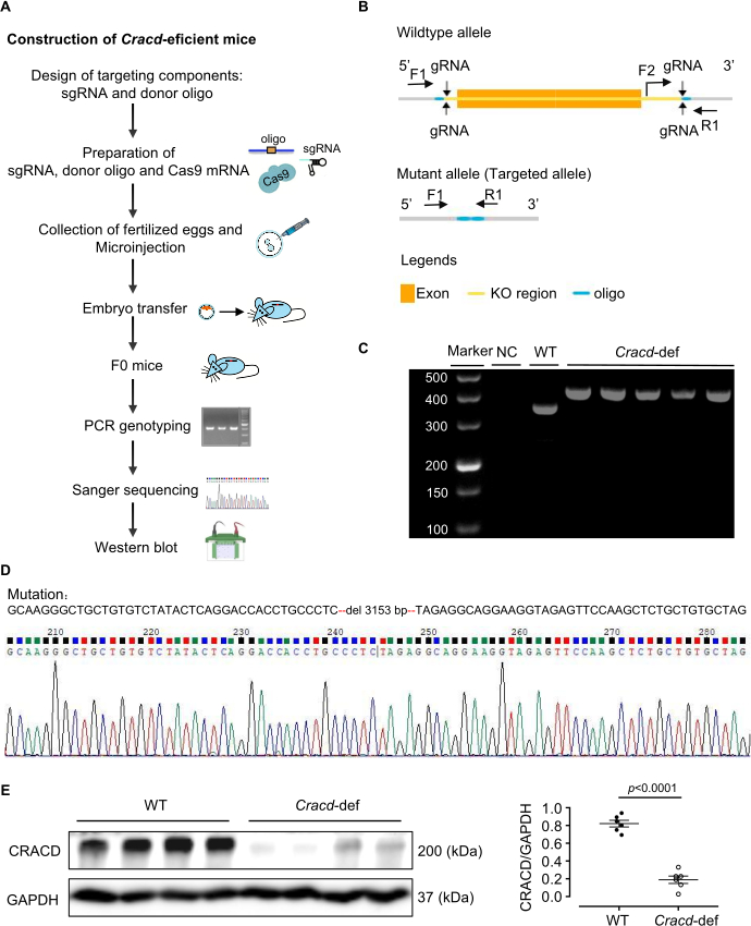
Figure 1: Generation of Cracd-deficient mice. (A) Construction of Cracd-deficient mice. (B) Schematic of the Cracd gene modification strategy. (C) PCR genotyping of tail genomic DNA. WT mice show a single 357 bp band, while homozygous (Cracd‑deficient) mice show a single 421 bp band. (D) Sanger sequencing of genomic DNA confirmed the predicted deletion in the Cracd locus of deficient mice compared to WT controls. (E) Western blot analysis showed significantly reduced CRACD protein levels in the hearts of Cracd-deficient mice compared to WT controls. n = 6 mice per group. Data are presented as mean ± SEM. Statistical significance was calculated with a two-tailed unpaired Student's t-test. Please click here to view a larger version of this figure.
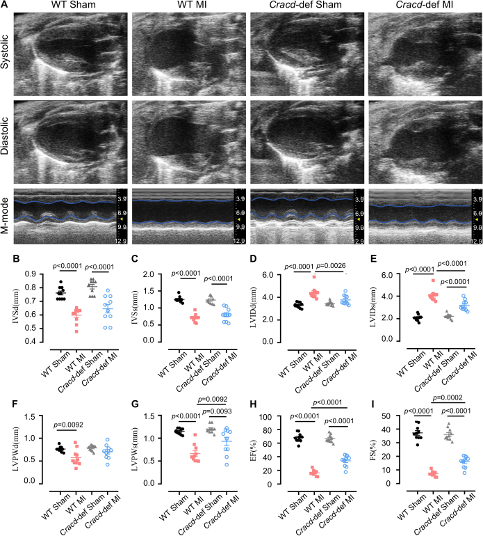
Figure 2: Echocardiographic assessment of cardiac function. (A) Representative B-mode and M-mode echocardiographic images of mouse hearts at diastole and systole. Quantitative analysis of (B) interventricular septum thickness at diastole (IVSd) and (C) systole (IVSs), (D) left ventricular internal diameter at diastole (LVIDd) and (E) systole (LVIDs), (F) left ventricular posterior wall thickness at diastole (LVPWd) and (G) systole (LVPWs), (H) ejection fraction (EF), and (I) fractional shortening (FS). n = 10 mice per group. Data are presented as mean ± SEM. Statistical significance was determined by one-way ANOVA followed by Tukey's multiple comparisons test. Please click here to view a larger version of this figure.
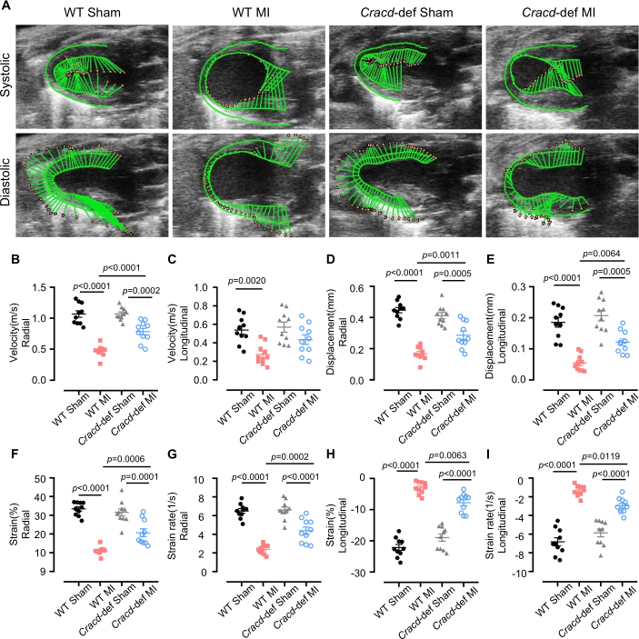
Figure 3: Speckle-tracking strain analysis. (A) Representative images of left ventricular wall motion and deformation at diastole and systole. Quantitative analyses of the following parameters: (B) radial velocity, (C) longitudinal velocity, (D) radial displacement, (E) longitudinal displacement, (F) radial strain, (G) radial strain rate, (H) longitudinal strain, and (I) longitudinal strain rate. n = 10 mice per group. Data are presented as mean ± SEM. Statistical significance was determined by one-way ANOVA followed by Tukey's multiple comparisons test. Please click here to view a larger version of this figure.
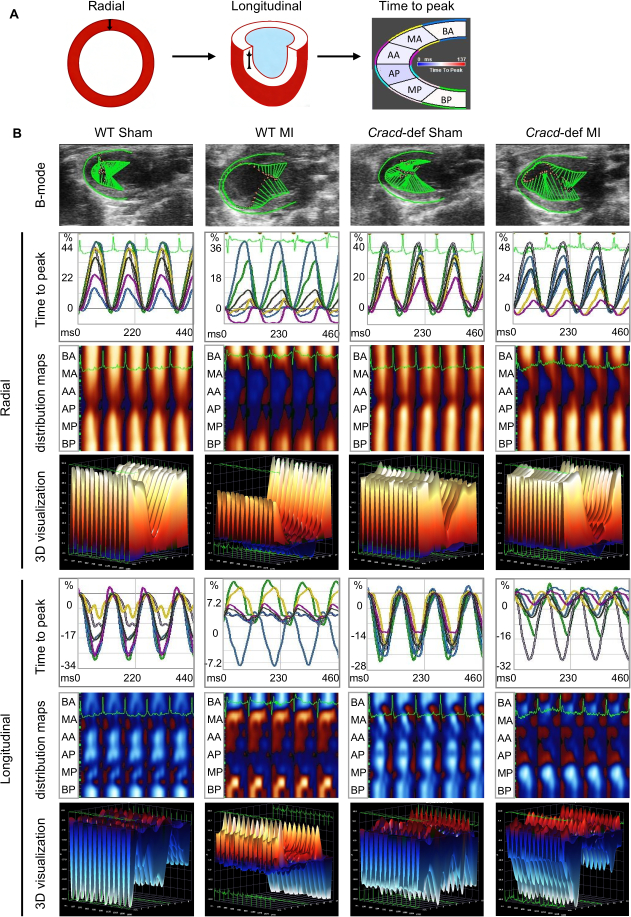
Figure 4: Speckle-tracking imaging in WT-Sham, WT-MI, Cracd-deficient Sham, and Cracd-deficient MI groups at 7 days post-MI. (A) Parasternal long‑axis B‑mode images. Left ventricular wall was segmented into six regions: BA (basal anterior), MA (mid anterior), AA (apical anterior), AP (apical posterior), MP (mid posterior), and BP (basal posterior). Color gradient from blue to red represents increasing strain magnitude. (B) Cardiac motion trajectories; time-to-peak curves for radial and longitudinal strain values along with corresponding peak times. Parametric distribution maps of strain synchrony (blue/red indicate direction: radial – away from/toward center; longitudinal – away from/toward apex). White dots = highest velocity regions, black dots = lowest velocity. Three-dimensional reconstructions (X: time, Y: spatial position, Z: strain magnitude). Please click here to view a larger version of this figure.
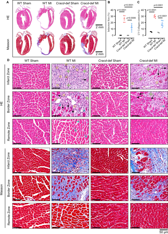
Figure 5: Histopathological analysis of cardiac remodeling after MI. (A) Whole-heart longitudinal sections with H&E and Masson's trichrome staining. Scale bars: 2 mm. (B) Quantitative analysis of infarct size and (C) collagen content. (D) High‑magnification views of infarct zone (IZ), border zone (BZ), and remote zone (RZ). H&E: yellow circles = inflammatory infiltration; black arrows = myocardial necrosis; green stars = fibrosis; blue arrows = hypertrophy. Masson’s trichrome: blue indicates collagen deposition. Scale bar: 50 μm. n = 5 mice per group. Data are presented as mean ± SEM. Statistical significance was determined by one-way ANOVA followed by Tukey's multiple comparisons test. Please click here to view a larger version of this figure.
Tartışma
Bu çalışmada, CRISPR/Cas9 ile düzenlenmiş Cracd eksik bir fare modeli oluşturduk ve konvansiyonel ekokardiyografi, speckle‑tracking strain görüntüleme ve histoloji kullanarak MI sonrası fenotipleme için bir iş akışı kurduk. Protokol, CRACD protein kaybı doğrulanmış nakavtlar üretmiştir. Cracd eksik fareler, MI sonrası WT kontrollere kıyasla daha az ventriküler dilatasyon, daha iyi EF ve azalmış fibrozis göstermiş, bu da modelin kardiyak yeniden şekillenmeyi incelemedeki faydasını doğrulamıştır.
Bu modelin başarısı birkaç kritik adıma bağlıdır. gRNA tasarımı ve in vitro doğrulama, zigot düzenleme verimliliğini doğrudan etkiler.23CRISPRater skorları ≥ 0,5 olan gRNA'ları seçmek için CRISPick veya Benchling kullanıyoruz ve ardından bir in vitro Cas9/gRNA analizi ile kesim verimliliğini test ediyoruz. Yalnızca şu başarıyı elde eden gRNA'lar... > Klivajların %50'si mikroenjekte edilir. Uygun LAD ligasyonu da aynı derecede kritiktir: Sağ ventrikülü delmeyi (çok derin) veya damarı tıkayamamayı (çok sığ) önlemek için 7-0 sütür, LAD başlangıcından 2-3 mm uzaklıkta ve 0,5-1 mm derinlikte geçirilmelidir; anterior duvarın anında beyazlaşması tıkanıklığı doğrular. Ekokardiyografi sırasında kalp hızının kontrol altında tutulması da esastır.24izofluranın (genellikle %1-2) ayarlanmasıyla 450–550 bpm değerinin korunması, konvansiyonel ve strain ölçümlerinin tekrarlanabilirliğini artırır. Son olarak, speckle‑tracking analizi için renk kodlu ağın görsel değerlendirmesi gereklidir25Ağ, sıçramalar olmadan miyokard sınırlarını takip etmelidir; aksi takdirde manuel iyileştirme gereklidir; üç denemeden sonra kalite hâlâ düşükse fare çalışma dışı bırakılır.
Protokol titizlikle takip edilse bile, bazı teknik zorluklar ortaya çıkabilir. Sistematik sorun giderme yöntemleri bunların çoğunu çözebilir. Düşük germ hattı iletim oranları (%10'un altı), genellikle zayıf donör oligonükleotid entegrasyonuna veya düşük gRNA aktivitesine işaret eder26. Sonuç olarak, donör oligonükleotidin 20 ng/µL'ye çıkarılması, homoloji kollarının 120–200 bp'ye uzatılması ve gRNA kesiminin (> %50) yeniden kontrol edilmesi iletimi artırabilir. MI sonrası yüksek mortalite (%30'un üzeri), sıklıkla pnömotoraks, kanama ve geniş enfarktüsten kaynaklanır27. Bu nedenle, farelerin uyanana kadar sürekli oksijen altında tutulması ve vücut sıcaklığını korumak için bir ısıtma pedi kullanılması ölüm oranlarını belirgin şekilde düşürür. Ligasyondan sonra ön duvar soluklaşmıyorsa, LAD muhtemelen yanlış tanımlanmıştır veya sütür çok yüzeyeldir. Bu durumda, LAD'nin (parlak kırmızı diagonal bir damar) yeniden konumlandırılması ve iğnenin, bazen bir diseksiyon mikroskobu eşliğinde, 0,5–1 mm derinliğe yeniden geçirilmesi bu sorunu çözer. Kötü speckle-tracking kalitesi için düşük kare hızı, bulanık sınırlar veya hareket artefaktları olağan nedenlerdir28. Buna göre, kare hızının artırılması, kazancın (gain) ince ayarının yapılması, endokardiyal izin manuel olarak düzeltilmesi ve aritmi içeren döngülerin dışlanması yardımcı olur.
Bu operasyonel hususların ötesinde, yöntemin belirgin sınırlamaları vardır. Cracd nakavtı globaldir, bu nedenle CRACD tüm dokularda eksiktir. Sonuç olarak, sistemik katkılar da rol oynayabileceği için gözlemlenen koruma yalnızca kardiyomiyositlere atfedilemez. Hücre tipine özgü rolleri birbirinden ayırmak için koşullu bir nakavt gerekecektir. Bir diğer sınırlama ise 7. gündeki tek zaman noktasıdır; kronik yeniden şekillenme hakkında herhangi bir bilgi edinilememektedir. Bu nedenle, uzun süreli çalışmalar için daha geç zaman noktaları (4 veya 8 hafta) eklenmelidir. Tekrarlanabilirlik ayrıca görüntü kalitesine bağımlılıktan etkilenmektedir: her ultrason sistemi temiz endokardiyal sınırlar sağlayamaz ve operatör deneyimi önem taşır. Bu nedenle, tüm cerrahi işlemlerin ve ölçümlerin profesyonelce eğitilmiş bir kişi tarafından gerçekleştirilmesi önerilir.
Bu kısıtlamalar göz önüne alındığında, yaklaşımımızı post-MI yeniden yapılanmasında gen fonksiyonunu incelemeye yönelik alternatif stratejilerle karşılaştırmak faydalıdır. AAV aracılı gen manipülasyonu ve antisense oligonükleotidler (ASO'lar) yaygın kullanılan iki alternatiftir29,30. AAV9, herhangi bir üretim gerektirmeden kalp spesifik, geçici overekspresyon veya knockdown sağlayabilir; ancak paketleme sınırı (~4.7 kb), değişken transdüksiyon verimliliği ve hedef dışı etkileri dezavantajlarıdır31. Buna karşılık, CRISPR/Cas9 knockout modelimiz, uzun süreli çalışmalar için avantaj sağlayan kalıcı ve tam fonksiyon kaybı sunar. ASO'lar, hızlı başlangıçla geri dönüşlü ve doz bağımlı knockdown sağlayarak onları akut müdahaleler için uygun kılar; ancak tekrarlayan dozlama gerektirirler ve doku spesifisitesinden yoksundurlar32. Bu nedenle, burada açıklanan konstitütif knockout, ilk keşif ve kronik fenotipleme için en uygun yöntemdir.
Son olarak, protokol CRACD'nin çok ötesine genişletilebilir. Aynı iş akışı —CRISPR/Cas9 ile nakavt farelerin oluşturulmasının ekokardiyografi, strain görüntüleme ve histoloji ile birleştirilmesi— MI sonrası remodeling sürecindeki herhangi bir aday geni değerlendirmek için kullanılabilir. Fenotipleme çerçevesi, transvers aortik konstriksiyon (basınç aşırı yüklenmesi) veya iskemi-reperfüzyon hasarı gibi diğer kardiyovasküler hastalık modellerine de uygundur. Ayrıca, bu protokolden elde edilen doku örnekleri, moleküler mekanizmaları ortaya çıkarmak amacıyla multi-omik analizler (transkriptomik, proteomik, metabolomik) için kullanılabilir. Cracd eksiklik modeli, ilaç testleri için bir araç olarak hizmet edebilir: CRACD yolu üzerinden etki ettiği düşünülen bileşikler değerlendirilebilir. Böylece bu protokol, kardiyovasküler araştırmalarda geniş bir yelpazedeki mekanistik ve translasyonel çalışmalara uyarlanabilir bir platform sunmaktadır.
Açıklamalar
Yazarlar, herhangi bir çıkar çatışması olmadığını beyan ederler.
Teşekkürler
Bu çalışma; Guangdong Temel ve Uygulamalı Temel Araştırmalar Vakfı (2023A1515011278), Guangdong Eyaleti Biyoteknoloji Araştırma Enstitüsü Öz Kaynaklı Projesi (GDBRI-ZL202602) ve Kardiyovasküler Hastalık Modelleri ve Patogenezi üzerine Çin-Kanada Akademik Değişim ve İşbirliği (MS202500058) tarafından desteklenmiştir.
Malzemeler
| Ad | Şirket | Katalog numarası | Yorumlar |
|---|---|---|---|
| CRISPR/Cas9 | |||
| Trizma Hidroklorür Çözeltisi | Sigma-Aldrich, USA | T2663 | DNA ekstraksiyon tamponu hazırlamak için kullanılır |
| Proteinaz K | Sigma-Aldrich, USA | 539480 | Fare kuyruğu sindirimi için kullanılır |
| Green Taq Mix | Vazyme, China | P131 | PCR amplifikasyonu için kullanılır |
| Agaroz | BioFroxx, Germany | 1110GR500 | 1–2% konsantrasyonda DNA jel elektroforezi için kullanılır |
| DNA Markörü | Thermo Scientific, USA | SM0242 | PCR ürünlerinin boyutlandırması için DNA merdiveni olarak kullanılır |
| TIANamp Genomik DNA Kiti | Tiangen Biotech, China | DP304 | Fare kuyruğu dokusundan genomik DNA ekstraksiyonu için kullanılır |
| MEGAshortscript™ T7 transkripsiyon kiti | Thermo Scientific, USA | AM1354 | gRNA'nın in vitro transkripsiyonu için kullanılır |
| RNA saflaştırma kiti | Zymo Research, USA | R1016 | Transkribe edilen gRNA'yı saflaştırmak için kullanılır |
| BeyoCRISPR™ sgRNA Tarama Kiti | Beyotime, China | D8412S | gRNA kesim aktivitesinin in vitro değerlendirmesi için kullanılır |
| RNaz inhibitörü | NEB, USA | M0314 | In vitro transkripsiyon sırasında RNA degradasyonunu önlemek için kullanılır |
| TaKaRa Taq | Takara Bio, Japan | R001A | Fare kuyruğu DNA'sının PCR genotiplemesi için kullanılır |
| FERTIUP® Fare Sperm Preinkübasyon Ortamı: PM | Cosmo Bio, Japan | KYD-002-EX | In vitro fertilizasyon öncesi sperm kapasitasyonu için kullanılır |
| T7 ARCA mRNA Kiti | NEB, USA | E2060 | Cas9 mRNA'sının in vitro transkripsiyonu için kullanılır |
| Gebe kısrak serum gonadotropini | MCE, USA | 9002-70-4 | Süperovülasyon için kullanılır, intraperitonal enjeksiyonla uygulanır |
| İnsan koryonik gonadotropini | ProSpec, Israel | HOR-250 | Ovülasyon indüksiyonu için kullanılır, intraperitonal enjeksiyonla uygulanır |
| RNaz içermeyen su | Aladdin, China | R665526 | RNA çözündürme ve PCR kurulumu için kullanılır |
| Primerler | Sangon Biotech, China | / | Genotipleme için kullanılır |
| Cerrahi | |||
| İzofluran | RWD Life Science, China | R510-22-10 | Cerrahi sırasında anestezinin indüksiyonu ve idamesi için kullanılır |
| Tüy dökücü krem | Veet, France | 1000207 | Cerrahi ve ultrason öncesi tüyleri temizlemek için göğüs bölgesine uygulanır |
| 7-0 Sütür | Lingqiao, China | LQ7022380206 | Sol anterior descendant (LAD) koroner arterin kalıcı ligasyonu için kullanılır |
| 4-0 Sütür | Lingqiao, China | LQ4022380412 | Cerrahi sonrası cilt insizyonunu kapatmak için kullanılır |
| Anestezi ventilatörü | Harvard Apparatus, USA | Tabletop | İzofluran iletimi ve solunum desteği için kullanılır |
| Küçük hayvan ventilatörü | Taimeng, China | HX-101E | Açık göğüs cerrahisi sırasında pozitif basınçlı ventilasyon sağlamak için kullanılır |
| Sabit sıcaklıklı ısıtma pedi | TigerGene, USA | TG-TP-GS | Cerrahi ve iyileşme sürecinde fare vücut sıcaklığını korumak için kullanılır |
| Kaburga retraktörü | Shenzhen Huayon Biotech, China | LN-18-4101 | Kaburgaları ayırmak ve LAD ligasyonu için kalbi açığa çıkarmak için kullanılır |
| Makas | Shenzhen Huayon Biotech, China | 18-0551 | Cilt insizyonları yapmak ve doku kesmek için kullanılır |
| Stereoskopik mikroskop | Nikon, Japan | SMZ 745 | LAD arter ligasyonu cerrahisi sırasında büyütülmüş bir görünüm sağlamak için kullanılır |
| Mikroskobik iğne tutucu | Shenzhen Huayon Biotech, China | 18-2211 | Mikroskop altında küçük 7-0 sütür iğnesini manipüle etmek için kullanılır |
| Kimyasallar ve reaktifler | |||
| Ultrasonik kuplaj ajanları | Tianjin Jinya Technology Development, China | TM-100 | Ekokardiyografi için göğse uygulanır, uygun prob temasını sağlar |
| Paraformaldehit | Sigma-Aldrich, USA | V900894 | Hasat sonrası kalp dokusu fiksasyonu için kullanılır |
| Ksilen | Macklin, China | 1330-20-7 | Parafin giderme ve boyama öncesi doku şeffaflaştırma için kullanılır |
| Hematoksilin | Sigma-Aldrich, USA | H3136 | Çekirdek boyası, HE boyamasında kullanılır |
| Eozin | Sigma-Aldrich, USA | E4009 | Sitoplazmik boya, HE boyamasında kullanılır |
| Ponceau S | Sigma-Aldrich, USA | P3504 | Masson boyamasında sitoplazma boyama için kullanılır |
| Fosfomolibdik asit | Sigma-Aldrich, USA | 221856 | Masson boyamasında mordan ve diferansiyasyon için kullanılır |
| Anilin mavisi çözeltisi | Sigma-Aldrich, USA | 415049 | Masson boyamasında kollajen lif boyama (mavi) için kullanılır |
| CRACD antikoru | Absea, China | KC-35356 | Western Blot ile CRACD proteinini saptamak için primer antikor, dilüsyon 1:2000 |
| GAPDH antikoru | CST, USA | 2118S | Western Blot için yükleme kontrol antikoru, dilüsyon 1:5000 |
| Etanol | Macklin, China | 64-17-5 | Dehidrasyon ve reaktif hazırlama için kullanılır (%70, %80, %95, %100 derecelerde) |
| Laboratuvar ekipmanları | |||
| Sorvall™ Legend™ Micro 17 Santrifüj | Thermo Scientific, USA | 75002541 | Rutin örnek hazırlama için kullanılır |
| PCR Termal Döngüleyici | Bio-Rad Laboratories, USA | 1861096 | DNA amplifikasyonu için kullanılır |
| Mikroenjeksiyon sistemi | Eppendorf, Germany | 5193000020 | CRISPR/Cas9 reaktiflerinin zigotlara mikroenjeksiyonu için kullanılır |
| Vevo 2100 Görüntüleme Sistemi | VisualSonics, Canada | Vevo2100 | MI sonrası kalp fonksiyonlarının ve yapısının non-invaziv değerlendirmesi için yüksek frekanslı görüntüleme sistemi |
| Işık Mikroskobu | Leica, Germany | DM2500 | HE ve Masson boyalı kalp kesitlerini gözlemlemek için kullanılır |
| Analiz yazılımları | |||
| ImageJ | National Institutes of Health, USA | / | Alan ölçümü, sinyal kantifikasyonu ve histolojik görüntülerin (HE ve Masson boyama) işlenmesi dahil olmak üzere görüntü analizi için kullanılır |
| VevoLab 3.2.0 / VevoStrain | VisualSonics, Canada | / | Kardiyak fonksiyon analizi için VevoLab yazılımı; MI sonrası kardiyak performansı değerlendirmek için miyokardiyal strain analizi yapan VevoStrain modülü |
| GraphPad Prism 8 | GraphPad Software, USA | / | İstatistiksel analiz ve grafik hazırlama için kullanılır |
Kaynaklar
- Fauconnier, J., Meli, A. C., Thireau, J., Roberge, S., Shan, J., et al. Ryanodine receptor leak mediated by caspase-8 activation leads to left ventricular injury after myocardial ischemia-reperfusion. Proc Natl Acad Sci U S A. 108 (32), 13258-13263 (2011).
- Cohn, J. N., Ferrari, R., Sharpe, N. Cardiac remodeling-concepts and clinical implications: a consensus paper from an international forum on cardiac remodeling. J Am Coll Cardiol. , (2000).
- Liu, D., Tian, X., Liu, Y., Song, H., Cheng, X., et al. CREG ameliorates the phenotypic switching of cardiac fibroblasts after myocardial infarction via modulation of CDC42. Cell Death Dis. 12 (4), (2021).
- Contessotto, P., Pandit, A. Therapies to prevent post-infarction remodelling: from repair to regeneration. Biomaterials. 275, (2021).
- Zhu, R., Sun, H., Yu, K., Zhong, Y., Shi, H., et al. Interleukin-37 and dendritic cells treated with interleukin-37 plus troponin I ameliorate cardiac remodeling after myocardial infarction. J Am Heart Assoc. 5 (12), (2016).
- Chang, Y., Zhang, J., Huo, X., Qu, X., Xia, C., et al. Substrate rigidity dictates colorectal tumorigenic cell stemness and metastasis via CRAD-dependent mechanotransduction. Cell Rep. 38 (7), (2022).
- Seo, Y., Jang, J., Ko, K. P., Zou, G., Huang, Y., et al. Actin dysregulation induces immune evasion via oxidative stress-activated PD-L1 in gastric cancer. bioRxiv. , (2024).
- Kim, B., Zhang, S., Huang, Y., Ko, K. P., Jung, Y. S., et al. CRACD loss induces neuroendocrine cell plasticity of lung adenocarcinoma. Cell Rep. 43 (6), (2024).
- Jung, Y. S., Wang, W., Jun, S., Zhang, J., Srivastava, M., et al. Deregulation of CRAD-controlled cytoskeleton initiates mucinous colorectal cancer via β-catenin. Nat Cell Biol. 20 (11), 1303-1314 (2018).
- Liu, Z., Cao, H., Shi, Y., Yang, R. KIAA1211 plays an oncogenic role in human non-small cell lung cancer. J Cancer. 10 (26), 6747-6753 (2019).
- Snider, P. L., Snider, E., Simmons, O., Lilly, B., Conway, S. J. Analysis of uncharacterized mKiaa1211 expression during mouse development and cardiovascular morphogenesis. J Cardiovasc Dev Dis. 6 (2), (2019).
- Lin, Y. H., Warren, C. M., Li, J., McKinsey, T. A., Russell, B. Myofibril growth during cardiac hypertrophy is regulated through dual phosphorylation and acetylation of the actin capping protein CapZ. Cell Signal. 28 (8), 1015-1024 (2016).
- Yang, F. H., Pyle, W. G. Reduced cardiac CapZ protein protects hearts against acute ischemia-reperfusion injury and enhances preconditioning. J Mol Cell Cardiol. 52 (3), 761-772 (2012).
- Tong, C., Huang, G., Ashton, C., Wu, H., Yan, H., et al. Rapid and cost-effective gene targeting in rat embryonic stem cells by TALENs. J Genet Genomics. 39 (6), 275-280 (2012).
- Cordes, S. P. N-ethyl-N-nitrosourea mutagenesis: boarding the mouse mutant express. Microbiol Mol Biol Rev. 69 (3), 426-439 (2005).
- Cong, L., Ran, F. A., Cox, D., Lin, S., Barretto, R., et al. Multiplex genome engineering using CRISPR/Cas systems. Science. 339 (6121), 819-823 (2013).
- Kanke, K. L., Rayner, R. E., Bozik, J., Abel, E., Venugopalan, A., et al. Single-stranded DNA with internal base modifications mediates highly efficient knock-in in primary cells using CRISPR-Cas9. Nucleic Acids Res. 52 (22), 13561-13576 (2024).
- Mihos, C. G., Liu, J. E., Anderson, K. M., Pernetz, M. A., O'Driscoll, J. M., et al. Speckle-tracking strain echocardiography for the assessment of left ventricular structure and function: a scientific statement from the American Heart Association. Circulation. 152 (10), (2025).
- Garg, P. K., Biggs, M. L., Kizer, J. R., Shah, S. J., Psaty, B., et al. Glucose dysregulation and subclinical cardiac dysfunction in older adults: the Cardiovascular Health Study. Cardiovasc Diabetol. 21 (1), (2022).
- Asanuma, T., Fukuta, Y., Masuda, K., Hioki, A., Iwasaki, M., et al. Assessment of myocardial ischemic memory using speckle tracking echocardiography. JACC Cardiovasc Imaging. 5 (1), 1-11 (2012).
- Chong, S. Y., Zharkova, O., Yatim, S. M. J. M., Wang, X., Lim, X. C., et al. Tissue factor cytoplasmic domain exacerbates post-infarct left ventricular remodeling via orchestrating cardiac inflammation and angiogenesis. Theranostics. 11 (19), 9243-9261 (2021).
- Hung, C. L., Verma, A., Uno, H., Shin, S. H., Bourgoun, M., et al. Longitudinal and circumferential strain rate, left ventricular remodeling, and prognosis after myocardial infarction. J Am Coll Cardiol. 56 (22), 1812-1822 (2010).
- Labuhn, M., Adams, F. F., Ng, M., Knoess, S., Schambach, A., et al. Refined sgRNA efficacy prediction improves large- and small-scale CRISPR-Cas9 applications. Nucleic Acids Res. 46 (3), 1375-1385 (2018).
- Wu, J., Bu, L., Gong, H., Jiang, G., Li, L., et al. Effects of heart rate and anesthetic timing on high-resolution echocardiographic assessment under isoflurane anesthesia in mice. J Ultrasound Med. 29 (12), (2010).
- Voigt, J. U., Pedrizzetti, G., Lysyansky, P., Marwick, T. H., Houle, H., et al. Definitions for a common standard for 2D speckle tracking echocardiography: consensus document of the EACVI/ASE/Industry Task Force to standardize deformation imaging. J Am Soc Echocardiogr. 28 (2), 183-193 (2015).
- Port, F., Chen, H. M., Lee, T., Bullock, S. L. Optimized CRISPR/Cas tools for efficient germline and somatic genome engineering in Drosophila. Proc Natl Acad Sci U S A. 111 (29), (2014).
- Gao, E., Lei, Y. H., Shang, X., Huang, Z. M., Zuo, L., et al. A novel and efficient model of coronary artery ligation and myocardial infarction in the mouse. Circ Res. 107 (12), 1445-1453 (2010).
- Rösner, A., Barbosa, D., Aarsæther, E., Kjønås, D., Schirmer, H., et al. The influence of frame rate on two-dimensional speckle-tracking strain measurements: a study on silico-simulated models and images recorded in patients. Eur Heart J Cardiovasc Imaging. 16 (10), 1137-1147 (2015).
- Argiro, A., Bui, Q., Hong, K. N., Ammirati, E., Olivotto, I., et al. Applications of gene therapy in cardiomyopathies. JACC Heart Fail. 12 (2), 248-260 (2024).
- Micheletti, R., Plaisance, I., Abraham, B. J., Sarre, A., Ting, C. C., et al. The long noncoding RNA Wisper controls cardiac fibrosis and remodeling. Sci Transl Med. 9 (395), (2017).
- Wu, Z., Yang, H., Colosi, P. Effect of genome size on AAV vector packaging. Mol Ther. 18 (1), 80-86 (2010).
- Juliano, R. L. The delivery of therapeutic oligonucleotides. Nucleic Acids Res. 44 (14), 6518-6548 (2016).
Yeniden basım ve izinler
