All animal procedures described here must be conducted in accordance with institutional animal ethics guidelines and approved by IACUC. All procedures must follow the principles of the 3Rs—Replacement, Reduction, and Refinement—and must be performed by trained personnel.
1. Footpad Injection

Figure 1. Footpad injection in mice and rats.
Key Terms and Definitions
Footpad injections should be scientifically justified, as they involve a weight-bearing surface and may impact mobility. Alternatively, hock injections administered in the tarsal region are often preferred because they target the popliteal lymph nodes while minimizing pain and preserving mobility compared with footpad injections. Despite the controversy, the use of the foot pad as an injection site is still required for some studies. It has been demonstrated that when injected via the foot pad, the antibody response in some mouse strains was significantly stronger than when injected into the hock. All animals must be closely monitored for signs of pain, level of food consumption, and for normal ambulation. Self-mutilation of the foot can occur to the extent that the foot is destroyed. This is a sign of chronic pain. Any animal demonstrating self-mutilation should be brought to the attention of the veterinary staff immediately.
Footpad measurements should be done daily as soon as obvious swelling has occurred. Endpoints must be in place according to IACUC guidelines. Generally, the animal must be euthanized when the lesion or tumor interferes with the animal's ability to ambulate or reach food and water. The maximum volume that can be injected into a footpad is 50 µL. A 29-30 gauge needle is recommended for the injection.
Questions that this video will help you answer
The administration of compounds into animals can have a significant effect on both the well-being of the animal and the outcome of the experimental data and scientific value. The proper method of delivery is essential to the success of the experiment. Many factors must be considered to determine the best route, including the scientific aim of the study, the pH of the substance, the required dosage volume, the viscosity of the substance, and the well-being of the animals. Technical expertise is also a requirement for all injection methods.
Source: Kay Stewart, RVT, RLATG, CMAR; Valerie A. Schroeder, RVT, RLATG. University of Notre Dame, IN
There are many commonly used routes for compound administration in laboratory mice and rats. Protocols may, however, require the use of the less commonly used routes: intracardiac, footpad, and retro-orbital injections. Specialized training is essential for these procedures to be performed successfully. Justification for these routes may need to be provided to gain Institutional Animal Care and Use Committee (IACUC) approval.
All animal procedures described here must be conducted in accordance with institutional animal ethics guidelines and approved by IACUC. All procedures must follow the principles of the 3Rs—Replacement, Reduction, and Refinement—and must be performed by trained personnel.
1. Footpad Injection

Figure 1. Footpad injection in mice and rats.
Key Terms and Definitions
Footpad injections should be scientifically justified, as they involve a weight-bearing surface and may impact mobility. Alternatively, hock injections administered in the tarsal region are often preferred because they target the popliteal lymph nodes while minimizing pain and preserving mobility compared with footpad injections. Despite the controversy, the use of the foot pad as an injection site is still required for some studies. It has been demonstrated that when injected via the foot pad, the antibody response in some mouse strains was significantly stronger than when injected into the hock. All animals must be closely monitored for signs of pain, level of food consumption, and for normal ambulation. Self-mutilation of the foot can occur to the extent that the foot is destroyed. This is a sign of chronic pain. Any animal demonstrating self-mutilation should be brought to the attention of the veterinary staff immediately.
Footpad measurements should be done daily as soon as obvious swelling has occurred. Endpoints must be in place according to IACUC guidelines. Generally, the animal must be euthanized when the lesion or tumor interferes with the animal's ability to ambulate or reach food and water. The maximum volume that can be injected into a footpad is 50 µL. A 29-30 gauge needle is recommended for the injection.
Questions that this video will help you answer
The administration of compounds into animals can have a significant effect on both the well-being of the animal and the outcome of the experimental data and scientific value. The proper method of delivery is essential to the success of the experiment. Many factors must be considered to determine the best route, including the scientific aim of the study, the pH of the substance, the required dosage volume, the viscosity of the substance, and the well-being of the animals. Technical expertise is also a requirement for all injection methods.
Footpad is one of the specialized injection methods that biomedical researchers use for experiments necessitating the delivery of compounds via these atypical routes.
A footpad injection involves subcutaneous administration of the compound into the animal's hind foot. This video will illustrate the considerations, procedures and applications of this special injection technique.
Footpad injection is used in certain studies, especially those involving inflammation and tumor growth. Note that injections should be performed on only one foot and never bilaterally.
Alternatively, hock injections administered in the tarsal region are often preferred, as they target the popliteal lymph nodes while minimizing pain and preserving mobility compared with footpad injections.
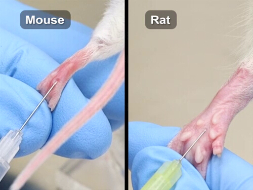
Footpad injection is associated with increased discomfort, as it involves a weight-bearing surface and may impact normal ambulation.
Footpad measurements should be taken daily once obvious swelling occurs.
Therefore, this technique should only be performed when scientifically justified and when alternative approaches are not suitable for the experimental objectives.
A 27-gauge or small-gauge needle is recommended for the injection, and the maximum volume recommended is 50 microliters or as specified in IACUC-approved institutional guidelines.
Following any injection, all animals must be closely monitored for signs of pain, level of food consumption, and for normal ambulation.
Humane endpoints must be established to ensure that animals are euthanized before mobility is significantly impaired or access to food and water is compromised.
Let’s learn this injection procedure in mice and rats. Attach the appropriate needle and fill the syringe with the correct volume. This procedure can be done in conscious animals.
Place the animal in a restraint tube with one hind foot isolated and extended by grasping the skin above the stifle.
Wipe the foot with water or alcohol to remove debris before injection.
To avoid the blood vessel that runs the length of the foot, the injection landmark is at the center, but just off the midline, closer to the toes.
Place the needle bevel up at the injection site, directing it towards the heel.
Inject the compound slowly and steadily to avoid rapid distention of the foot tissues. This will cause the footpad to swell as the injected material fills the subcutaneous space.
In a small animal's foot, the swelling from the injection can extend to the heel, whereas in a larger animal, it will be more localized.
After the injection, observe the animals daily, and if persistent swelling is present or if there are lesions or tumors as a result of the experimental protocol, then, using a caliper, perform the footpad measurement.
This instrument measures foot thickness in millimeters and helps in the quantification of swelling.
Now, let's discuss a few example experiments that use footpad injections.
Investigators often use footpad injection to analyze inflammatory responses. In this experiment, researchers isolated peripheral blood mononuclear cells from human blood samples, mixed them with different test antigens and injected the solutions into an animal's footpad.
Finally, they measured foot thickness to quantify the swelling response due to different antigens.
You've just watched JoVE's final installment on the common and specialized compound administration techniques.
Just to recap, in the first part, we reviewed the most common parenteral routes. In the second chapter, we discussed the enteral and topical procedures. The third installment covered with the first set of atypical procedures, like intradermal, intranasal, and intracranial administration in neonates.
Lastly, here we discussed one additional atypical route that biomedical researchers use in labs for specific purposes.
After watching this series, you should have a much better understanding of different administration techniques, and you should also know the general and specific considerations related to these protocols of compound administration.
Whenever scientifically appropriate, non-animal methods, including cell-based models, organoids, organ-on-chip platforms, and other in vitro assays, should be used to reduce or replace animal use in studies involving compound administration.
Chapters in this video
0:00
Overview
0:44
Considerations for the Specialized Injections
2:16
Footpad Injection
3:42
Application
4:16
Summary
Videos from this collection: