Compound Administration III

Source: Kay Stewart, RVT, RLATG, CMAR; Valerie A. Schroeder, RVT, RLATG. University of Notre Dame, IN
There are many commonly used routes for compound…
All animal procedures described here must be conducted in accordance with institutional animal ethics guidelines and approved by IACUC. All procedures must follow the principles of the 3Rs—Replacement, Reduction, and Refinement—and must be performed by trained personnel.
1. Intradermal administration
- Most intradermal injections are aqueous-based compounds. The solutions must be physiologically buffered to have a neutral pH, in order to avoid tissue necrosis at the injection site.
- The needle size range is 25-27 gauge, the smallest possible.
- The dose range per injection site is 50-100 µL. Injecting excessive volumes can result in necrosis at the injection site or leakage of the compound out of the site due to pressure.
- For accurate placement of the needle into the intradermal space, it is necessary to anesthetize both mice and rats. Inhalation anesthesia allows for a rapid induction and recovery; however, injectable anesthesia has the advantage of providing ample time for preparation of the area and performing the injection.
- Remove the hair at the injection site through the use of a depilatory cream or by shaving the area.
- Thoroughly remove any residual depilatory cream or hair debris.
- Apply a topical antiseptic, such as an iodine solution, chlorhexidine solution, or alcohol.
- Administration procedures
- Stretch the skin taut between the thumb and index finger. This provides stability to the skin when positioning the needle.
- Place the needle bevel up on the skin.
- Gently insert the needle into the skin between the epidermis and the dermis. Advance the needle just beyond the bevel.
- Inject the substance slowly. Injection of the compound will create a bleb, or small blister, in the skin.
- Pause after the injection to allow the skin to stretch and adjust before withdrawing the needle.
- Precautions
- Pulling back on the plunger is not necessary.
- If the needle is inserted into the subcutaneous space, no bleb is formed. Injecting too deeply results in a subcutaneous injection.
- Avoid blotting or wiping the area, as that can cause the compound to leak from the injection site.
- When performing multiple injections, space them so that the blebs do not overlap.
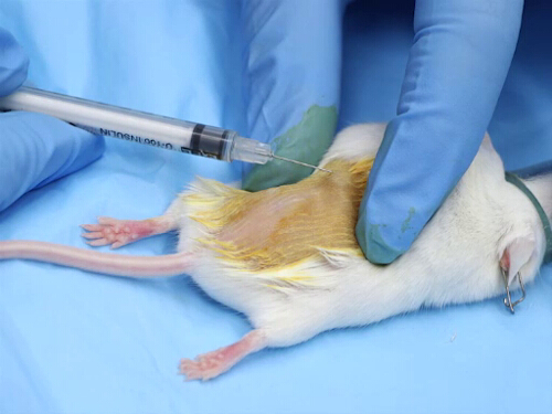
Figure 1. Intradermal injection in mice.
2. Intranasal administration
- Equipment
- Use a micropipetting unit pipette that can be calibrated to deliver an accurate volume.
- Disposable pipette tips should be used to avoid cross-contamination. TB syringes, blunt needles, and flexible tubing can also be used for dosing.
- Total injection volumes for rats should not exceed 50 µL. For mice, the maximum total volume is less than 20 µL.
- Administration in conscious animals
- Manual restraint in conscious animals requires that the head be relatively immobile so that the pipette tip or blunt needle can be placed close enough to the nares to deliver the compound, but not so close as to poke or lacerate the nasal tissue.
- Restrain the animal and hold it in a vertical position.
- Place a small drop of a liquid compound at the nasal opening. The animal should inhale the droplet.
- Administer an additional volume, alternating nares until the entire volume has been given.
- When administering larger volumes, it is important to not constrict the chest during restraint. Chest compression impedes the ability of the animal to take sufficiently deep breaths to draw the liquid into the bronchi and lungs.
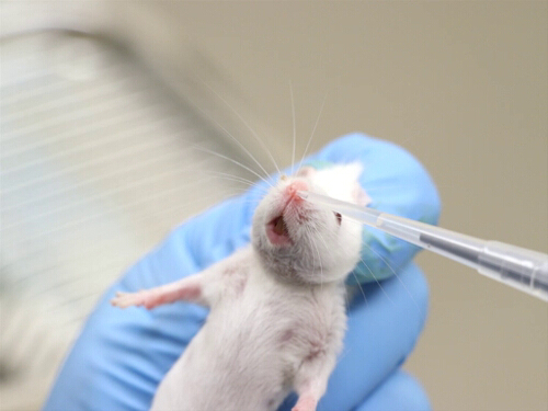
Figure 2. Intranasal administration in conscious mice.
- Administration in unconscious animals
- Anesthetize the animal using the appropriate protocol for this procedure, as inhalant anesthesia requires precise timing and may need adjustment during delivery. Using inhalation anesthetics allows for an immobilized animal during the approach and delivery of the compound. This eliminates the possibility of the animal biting the dosing equipment, the loss of the compound due to jerking of the head, and injury to the animal's nasal tissue, eyes, or facial skin. The animal is also less likely to snort and spray the compound from the nares upon administration.
- Place the animal in the supine position. The position of the head influences the placement of the solution in the nares. It has been demonstrated that the ideal position for CNS delivery is with the animal supine; this allows for better absorption.
- Administer half of the compound directly into one side of the nasal opening, timing it with inhalation. Then, turn the animal over.
- As above, administer the other half of the volume into the other nasal opening.
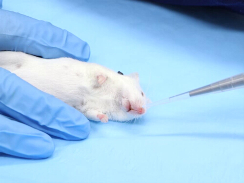
Figure 3. Intranasal administration in unconscious mice.
3. Intracranial administration in neonatal mice and rats
- Mice or rats must be anesthetized for intracranial injections.
- Equipment
- Determine the correct needle gauge and maximum volume of administration as per institutional guidelines.
- Prepare a needle guard prior to anesthetizing the animal.
NOTE: The injection depth of the needle is controlled through the use of a guard that is created with the needle cap.- To create the guard, a needle is measured against the needle cap, and a mark is placed on the cap to indicate where to cut. The cut should be made so that 2 to 5 mm of the needle is exposed when the cap is replaced on the needle.
- The length of the exposed needle must be long enough to penetrate the skin and skull, and reach the desired depth in the brain.
- The needle lengths needed for mice and rats need to be used as per institutional guidelines.
- A heating source is required to prevent hypothermia in the pups. There are several types: an electric heating pad set on low, a circulating water blanket, or a reusable chemical reaction heat pouch.
- Restraint
- Mouse and rat pups 10 days of age or younger do not require anesthesia for this procedure. Restrain them manually by holding them just behind the head and pulling the skin caudally to put slight pressure over the shoulders.
- Anesthetize pups over 10 days of age with isoflurane inhalation. Attach an induction chamber to a precision vaporizer with controlled delivery of isoflurane. Once the pup is immobilized, the anesthesia is effective for about 40 seconds, which provides ample time for the injection.
- Injection technique
- Draw the substance into the syringe and place the cap/needle guide over the needle.
- The injection volume recommended must be species-, age-, and target-specific and must match the approved protocol.
- As the maximum volume per mouse (for a 25g mouse) or rat (for a 200g rat) is 1 and 2 µL/per injection site.
- To inject into the cerebral cortex of neonates, insert the needle 5 mm behind the eye, approximately 3 mm off the midline of the skull.
- The weanling mouse injection site is approximately halfway between the eye and ear, and just off the midline.
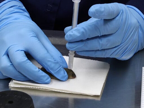
Figure 4. Intracranial administration in a mouse pup.
Key Terms and Definitions
Intradermal injections are delivered into the outer layers of the dermis, underneath the upper skin layer (the epidermis). This injection route is usually reserved for the assessment of inflammation, cutaneous blood flow diagnostics, or allergenic reactions to an antigen.
Although often used for local delivery of vaccinations or decongestant spray, intranasal administration can also be used for systemic and central nervous system (CNS) delivery. The mucosa that lines the nasal cavity has a rich supply of blood vessels and nerves that allow for rapid systemic absorption and direct targeting to the CNS. Substances comprised of small lipophilic molecules have a far greater absorption rate than those containing larger molecules.
Although anesthesia is not required for this procedure, it can facilitate proper placement of the compound at the nares, ensuring accurate dosing. It has been demonstrated that anesthetized animals have fivefold greater delivery of a drug to the brain compared to a conscious animal. Unanesthetized rats are very resistant to intranasal administration. However, there is a study that demonstrated effective delivery techniques for intranasal administration in freely moving rats. Additionally, alert animals may try to bite at the pipette tip or needle, making delivery of the substance difficult.
Advantages of intranasal administration are that this technique requires minimal training and skill, and it is noninvasive to the animal. However, due to the possibility of aerosolization, the use of a biosafety cabinet and eye protection is advised, especially when working with a conscious animal. Furthermore, to prevent aspiration or respiratory distress associated with excessive intranasal administration, the smallest effective volume should be used. If at any time during this procedure cyanosis, mouth breathing, or other signs of distress are seen in the animal, the procedure should be aborted immediately.
Intracranial injections in adult mice and rats employ the use of stereotaxic equipment to ensure the proper positioning and depth of the injection. In neonatal rodents, the skull is sufficiently thin to allow direct intracranial injection without stereotaxic support. However, the approach should be selected based on species, age, and protocol-specific considerations. The mouse or rat pups should be left with the mother until the procedure, and they should be returned to her as soon as possible once the procedure is completed. Postinjection care includes continuous monitoring of the heat source until normal movement and behaviors, including nursing, are observed. The primary reason to use this technique is to provide pharmacologic agents to the central nervous system that require the blood-brain barrier to be crossed, or to avoid the effects involved in the systemic route.
Questions that this video will help you answer
The administration of compounds into animals can have a significant effect on both the well-being of the animal and the outcome of the experimental data and scientific value. The proper method of delivery is essential to the success of the experiment. Many factors must be considered to determine the best route, including the scientific aim of the study, the pH of the substance, the required dosage volume, the viscosity of the substance, and the well-being of the animals. Technical expertise is also a requirement for all injection methods.
At times, different experimental approaches necessitate the use of less commonly employed routes of compound administration in rodents. Intradermal, intranasal, and intracranial are three of such alternate routes that biomedical researchers use in labs today.
As their name suggests, intradermal delivers compounds into the outer layers of the dermis. Intranasal is placing the solution in the animal's nares. Intracranial involves inserting the needle directly into the rodent's brain.
Specialized training is essential to perform these procedures successfully. Here, we'll first illustrate the considerations for each of these methods, and then we will demonstrate the techniques that will help you learn the procedures while ensuring the safety of the animal and the success of the experiment.
Let's begin with a discussion of when these routes are used and things one should bear in mind before beginning to perform these specialized administration techniques.
Intradermal injections are used to deliver an article into the space between the epidermis and the dermis.
This route is usually reserved for the assessment of inflammation, cutaneous blood flow diagnostics, or allergenic reactions to an antigen.
Similar to other routes, the intradermal solution should also be prepared using sterile technique. And it must be physiologically buffered to a neutral pH to avoid tissue necrosis at the injection site.
A hub-less system with a 25-27-gauge needle is often used for this injection, or as per institutional guidance.
This system helps in preserving the volume of administration, which is in the range of 50-100 microliters per injection site, or as per institutional guidance. Injecting excess can result in necrosis or undesirable compound leakage due to pressure.
The intranasal route is often chosen for local delivery of vaccinations or decongestant spray, as well as systemic and central nervous system or CNS delivery.
The mucosa that lines the nasal cavity has a rich supply of blood vessels and nerves that allow for rapid systemic absorption and direct targeting to the CNS.
This is a non-invasive method that requires minimal training and skill, and simple equipment - a calibrated micropipette and some disposable tips.
The administration volumes for rats should not exceed 50 microliters. And for mice, the maximum total volume is less than 20 microliters.
Although anesthesia is not required for this procedure, it has some advantages over intranasal administration in conscious animals 1) it facilitates proper placement of the compound at the nares, ensuring accurate dosing 2) eliminates the possibility of the animal biting the dosing equipment 3) ensures that there is no injury to the animal's nasal tissue, eyes, or facial skin due to jerking of the head, and 4) animal is less likely to snort and spray the compound from the nares upon administration.
Intracranial injections in adult mice and rats employ the use of stereotaxic equipment, which is described in a video in the "Essentials of Neuroscience" collection. The equipment assures proper positioning and correct depth of injection.
Here, we will focus on intracranial delivery in neonatal mice and rats, in whom the skull is thin enough to inject directly through it, and may be too fragile to support the stereotaxic device.
The primary purposes of this technique are to deliver CNS pharmacologic agents directly into the CNS and to avoid the effects encountered via any systemic route.
Injection coordinates must be validated for the target structure, species, age, and body size, and must match the approved protocol or stereotaxic/neonatal atlas method used by the laboratory.
With this background information in mind, let's delve into the procedures of these injection methods.
First up is the intradermal administration technique. This procedure has to be performed in anesthetized animals.
Review another video in this collection to understand the procedures for anesthesia induction and maintenance.
Once the animal is anesthetized, shave the injection site using an electric razor or a depilatory cream. With a water-dampened gauze, thoroughly remove the lingering hair from the site.
Then, with another gauze pad, apply a topical antiseptic solution to the shaved area.
For administration, first stabilize the skin at the injection site by stretching it between your thumb and index finger.
Use a species-appropriate needle and the smallest feasible intradermal volume.
Now place the needle bevel up on the skin and gently insert it just beyond the bevel so that the opening is between the epidermis and the dermis layers.
Then inject slowly and note that it creates a bleb in the skin. If the needle is inserted too deeply, then no bleb will be formed.
After injecting, pause to allow the skin to stretch and adjust, and then withdraw the needle slowly. Do not pull back on the plunger at any time, as you would draw up the tissue and cause trauma at the injection site.
Also, do not wipe or blot the injection site, as this may cause the injected substance to leak. When performing multiple injections, be sure to space them apart wide enough so that the blebs do not overlap with one another.
Next, let's learn the intranasal administration procedure in conscious and anesthetized animals.
For awake animals, restrain them by scruffing the skin at the nape of the neck and then hold the animal in a vertical position with its head immobilized.
Be careful not to constrict the chest, as that might impede the animal's ability to take sufficiently deep breaths to draw the liquid into the lungs.
Using a micropipette, administer part of the solution by placing a small drop of liquid at the nasal opening. The animal will inhale the droplet.
With a brief pause, repeat this process, alternating between the two nasal openings until the entire volume to be administered has been recommended for the specific experiment or in accordance with IACUC guidelines.
For adult mice and rats, intradermal administration should generally be less than 20 µL and not exceed 50 µL per site, respectively, unless otherwise justified and IACUC-approved.
For anesthetized mice, place the animal in a dorsal recumbency position and administer the material dropwise into each nostril using a pipette, allowing inhalation and monitoring respiration throughout.
For rats, use species-appropriate restraint and administer the material dropwise into each nostril in small aliquots.
Use only IACUC-approved species-specific volumes, monitor for distress between nostril administrations, and return the animal to its cage only after normal breathing is confirmed.
Next, let's review the intracranial administration procedure for neonatal mice and rats. Before beginning the procedure, place the cage with the pups and the dam on an electric heating pad set to low.
Ensure that part of the cage remains off the heating pad to maintain a temperature gradient. This prevents overheating while allowing the mother to self-regulate her exposure to heat.
Next, select a needle gauge appropriate for the age of the animal. The needle gauge, needle length used to control injection depth and administration volume vary according to the animal’s age and species.
The length is adjusted using a guard. To prepare this guard, measure the correct needle against its cap and make a mark.
Next, place a second mark on the cap to indicate where it will be cut. The distance between the two marks is the desired needle length.
Then, cut the cap with a razor blade. Do not use scissors as they will crush the cap and will not produce a clean, level cut. This is the "needle guard."
Any custom device used to control depth must be prepared aseptically and used only by trained personnel.
Dispose of the needle used for creating the guard, as it is no longer sterile, and instead insert a new needle into the guard and ensure that the correct length is exposed.
A separate needle may be used to draw the test article to avoid dulling the injection needle.
A different needle is used to do this because placement into the stopper will significantly dull these fine-gauge needles, which is not ideal for intracranial administration.
Then, place the filled syringe on the needle with the guard. Now the system is ready for an injection.
The injection site and depth must be species-, age-, and target-specific and must match the approved protocol.
Insert the needle only to the validated depth according to the institutional guidelines.
Then, inject in a slow, steady manner to avoid trauma to the brain.
Remove the needle immediately and with great care to prevent injury to the brain tissue. Finally, place the animal back in with the dam to allow for proper recovery.
Injections can be administered through a surgically implanted cerebral cannula or an osmotic pump catheter.
Now let's review some experiments being conducted in labs today that utilize these uncommon routes of administration.
An intradermal injection is often used to study skin inflammatory reactions. In this experiment, the researchers used this method to inject an allergen into one ear and a neutral substance into the opposite ear of a pre-sensitized mouse. Next, they delivered a blue dye into the animal's circulatory system to examine the changes in vascular permeability due to allergen injection.
As mentioned earlier, one of the applications of intranasal administration is to administer vaccines. Here, scientists used this route to deliver a genetically modified, live attenuated influenza vaccine into the wild type and transgenic mice and studied mucosal immunity via production of a specific type of T-cells.
Lastly, these biomedical researchers used intracranial administration to implant cancer cells in immunocompromised mice in order to create a human brain tumor model. The efficacy of the injection was then analyzed using an in vivo imaging system.
You have just watched JoVE's video on some of the special methods of compound administration in laboratory mice and rats. You should now understand when these procedures are helpful, the considerations you should bear in mind before and while performing these techniques, and the essential procedural steps to ensure that the administration has minimal impact on the animal's health and on the experimental data to be collected.
View the full transcript and gain access to JoVE Science Education videos
Q1: When is intradermal injection used in rodent research?
Intradermal injection delivers compounds into the space between the epidermis and dermis layers. This route is typically used to assess inflammation, measure cutaneous blood flow, or evaluate allergenic reactions to antigens. The procedure requires anesthesia and specialized training to ensure accurate placement and minimize tissue damage at the injection site.
Q2: What are the key advantages of intranasal administration in laboratory animals?
Intranasal administration is non-invasive and requires minimal training and simple equipment like a calibrated micropipette. The nasal mucosa's rich blood vessel and nerve supply enables rapid systemic absorption and direct central nervous system targeting. This route is commonly used for vaccine delivery and local decongestant applications in rodents.
Q3: Why is anesthesia recommended for intranasal dosing in conscious rodents?
Anesthesia during intranasal administration ensures proper compound placement at the nares for accurate dosing, prevents animals from biting equipment, and eliminates head jerking that could injure nasal tissue or eyes. Anesthesia also reduces the likelihood of the animal snorting and spraying the compound from the nares upon administration.
Q4: What volume limits apply to intranasal administration in different rodent species?
For rats, intranasal administration should not exceed 50 microliters per administration. For mice, the maximum total volume is less than 20 microliters. These volume restrictions prevent complications and ensure compliance with institutional guidelines and IACUC-approved protocols for safe compound delivery.
Q5: How is needle depth controlled during neonatal intracranial injection?
A needle guard is created by measuring the correct needle against its cap, marking the desired length, and cutting the cap with a razor blade to produce a clean, level cut. This custom depth-control device is prepared aseptically and ensures the needle penetrates only to the validated depth appropriate for the target brain structure and animal age.
Q6: What preparation steps are essential before intradermal injection in rodents?
The injection site must be shaved using an electric razor or depilatory cream, then thoroughly cleaned with water-dampened gauze to remove lingering hair. A topical antiseptic solution is applied to the shaved area. The skin is stabilized by stretching it between thumb and index finger before needle insertion to ensure accurate bleb formation.
Q7: Why is a separate needle used to draw test articles for intracranial injection?
A separate needle is used to draw the test article because insertion into the stopper significantly dulls fine-gauge needles, which compromises injection quality. Using a fresh needle for intracranial administration preserves needle sharpness and ensures precise, trauma-free delivery directly into the neonatal rodent brain.