Considerations for Rodent Surgery

All animal procedures described here must be conducted in accordance with institutional animal ethics guidelines and approved by IACUC. All procedures must follow the principles of the 3Rs—Replacement, Reduction, and Refinement—and must be performed by trained personnel.
1. Presurgical Planning
Each laboratory animal procedure should be developed in consultation with a veterinarian to establish appropriate surgical protocols, anesthesia, and postoperative analgesia plans. These considerations should guide the setup of the surgical environment and the preparation of the animal. Although the guidelines do not require that rodent surgeries be performed in a dedicated surgical facility, the area used must be sanitized with an appropriate hard surface disinfectant, which should be used in accordance with the manufacturer's listed concentrations and contact times. The area should also be kept free of clutter, and not be in the direct line of the supply and exhaust ducts, as the drafts could contribute to hypothermia of the animal. Access to the room should be limited when surgical procedures are conducted. An area for surgical prep (especially for the removal of the animal's hair), and for postoperative recovery and care, should also be designated and in close proximity, if not within the surgery room. In general, a designated space must be reserved exclusively for surgical procedures and related activities during the time of surgery. This area requires strict control to prevent cross-contamination from unrelated activities conducted in the same room before or after the procedure.
Preoperative preparations should include a physical examination of the surgical patient to identify any underlying health conditions that may interfere with the surgery. Since rodents have such a high metabolic rate, and very limited fat reserves, they should not be fasted prior to surgery. The animal's hydration status should be evaluated by a skin elasticity test. The skin above the shoulders is gently lifted. In a normally hydrated animal, the skin will quickly fall back into place, whereas in a dehydrated animal, the skin will not immediately go back to its normal position. Overall appearance, such as posture and the condition of the hair coat, should also be noted. An animal that is displaying a hunched posture, or has an unkempt hair coat, may be harboring a disease. The examination should be performed just prior to the administration of anesthesia, and any abnormal conditions should be noted on the animal's chart. Lastly, considerations for the anatomy and physiology of the rat or mouse must be made when preparing them for surgery.
After setting up the surgery area and confirming that the animal is in good health, begin preparation for the procedure. The first step is induction of anesthesia. For detailed guidance on anesthetic induction and maintenance, refer to the corresponding protocol. Once the animal is anesthetized, remove it from the induction chamber, if applicable, and place it on a preparation surface for hair removal. Confirm adequate anesthetic depth using reflex checks, such as the pedal withdrawal reflex, before proceeding. Shaving should be performed in an area separate from the surgical platform to minimize contamination. Apply ophthalmic lubricant to prevent corneal drying, particularly during prolonged procedures.
2. Platform
Both species have a high surface area to body volume ratio making them susceptible to hypothermia during surgical procedures, especially when surgery exposes the body cavity. Efforts to prevent hypothermia include the use of a heated surgical platform.
The platform used for rodent surgery, which is usually constructed of stainless steel or hard plastic, must be covered with an insulating material or supplemental heat source to prevent the animal's body heat from escaping during the procedure. Supplemental heat sources include water circulating heating pads, forced air-warming blankets, heat lamps, or a layer of foam padding covering the surgical platform. Surgical platforms with a built in heat source are commercially available. All platforms must be of a material that is easily disinfected and impervious to moisture.
Other methods of preventing hypothermia include the use of mass insulators, placing insulating materials between the animal and the surgical platform, and using external heat sources. Mass insulators entrap air within a fiber matrix, producing "still air" that surrounds the animal. Circulating hot water blankets can be used beneath the patient. This equipment is available in various sizes from rodent to equine, and allows precise thermal support with built-in thermostats.
Chemically activated heating sources can be either onetime use or reusable. One type consists of a plastic pouch filled with a chemical solution and a metal disc, which when pressed creates an exothermal reaction. This causes the liquid to solidify and release heat. Generally, they have a limited amount of heat and are only suitable for short procedures. Other chemical heating sources are available as solids at room temperature, but when heated, they become liquid. As an animal is placed on the pad, the liquid releases heat, and the pad contents solidify as they cool. These can release heat over a much longer period. As a benefit, they cannot exceed the temperature of activation (~39°C), thus eliminating the need for a thermostat.
Water packs are available as hot water bottles consisting of a rubber or silicone bag with a stopper. The packs are filled with hot water that then emits heat on the outside surface. The pack will gradually lose heat as the water cools down. A more modern version consists of a plastic sheet with water-permeable fabric adhered to the top. The space between is filled with a hydrophilic powder, which absorbs water and swells. It can be used as either a heat source or as a cooling source. Depending on the quality of the materials, it can be reused and reheated in a microwave or soaked in hot water.
Precautions must be taken when utilizing external heat sources. Body temperature should be monitored either with a rectal probe or a thermometer placed next to the animal on the heat source. All external heat sources must be checked for defects prior to use.
3. Removal of hair
The surgical site must be prepped to minimize contamination of the incision. The hair should be closely clipped or removed with a chemical depilatory cream, which dissolves the hair at the follicle. Although the hair clipping can sometimes be performed on a conscious animal with manual restraint, the application of the depilatory cream should only be done on an anesthetized animal to prevent the ingestion of the product, eye damage, and removal of excess hair. Shaving with a razor is an option if there is no alternative. This method requires technical skill, extra time, and patience to prevent lacerations to the skin. The surgical field should be sufficiently large to allow for incision and suturing without inclusion of fur into the surgical wound, but as small as possible as to avoid the exacerbation of hypothermia.
- Clipping
- Hair can be clipped using corded-electric or battery-operated clippers, preferably with a surgical A40 blade. The width of the blade should be considered. A standard 2" blade can be used for rats, whereas a ½-1" blade is more appropriate for mice.
- The hair is clipped against the direction of growth. Stretch the skin to stabilize it, as rodents have loose attachment of the skin to the underlying muscle.
- Care must be taken to avoid nicking or cutting the skin. The flat end of the blade is placed on the skin when clipping the hair. The blade should never be used with the teeth perpendicular to the skin.
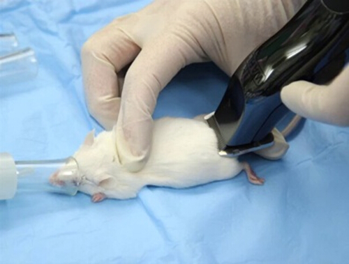
Figure 1. The correct position of a hair clipper while shaving.
- Chemical depilatory creams or lotions
- Apply the product to the surgical site area.
- After 10 minutes, the skin must be completely rinsed and cleaned of all traces of the depilatory to avoid irritation or chemical burns to the skin.
4. Surgical scrub
Surgical scrub solutions to be used should: 1) substantially reduce microbes present on the skin and contain a nonirritating antimicrobial preparation; 2) possess a broad-spectrum of antimicrobial properties; 3) be fast-acting; and 4) have persistent, cumulative activity.
The two commonly used cleansing scrub solutions are chlorhexidines and iodophors. Chlorhexidine solutions are effective against bacteria and viruses even in the presence of organic matter. In contrast, iodophors have a wide range of microbicidal action, but their efficacy is reduced in the presence of organic material; the residual activity is less than that of the chlorhexidines.
Rinses used between the scrubs are either sterile water or alcohol. Alcohol-based solutions containing 60-95% alcohol have great antimicrobial actions through the denaturing of proteins. However, alcohol can be a strong skin irritant. Sterile water is effective in rinsing the area, yet it does not have any antimicrobial properties.
- Use a moistened gauze sponge to remove gross debris including hair and dander.
- Antiseptic cleansing solution soaked onto a gauze pad is applied to the skin starting at the incision site. Disinfection should begin along the incision line and extend outward in a circular pattern.
- The antiseptic rinse is then applied beginning at the incision site. The skin is wiped in a circular pattern spiraling from the incision line to the periphery of the surgical field to remove the cleansing solution.
- This is repeated three times.
- After a final rinse, a sterile gauze pad is placed over the surgical field. This gauze may be wet with either alcohol or iodine. The gauze will be removed once the animal is transported and placed in position on a surgical platform or the surgical table/bench.
Areas of the body where the standard surgical scrubbing methods are not used include the eyes, the mouth, and the anal area. Because the surface of the eye would be damaged if scrub solutions were used, a surgical scrub is done only to the eyelids after instilling a protective ointment into the eye. In some situations, a physiologically balanced saline solution is used to flush the eye to remove gross debris and to dilute any bacteria to a level acceptable for a surgery to occur. The mouth also proves difficult to clean sufficiently for surgery. It can be rinsed with a physiologically balanced saline solution to dilute any bacteria; however, it is important to avoid using too much saline, which could cause aspiration of the fluid. Gums, teeth, and the tongue can be wiped with a nontoxic antiseptic. However, application of solutions to the mucous membranes can result in systemic absorption. Surgeries in the anal area, such as the surgical reduction of rectal prolapses, are not considered clean surgeries. The use of some antiseptic solutions can increase tissue damage and prevent or prolong healing. The use of a physiologically balanced saline solution to wash the area clean of gross debris is the preferred method of surgical preparation.
5. Positioning
Patient positioning for abdominal procedures involves the securing of the limbs of the prone animal onto the platform with tape or a ligature. When using a ligature to extend the limbs, care must be taken to prevent circulation to the feet being compromised, to avoid excessive tension on the limbs and extreme stretching of the limbs that could impair the joints, and to avoid the impediment of breathing. The ties should be a quick release with only a half hitch loop over the limb. Some commercially available platforms come with built-in limb retraction that consists of hooks or loops of stainless steel wire or ball chain, which can be adjusted according to the size of the animal. If tape is used, it must be adhered to dry surfaces.
6. Draping
Once the animal is prepped and positioned onto the surgical platform, surgical drapes are used to prevent contamination of suture material and to maintain a sterile field at the surgical site. Drapes can be a reusable cloth material, a paper disposable material, or a disposable plastic adhesive material.
Disposable paper drapes have a woven fiber matrix for strength that allows cutting into any shape or size, including cutting a fenestration or opening in the drape, without tearing or fraying of the cut edges. They are also moisture repellent. The disposable drapes can be purchased prepackaged and presterilized in a variety of sizes and shapes. Cloth drapes are not designed to be cut by the surgeon to create a fenestration. They are purchased with a precut and bound edge fenestration. Cloth drapes require laundering and sterilization. When cared for well, cloth drapes can last for years, which makes them an economical investment.
Paper and cloth drapes may be secured using sterile technique; however, in small rodents, mechanical fixation, such as towel clamps, is generally avoided due to the risk of tissue trauma. Adhesive drapes are preferred, particularly for larger rodents such as adult rats, as they help maintain a stable sterile field. For smaller rodents, careful handling is required to prevent dislodging or shifting of the drape once positioned.
Adhesive drapes are either clear or opaque. The clear drapes are preferred for rodent surgeries, as they allow for direct visualization of the animal. Some plastic drapes are a combination of plastic and paper, with the plastic area being directly over the animal and the paper area defining the extended sterile field. The portion of the drape that is directly over the surgical incision site is designed to adhere to the incision area. The surgeon can then cut directly through the plastic when making the skin incision. Sterilized plastic wrap has been accepted as a cost-effective and useful material for rodent surgeries. Care must be taken to avoid constriction of movement for breathing when the wrap is placed around the patient. The wrap will conserve body heat, allow visualization of the patient, and provide a moisture barrier between the sterile field and the animal. It can also serve to assist in the positioning and holding of the animal for the surgery in lieu of limb fixation.
Drapes of any type should be carefully unfolded to avoid contact with nonsterile areas, equipment, and personnel; they should never be unfolded by shaking or waving.
- Paper drapes: In the Single Drape Method, the drape is unfolded to allow for the cutting of the fenestration if one is not precut.
- The surgeon will place the drape over the animal keeping the hands on the side of the drape that will not touch the patient.
- The drape is adjusted so that the surgical field is visible through the fenestration.
- The drape is held in place with towel clamps through the skin of the animal in the case of larger rats.
- Paper or plastic with an adhesive window
- A drape with an adhesive window requires peeling of the paper area to allow the adhesive area to stick to the surgical field.
- When unfolding the sterile drape, the adhesive area is generally the uppermost region and is easily accessible to the surgeon.
- Once the adhesive is uncovered, the drape is carefully unfolded and turned so that the sticky side is facing the animal.
- It is imperative that the drape is placed correctly, as once the adhesive contacts the animal it will not be able to be adjusted.
- The surgeon should gently press the adhesive to the surgical field to create a seal with the skin.
- Cloth drapes
- Fenestrated cloth drapes are not to be cut, therefore it is the surgeon's responsibility to select a drape with a sufficiently large opening to adequately expose the surgical area, but not so large to allow exposure of any unshaved and unprepared body surfaces.
- The drape is carefully unfolded to reveal the fenestration.
- The surgeon will place the drape over the animal, keeping the hands on the side of the drape that will not touch the patient.
- The drape is adjusted so that the surgical field is visible through the fenestration.
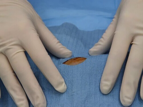
Figure 2. Surgical field visible through the fenestration of a correctly placed drape.
- Adhesive drapes: a cost-effective and useful material for rodent surgeries.
- The drape must be pulled from the roll in a manner to ensure the section being used remains sterile.
- The surgical assistant opens the box and pulls a length of the wrap out, being careful to avoid touching it to the box or any other surface.
- The surgeon grasps the wrap on each side, and the assistant cuts the end they are holding away (approximately 3-4 inches).
- After discarding the cut edge, the assistant also cuts the section from the rest of the roll.
- The surgeon grasps one side of the wrap and places it over the animal.
- The properties of the wrap allow it to adhere to all surfaces.
- The surgeon presses the film to the animal and creates a sterile field.
- There is no need to use towel clamps or worry that the drape will shift.
- Care must be taken to avoid constriction of movement for breathing. The wrap will conserve body heat, allow visualization of the patient, and provide a moisture barrier between the sterile field and the animal.
7. Intraoperative monitoring
Anesthetized patients must be monitored for body temperature, respirations, and heart rate until they are fully recovered.
Body temperature can be monitored directly or indirectly. For direct monitoring, a rectal probe designed specifically for rodents must be used. Small animal rectal thermometers, either mercury or digital, are too large for use in mice and rats without damage to the anal sphincter and rectal tissues. In susceptible strains, their use could precipitate rectal prolapse. Indirect monitoring involves placing a thermometer next to the animal or under the body on an external heating source. Although this will not give an exact body temperature, it can indicate the effectiveness of the heat source and allow adjustments to reduce or increase the heat as needed.
It is difficult to auscultate the heart rate and count the respirations on small rodents without specialized equipment.
Most monitoring is visual and will only indicate the presence or absence of chest or abdominal respirations. Heart rates are evaluated as present or absent by palpation or visual observation of fine movement of the chest wall. This may not be possible during a surgery due to draping and the small size of the animal.
Additional monitoring can be done through the use of electrocardiograms (ECG) and pulse oximeters. ECG's evaluate the cardiac status of a rodent during anesthesia and surgery. The pulse oximeter uses red and infrared light refraction to measure oxygen in arterial blood. This technology has been adapted for use in rodents using the tail or a paw. Both types of noninvasive ongoing measurements of the patient's vital signs are easily accessed with minimal disruption of the surgical field.
8. Postoperative monitoring
One should consider using a heating pad under the recovery cage postsurgery. Current scientific literature and regulatory guidelines strongly advocate for social housing as the default for laboratory animals, including those undergoing surgery with implants. Single housing is increasingly viewed as a significant stressor that should only be implemented when there is a robust scientific or veterinary justification.c In addition, pre-emptive and postoperative analgesia should be provided as a mandatory requirement. Exceptions are permitted only when withholding analgesia is scientifically justified in writing and specifically approved by the IACUC. Analgesia provisions are most effective at reducing the intensity of painful stimulation when given prior to the painful event. Advantages of the pre-emptive use of analgesics include the reduction of the intensity of painful stimulation, improvement of the animal's comfort level postsurgery, the reduction in the amount of anesthesia required to maintain a surgical plane, and a smoother recovery from anesthesia once the procedure is concluded. Commonly used pre-emptive and postoperative drugs are shown in Table 1.
| Drug Class | Name | Dosage | Frequency |
| Non-steroidal anti-inflammatory drug (Noncontrolled Substance) | Ketoprofen | 2-5 mg/kg SC mice 5 mg/kg SC rats |
every 12–24 hours every 12–24 hours |
| Non-steroidal anti-inflammatory drug (Noncontrolled Substance) | Flunixin meglumine | 2.5 mg/kg SC mice | every 12–24 hours |
| Non-steroidal anti-inflammatory drug (Noncontrolled Substance) | Meloxicam | 5-10 mg/kg PO mice or 1-2 mg/kg SC mice 5-10 mg/kg PO or 1-2 mg/kg SC or PO rats |
every 12–24 hours every 24 hours every 12–24 hours every 24 hours |
| Non-steroidal anti-inflammatory drug (Sustained release) | Meloxicam | 4 mg/kg SC rats | every 72 h |
| Non-steroidal anti-inflammatory drug (Noncontrolled Substance) | Acetaminophen | 50 mg/kg SC/IP or 100 mg/kg PO rats | every 8–12 hours |
| Opioid (Controlled Substance) | Butorphanol | 0.5-3.0 mg/kg SC or 0.2-2 mg/kg IP mice 2.0 mg/kg SC 0.2-2 mg/kg IP rats |
every 4 hours every 2–4 hours every 4 hours every 2–4 hours |
| Opioid (Controlled Substance) | Buprenorphine | 0.05-2.5 mg/kg SC or IP mice 0.01 – 0.5 mg/kg SC rats |
every 6–12 hours every 8-12 hours |
| Opioid (Sustained release) | Buprenorphine | 0.5 - 1 mg/kg SC mice 1 - 1.2 mg/kg SC rats | every 72 hours |
| Opioid (Controlled Substance) | Oxymorphone | 0.2-0.5 mg/kg SC mice 0.2-0.5 mg/kg SC rats | every 6–12 hours every 6–12 hours |
| Amide-type local anesthetic | Bupivacaine | Do not exceed 8 mg/kg Rats and mice |
Table 1. Commonly used pre-emptive and postoperative drugs.
Key Terms and Definitions
Surgical procedures are categorized as survival or nonsurvival and major or minor. Survival surgery is a surgical procedure performed on a live, fully anesthetized animal that is expected to fully recover from both the anesthesia and the procedure. Nonsurvival or terminal surgery is a surgical procedure that concludes with the euthanization of the animal before its recovery from anesthesia. Major surgery involves the exposure or penetration of the skull or a body cavity—such as the thoracic or abdominal cavity—or procedures that result in permanent physical or physiological impairment, such as limb amputation. Minor surgery does not penetrate a body cavity or cause permanent deficiency, such as subcutaneous implantation of a transponder. All surgeries require that the animal be properly anesthetized and treated humanely.
Questions that this video will help you answer
Through the use of aseptic technique, the incidence of postsurgical infection is greatly curtailed. Minimizing tissue trauma during the procedure, taking precautions to prevent hypothermia, controlling postoperative pain and discomfort, and the use of nutritional supplements until the animal is able to ambulate normally will all reduce the extent of negative metabolic responses to the surgical process and increase the probability of a successful survival surgery.
Source: Kay Stewart, RVT, RLATG, CMAR; Valerie A. Schroeder, RVT, RLATG. University of Notre Dame, IN
The Guide for the Care and Use of Laboratory Ani…
All animal procedures described here must be conducted in accordance with institutional animal ethics guidelines and approved by IACUC. All procedures must follow the principles of the 3Rs—Replacement, Reduction, and Refinement—and must be performed by trained personnel.
1. Presurgical Planning
Each laboratory animal procedure should be developed in consultation with a veterinarian to establish appropriate surgical protocols, anesthesia, and postoperative analgesia plans. These considerations should guide the setup of the surgical environment and the preparation of the animal. Although the guidelines do not require that rodent surgeries be performed in a dedicated surgical facility, the area used must be sanitized with an appropriate hard surface disinfectant, which should be used in accordance with the manufacturer's listed concentrations and contact times. The area should also be kept free of clutter, and not be in the direct line of the supply and exhaust ducts, as the drafts could contribute to hypothermia of the animal. Access to the room should be limited when surgical procedures are conducted. An area for surgical prep (especially for the removal of the animal's hair), and for postoperative recovery and care, should also be designated and in close proximity, if not within the surgery room. In general, a designated space must be reserved exclusively for surgical procedures and related activities during the time of surgery. This area requires strict control to prevent cross-contamination from unrelated activities conducted in the same room before or after the procedure.
Preoperative preparations should include a physical examination of the surgical patient to identify any underlying health conditions that may interfere with the surgery. Since rodents have such a high metabolic rate, and very limited fat reserves, they should not be fasted prior to surgery. The animal's hydration status should be evaluated by a skin elasticity test. The skin above the shoulders is gently lifted. In a normally hydrated animal, the skin will quickly fall back into place, whereas in a dehydrated animal, the skin will not immediately go back to its normal position. Overall appearance, such as posture and the condition of the hair coat, should also be noted. An animal that is displaying a hunched posture, or has an unkempt hair coat, may be harboring a disease. The examination should be performed just prior to the administration of anesthesia, and any abnormal conditions should be noted on the animal's chart. Lastly, considerations for the anatomy and physiology of the rat or mouse must be made when preparing them for surgery.
After setting up the surgery area and confirming that the animal is in good health, begin preparation for the procedure. The first step is induction of anesthesia. For detailed guidance on anesthetic induction and maintenance, refer to the corresponding protocol. Once the animal is anesthetized, remove it from the induction chamber, if applicable, and place it on a preparation surface for hair removal. Confirm adequate anesthetic depth using reflex checks, such as the pedal withdrawal reflex, before proceeding. Shaving should be performed in an area separate from the surgical platform to minimize contamination. Apply ophthalmic lubricant to prevent corneal drying, particularly during prolonged procedures.
2. Platform
Both species have a high surface area to body volume ratio making them susceptible to hypothermia during surgical procedures, especially when surgery exposes the body cavity. Efforts to prevent hypothermia include the use of a heated surgical platform.
The platform used for rodent surgery, which is usually constructed of stainless steel or hard plastic, must be covered with an insulating material or supplemental heat source to prevent the animal's body heat from escaping during the procedure. Supplemental heat sources include water circulating heating pads, forced air-warming blankets, heat lamps, or a layer of foam padding covering the surgical platform. Surgical platforms with a built in heat source are commercially available. All platforms must be of a material that is easily disinfected and impervious to moisture.
Other methods of preventing hypothermia include the use of mass insulators, placing insulating materials between the animal and the surgical platform, and using external heat sources. Mass insulators entrap air within a fiber matrix, producing "still air" that surrounds the animal. Circulating hot water blankets can be used beneath the patient. This equipment is available in various sizes from rodent to equine, and allows precise thermal support with built-in thermostats.
Chemically activated heating sources can be either onetime use or reusable. One type consists of a plastic pouch filled with a chemical solution and a metal disc, which when pressed creates an exothermal reaction. This causes the liquid to solidify and release heat. Generally, they have a limited amount of heat and are only suitable for short procedures. Other chemical heating sources are available as solids at room temperature, but when heated, they become liquid. As an animal is placed on the pad, the liquid releases heat, and the pad contents solidify as they cool. These can release heat over a much longer period. As a benefit, they cannot exceed the temperature of activation (~39°C), thus eliminating the need for a thermostat.
Water packs are available as hot water bottles consisting of a rubber or silicone bag with a stopper. The packs are filled with hot water that then emits heat on the outside surface. The pack will gradually lose heat as the water cools down. A more modern version consists of a plastic sheet with water-permeable fabric adhered to the top. The space between is filled with a hydrophilic powder, which absorbs water and swells. It can be used as either a heat source or as a cooling source. Depending on the quality of the materials, it can be reused and reheated in a microwave or soaked in hot water.
Precautions must be taken when utilizing external heat sources. Body temperature should be monitored either with a rectal probe or a thermometer placed next to the animal on the heat source. All external heat sources must be checked for defects prior to use.
3. Removal of hair
The surgical site must be prepped to minimize contamination of the incision. The hair should be closely clipped or removed with a chemical depilatory cream, which dissolves the hair at the follicle. Although the hair clipping can sometimes be performed on a conscious animal with manual restraint, the application of the depilatory cream should only be done on an anesthetized animal to prevent the ingestion of the product, eye damage, and removal of excess hair. Shaving with a razor is an option if there is no alternative. This method requires technical skill, extra time, and patience to prevent lacerations to the skin. The surgical field should be sufficiently large to allow for incision and suturing without inclusion of fur into the surgical wound, but as small as possible as to avoid the exacerbation of hypothermia.
- Clipping
- Hair can be clipped using corded-electric or battery-operated clippers, preferably with a surgical A40 blade. The width of the blade should be considered. A standard 2" blade can be used for rats, whereas a ½-1" blade is more appropriate for mice.
- The hair is clipped against the direction of growth. Stretch the skin to stabilize it, as rodents have loose attachment of the skin to the underlying muscle.
- Care must be taken to avoid nicking or cutting the skin. The flat end of the blade is placed on the skin when clipping the hair. The blade should never be used with the teeth perpendicular to the skin.
Figure 1. The correct position of a hair clipper while shaving.
- Chemical depilatory creams or lotions
- Apply the product to the surgical site area.
- After 10 minutes, the skin must be completely rinsed and cleaned of all traces of the depilatory to avoid irritation or chemical burns to the skin.
4. Surgical scrub
Surgical scrub solutions to be used should: 1) substantially reduce microbes present on the skin and contain a nonirritating antimicrobial preparation; 2) possess a broad-spectrum of antimicrobial properties; 3) be fast-acting; and 4) have persistent, cumulative activity.
The two commonly used cleansing scrub solutions are chlorhexidines and iodophors. Chlorhexidine solutions are effective against bacteria and viruses even in the presence of organic matter. In contrast, iodophors have a wide range of microbicidal action, but their efficacy is reduced in the presence of organic material; the residual activity is less than that of the chlorhexidines.
Rinses used between the scrubs are either sterile water or alcohol. Alcohol-based solutions containing 60-95% alcohol have great antimicrobial actions through the denaturing of proteins. However, alcohol can be a strong skin irritant. Sterile water is effective in rinsing the area, yet it does not have any antimicrobial properties.
- Use a moistened gauze sponge to remove gross debris including hair and dander.
- Antiseptic cleansing solution soaked onto a gauze pad is applied to the skin starting at the incision site. Disinfection should begin along the incision line and extend outward in a circular pattern.
- The antiseptic rinse is then applied beginning at the incision site. The skin is wiped in a circular pattern spiraling from the incision line to the periphery of the surgical field to remove the cleansing solution.
- This is repeated three times.
- After a final rinse, a sterile gauze pad is placed over the surgical field. This gauze may be wet with either alcohol or iodine. The gauze will be removed once the animal is transported and placed in position on a surgical platform or the surgical table/bench.
Areas of the body where the standard surgical scrubbing methods are not used include the eyes, the mouth, and the anal area. Because the surface of the eye would be damaged if scrub solutions were used, a surgical scrub is done only to the eyelids after instilling a protective ointment into the eye. In some situations, a physiologically balanced saline solution is used to flush the eye to remove gross debris and to dilute any bacteria to a level acceptable for a surgery to occur. The mouth also proves difficult to clean sufficiently for surgery. It can be rinsed with a physiologically balanced saline solution to dilute any bacteria; however, it is important to avoid using too much saline, which could cause aspiration of the fluid. Gums, teeth, and the tongue can be wiped with a nontoxic antiseptic. However, application of solutions to the mucous membranes can result in systemic absorption. Surgeries in the anal area, such as the surgical reduction of rectal prolapses, are not considered clean surgeries. The use of some antiseptic solutions can increase tissue damage and prevent or prolong healing. The use of a physiologically balanced saline solution to wash the area clean of gross debris is the preferred method of surgical preparation.
5. Positioning
Patient positioning for abdominal procedures involves the securing of the limbs of the prone animal onto the platform with tape or a ligature. When using a ligature to extend the limbs, care must be taken to prevent circulation to the feet being compromised, to avoid excessive tension on the limbs and extreme stretching of the limbs that could impair the joints, and to avoid the impediment of breathing. The ties should be a quick release with only a half hitch loop over the limb. Some commercially available platforms come with built-in limb retraction that consists of hooks or loops of stainless steel wire or ball chain, which can be adjusted according to the size of the animal. If tape is used, it must be adhered to dry surfaces.
6. Draping
Once the animal is prepped and positioned onto the surgical platform, surgical drapes are used to prevent contamination of suture material and to maintain a sterile field at the surgical site. Drapes can be a reusable cloth material, a paper disposable material, or a disposable plastic adhesive material.
Disposable paper drapes have a woven fiber matrix for strength that allows cutting into any shape or size, including cutting a fenestration or opening in the drape, without tearing or fraying of the cut edges. They are also moisture repellent. The disposable drapes can be purchased prepackaged and presterilized in a variety of sizes and shapes. Cloth drapes are not designed to be cut by the surgeon to create a fenestration. They are purchased with a precut and bound edge fenestration. Cloth drapes require laundering and sterilization. When cared for well, cloth drapes can last for years, which makes them an economical investment.
Paper and cloth drapes may be secured using sterile technique; however, in small rodents, mechanical fixation, such as towel clamps, is generally avoided due to the risk of tissue trauma. Adhesive drapes are preferred, particularly for larger rodents such as adult rats, as they help maintain a stable sterile field. For smaller rodents, careful handling is required to prevent dislodging or shifting of the drape once positioned.
Adhesive drapes are either clear or opaque. The clear drapes are preferred for rodent surgeries, as they allow for direct visualization of the animal. Some plastic drapes are a combination of plastic and paper, with the plastic area being directly over the animal and the paper area defining the extended sterile field. The portion of the drape that is directly over the surgical incision site is designed to adhere to the incision area. The surgeon can then cut directly through the plastic when making the skin incision. Sterilized plastic wrap has been accepted as a cost-effective and useful material for rodent surgeries. Care must be taken to avoid constriction of movement for breathing when the wrap is placed around the patient. The wrap will conserve body heat, allow visualization of the patient, and provide a moisture barrier between the sterile field and the animal. It can also serve to assist in the positioning and holding of the animal for the surgery in lieu of limb fixation.
Drapes of any type should be carefully unfolded to avoid contact with nonsterile areas, equipment, and personnel; they should never be unfolded by shaking or waving.
- Paper drapes: In the Single Drape Method, the drape is unfolded to allow for the cutting of the fenestration if one is not precut.
- The surgeon will place the drape over the animal keeping the hands on the side of the drape that will not touch the patient.
- The drape is adjusted so that the surgical field is visible through the fenestration.
- The drape is held in place with towel clamps through the skin of the animal in the case of larger rats.
- Paper or plastic with an adhesive window
- A drape with an adhesive window requires peeling of the paper area to allow the adhesive area to stick to the surgical field.
- When unfolding the sterile drape, the adhesive area is generally the uppermost region and is easily accessible to the surgeon.
- Once the adhesive is uncovered, the drape is carefully unfolded and turned so that the sticky side is facing the animal.
- It is imperative that the drape is placed correctly, as once the adhesive contacts the animal it will not be able to be adjusted.
- The surgeon should gently press the adhesive to the surgical field to create a seal with the skin.
- Cloth drapes
- Fenestrated cloth drapes are not to be cut, therefore it is the surgeon's responsibility to select a drape with a sufficiently large opening to adequately expose the surgical area, but not so large to allow exposure of any unshaved and unprepared body surfaces.
- The drape is carefully unfolded to reveal the fenestration.
- The surgeon will place the drape over the animal, keeping the hands on the side of the drape that will not touch the patient.
- The drape is adjusted so that the surgical field is visible through the fenestration.
Figure 2. Surgical field visible through the fenestration of a correctly placed drape.
- Adhesive drapes: a cost-effective and useful material for rodent surgeries.
- The drape must be pulled from the roll in a manner to ensure the section being used remains sterile.
- The surgical assistant opens the box and pulls a length of the wrap out, being careful to avoid touching it to the box or any other surface.
- The surgeon grasps the wrap on each side, and the assistant cuts the end they are holding away (approximately 3-4 inches).
- After discarding the cut edge, the assistant also cuts the section from the rest of the roll.
- The surgeon grasps one side of the wrap and places it over the animal.
- The properties of the wrap allow it to adhere to all surfaces.
- The surgeon presses the film to the animal and creates a sterile field.
- There is no need to use towel clamps or worry that the drape will shift.
- Care must be taken to avoid constriction of movement for breathing. The wrap will conserve body heat, allow visualization of the patient, and provide a moisture barrier between the sterile field and the animal.
7. Intraoperative monitoring
Anesthetized patients must be monitored for body temperature, respirations, and heart rate until they are fully recovered.
Body temperature can be monitored directly or indirectly. For direct monitoring, a rectal probe designed specifically for rodents must be used. Small animal rectal thermometers, either mercury or digital, are too large for use in mice and rats without damage to the anal sphincter and rectal tissues. In susceptible strains, their use could precipitate rectal prolapse. Indirect monitoring involves placing a thermometer next to the animal or under the body on an external heating source. Although this will not give an exact body temperature, it can indicate the effectiveness of the heat source and allow adjustments to reduce or increase the heat as needed.
It is difficult to auscultate the heart rate and count the respirations on small rodents without specialized equipment.
Most monitoring is visual and will only indicate the presence or absence of chest or abdominal respirations. Heart rates are evaluated as present or absent by palpation or visual observation of fine movement of the chest wall. This may not be possible during a surgery due to draping and the small size of the animal.
Additional monitoring can be done through the use of electrocardiograms (ECG) and pulse oximeters. ECG's evaluate the cardiac status of a rodent during anesthesia and surgery. The pulse oximeter uses red and infrared light refraction to measure oxygen in arterial blood. This technology has been adapted for use in rodents using the tail or a paw. Both types of noninvasive ongoing measurements of the patient's vital signs are easily accessed with minimal disruption of the surgical field.
8. Postoperative monitoring
One should consider using a heating pad under the recovery cage postsurgery. Current scientific literature and regulatory guidelines strongly advocate for social housing as the default for laboratory animals, including those undergoing surgery with implants. Single housing is increasingly viewed as a significant stressor that should only be implemented when there is a robust scientific or veterinary justification.c In addition, pre-emptive and postoperative analgesia should be provided as a mandatory requirement. Exceptions are permitted only when withholding analgesia is scientifically justified in writing and specifically approved by the IACUC. Analgesia provisions are most effective at reducing the intensity of painful stimulation when given prior to the painful event. Advantages of the pre-emptive use of analgesics include the reduction of the intensity of painful stimulation, improvement of the animal's comfort level postsurgery, the reduction in the amount of anesthesia required to maintain a surgical plane, and a smoother recovery from anesthesia once the procedure is concluded. Commonly used pre-emptive and postoperative drugs are shown in Table 1.
| Drug Class | Name | Dosage | Frequency |
| Non-steroidal anti-inflammatory drug (Noncontrolled Substance) | Ketoprofen | 2-5 mg/kg SC mice 5 mg/kg SC rats | every 12–24 hours every 12–24 hours |
| Non-steroidal anti-inflammatory drug (Noncontrolled Substance) | Flunixin meglumine | 2.5 mg/kg SC mice | every 12–24 hours |
| Non-steroidal anti-inflammatory drug (Noncontrolled Substance) | Meloxicam | 5-10 mg/kg PO mice or 1-2 mg/kg SC mice 5-10 mg/kg PO or 1-2 mg/kg SC or PO rats | every 12–24 hours every 24 hours every 12–24 hours every 24 hours |
| Non-steroidal anti-inflammatory drug (Sustained release) | Meloxicam | 4 mg/kg SC rats | every 72 h |
| Non-steroidal anti-inflammatory drug (Noncontrolled Substance) | Acetaminophen | 50 mg/kg SC/IP or 100 mg/kg PO rats | every 8–12 hours |
| Opioid (Controlled Substance) | Butorphanol | 0.5-3.0 mg/kg SC or 0.2-2 mg/kg IP mice 2.0 mg/kg SC 0.2-2 mg/kg IP rats | every 4 hours every 2–4 hours every 4 hours every 2–4 hours |
| Opioid (Controlled Substance) | Buprenorphine | 0.05-2.5 mg/kg SC or IP mice 0.01 – 0.5 mg/kg SC rats | every 6–12 hours every 8-12 hours |
| Opioid (Sustained release) | Buprenorphine | 0.5 - 1 mg/kg SC mice 1 - 1.2 mg/kg SC rats | every 72 hours |
| Opioid (Controlled Substance) | Oxymorphone | 0.2-0.5 mg/kg SC mice 0.2-0.5 mg/kg SC rats | every 6–12 hours every 6–12 hours |
| Amide-type local anesthetic | Bupivacaine | Do not exceed 8 mg/kg Rats and mice |
Table 1. Commonly used pre-emptive and postoperative drugs.
Key Terms and Definitions
Surgical procedures are categorized as survival or nonsurvival and major or minor. Survival surgery is a surgical procedure performed on a live, fully anesthetized animal that is expected to fully recover from both the anesthesia and the procedure. Nonsurvival or terminal surgery is a surgical procedure that concludes with the euthanization of the animal before its recovery from anesthesia. Major surgery involves the exposure or penetration of the skull or a body cavity—such as the thoracic or abdominal cavity—or procedures that result in permanent physical or physiological impairment, such as limb amputation. Minor surgery does not penetrate a body cavity or cause permanent deficiency, such as subcutaneous implantation of a transponder. All surgeries require that the animal be properly anesthetized and treated humanely.
Questions that this video will help you answer
Through the use of aseptic technique, the incidence of postsurgical infection is greatly curtailed. Minimizing tissue trauma during the procedure, taking precautions to prevent hypothermia, controlling postoperative pain and discomfort, and the use of nutritional supplements until the animal is able to ambulate normally will all reduce the extent of negative metabolic responses to the surgical process and increase the probability of a successful survival surgery.
Guidelines for the care and use of laboratory animals dictate that rodent survival surgery be performed aseptically and in a humane manner. Each procedure should be developed in consultation with a veterinarian to establish appropriate surgical, anesthesia, and post-operative analgesia plans.
This is to ensure that there is minimal trauma to the animal; there is no infection due to the surgical procedure itself; to prevent hypothermia; and to decrease the pain and discomfort induced by the invasive steps.
This video will review the general considerations for pre-surgical planning, intraoperative monitoring, and post-operative care. This will be followed by a demonstration of a few types of surgeries performed in biomedical research.
First, let's review the preparatory steps that a scientist should perform prior to any rodent surgery. The area where the surgery would be performed should be free of clutter and must be sanitized with an appropriate hard surface disinfectant.
Note that the area should not be in direct line with the ventilation, as the drafts could contribute to hypothermia of the patient. To further prevent hypothermia, the surgery platform should be insulated with a supplemental heat source.
The surgical instruments should be sterilized beforehand. Wrap them in individually autoclavable pouches and use validated autoclave cycles and loading patterns, and include a sterilization indicator in each pack.
When performing serial surgeries, instruments should be sterilized between each animal in accordance with institutional guidelines and allowed to cool before use on the next animal.
Several days prior to surgery, observe the animal to evaluate its body condition, appetite, water intake, and urine and feces output. Additionally, note the body posture and the condition of the hair coat.
An animal displaying a hunched posture or an unkempt hair coat may be harboring a disease and may not be a good candidate for surgery.
Because rodents have such a high metabolic rate and very limited fat reserves, they should not be fasted prior to surgery. Evaluate the animal's hydration status by performing the skin elasticity test.
To do this, gently lift the skin above the shoulders. In a normally hydrated animal, the skin will quickly fall back into place, whereas in a dehydrated animal, the skin will not immediately go back to its normal position.
After setting-up the surgery area and making sure that the animal is in good health, you can begin preparing the animal for the procedure. The first step is anesthetization. In order to understand how to induce and maintain anesthesia refer to another video in this collection.
After the animal is sedated, remove it from the chamber and place it on the shaving pad. Confirm adequate anesthetic depth using reflex checks, such as pedal withdrawal or corneal reflex, before proceeding. Note that shaving should be performed in an area away from the surgical platform to prevent any contamination.
Apply lubricant eye ointment to prevent drying of the eyes, especially for relatively long surgeries.
Shaving is commonly done using electric clippers, preferably with a surgical A40 blade. A standard two-inch blade can be used for rats, whereas a half- to one-inch blade is more appropriate for mice. To avoid nicks and cuts, first stretch the skin to stabilize it.
Then place the flat of the blade on the skin and move it against the direction of the hair growth. The shaved surgical field should be sufficiently large to allow for incision and suturing without inclusion of fur into the surgical wound, but as small as possible so as to avoid the exacerbation of hypothermia.
Alternatively, hair may be removed using depilatory cream, which dissolves the hair at the follicle. First, apply the product to the surgical site area using a swab.
After approximately three minutes, rinse and clean the skin with sterile water to remove all traces of the cream in order to avoid irritation or chemical burns.
Following hair removal, use a moistened gauze sponge to remove any debris including hair and dander. Then, using a gauze pad pre-soaked in antiseptic or cleansing solution, scrub the area starting at the incision site and extending outward in a circular pattern.
Next, apply the rinsing liquid in the same circular pattern - spiraling from the incision line to the periphery of the surgical field. Repeat the cleansing and rinsing cycle three times or follow manufacturer instructions for single-application antiseptics.
After the final rinse, place a sterile gauze pad - wet with either alcohol or iodine - over the surgical field. The gauze will remain in place while the animal is transported and positioned on the surgical platform.
The next step is to position the animal. For dorsal procedures, place the animal in a prone position. Secure the limbs onto the platform using ligature or tape. Care must be taken not to: compromise circulation to the feet, cause excessive tension on the limbs, impair the joints due to overstretching, or impede the animal's breathing.
Once the animal is prepped and positioned onto the surgical platform, remove the gauze covering the incision area and now the animal is ready for draping.
Remember that draping is an aseptic technique, so the surgeon ought to be wearing sterile gloves, a bouffant cap, a surgical mask, and a clean lab coat or a sterile surgical gown.
Drapes can be made of cloth, which is reusable, or disposable paper or plastic adhesive material. Drapes of any type should never be shaken or waved to unfold.
They should be carefully opened to avoid contact with non-sterile areas, equipment, and personnel. If the drape is not precut, then unfold enough so that you can create an opening using sterile scissors that will be large enough to adequately expose the surgical area, but not so large to allow exposure of unshaved and unprepared body surfaces.
Next, place the drape over the animal keeping the hands on the side of the drape that will not touch the patient. And lastly, adjust it so that the surgical field is visible through the opening. This allows one to maintain a sterile field at the surgical site for the entire procedure.
If using a paper drape with an adhesive window, first peel the paper from the adhesive area. Then, unfold the drape carefully, turn it so that the sticky side is facing the animal, and place it over the surgical area. Then gently press the adhesive to the surgical field to create a seal with the skin.
After prepping, the next step is surgery. So let's take a look at a few things that the surgeon should bear in mind during and post surgery. While under anesthesia, animals must be monitored for body temperature, respirations, and heart rate until they are fully recovered.
For direct monitoring of body temperature, a rectal probe designed specifically for rodents must be used. The normal range is 35.3 to 38 oC.
Alternatively, for indirect monitoring, place the thermometer next to or under the animal. Although this will not give an exact body temperature, it can indicate the effectiveness of the heat source and allow adjustment to reduce or increase the heat as needed.
Meanwhile, throughout the procedure visually monitor the heart rate, respirations, and the color of the tail. Occasionally, additional monitoring can be done through the use of electrocardiograms, which evaluates the cardiac status of a rodent during anesthesia and surgery.
Post surgery, the animal should be returned to a recovery cage placed partially over a heating pad and food should be made easily accessible. The partial placement on the heat pad allows the animal to move off of the heat source after it has regained consciousness.
Once fully recovered, animals should be returned to group housing whenever possible. Single housing should be used only if scientifically justified or required for post-surgical monitoring.
In addition, pre-emptive and postoperative analgesia must be provided unless scientifically justified and approved otherwise. Multimodal analgesia, including sustained-release formulations and local anesthetic techniques such as incision-site blocks can be considered as per the study need. See text below for the list of recommended analgesics, their dosage, route and frequency of administration.
Now that you're well versed with the considerations, let's look at some of the survival surgeries performed by biomedical researchers.
In order to manipulate the brain in living animals, scientists often perform stereotaxic surgeries using a specialized equipment called the "stereotaxic frame". Using this tool and a three-dimensional coordinate system, one can target specific locations to measure brain activity, induce lesions, or perform genetic manipulations.
Scientists also perform surgery to create animal models of human disorders. Here, the researchers created a "skin flap" model to study ischemia induced tissue damage. By implanting a metal window in the shaved skin, the investigators were able to visualize the region's microcirculation in a live animal for several days.
In another experiment, researchers performed surgery to induce bone defect by drilling a plate into a rat's femur, and then studied in vivo bone healing process with the help of X-rays.
You've just watched JoVE's video on considerations that one should bear in mind before, during and after performing rodent surgery. Together, these measures would reduce the probability of infection and hypothermia, and help in ameliorating pain and discomfort to the animal, which will in turn increase the probability of a successful survival surgery.
View the full transcript and gain access to JoVE Science Education videos
Q1: Why should rodents not be fasted before surgery?
Rodents have a high metabolic rate and very limited fat reserves, making them vulnerable to complications from fasting. Pre-surgical fasting can compromise their health status and recovery outcomes. Instead, evaluate the animal's hydration using the skin elasticity test before proceeding with the surgical procedure.
Q2: What is the proper technique for preparing the surgical site on a rodent?
After anesthesia induction and maintenance, shave the surgical area using electric clippers with appropriate blade size. Remove debris with moistened gauze, then scrub with antiseptic solution in a circular pattern from the incision site outward. Repeat the cleansing and rinsing cycle at least three times, then cover with a sterile gauze pad soaked in alcohol or iodine until positioning.
Q3: How should surgical instruments be sterilized for rodent surgery?
Wrap instruments in individually autoclavable pouches and place them in a tented configuration inside the autoclave to allow complete steam penetration. For serial surgeries, sterilize instruments between each animal using a bead sterilizer for 10-15 seconds. Always allow instruments to cool completely before using them on the next animal to prevent thermal injury.
Q4: What vital signs should be monitored during rodent surgery?
Monitor body temperature using a rectal probe designed for rodents, maintaining the normal range of 35.3 to 38°C. Visually observe heart rate, respirations, and tail color throughout the procedure. Electrocardiograms can provide additional cardiac monitoring. Continue monitoring until the animal is fully recovered from anesthesia.
Q5: How should a rodent be positioned and secured for dorsal surgical procedures?
Place the animal in prone position on an insulated, heated surgical platform. Secure the limbs using ligature or tape while avoiding circulation compromise, excessive limb tension, joint overstretching, or impeded breathing. Proper positioning maintains the sterile field and ensures animal safety throughout the procedure.
Q6: What post-operative care should be provided after rodent survival surgery?
Return the animal to a recovery cage placed partially over a heating pad, allowing it to move away from heat after regaining consciousness. Make food easily accessible and provide pre-emptive and postoperative analgesia whenever possible. This approach reduces pain, prevents hypothermia, and supports successful recovery.
Q7: What environmental conditions must be maintained in the surgical area?
The surgical area must be free of clutter and sanitized with appropriate hard surface disinfectant. Position the area away from direct ventilation drafts to prevent hypothermia. Use an insulated surgery platform with supplemental heat source. These conditions minimize infection risk, prevent hypothermia, and create an optimal environment for aseptic technique.
Chapters in this video
0:00
Overview
1:06
Preoperative Preparations: Area and Animal Health
3:08
Prepping Steps: Shaving, Scrubbing, Positioning, and Draping
8:13
Intra and Postoperative Procedures
10:31
Applications
11:46
Summary
Videos from this collection: