The goal of this project is to detail a thoroughly scrutinized protocol of the surgical placement of an ameroid constrictor around the proximal left circumflex coronary artery (LCx), thereby inducing chronic coronary artery disease and precisely mapping the ischemic territory using isotope-labeled microspheres.
Method Article
A Clinically Relevant Swine Model of Chronic Myocardial Ischemia by Ameroid Constrictor Placement with Simultaneous Perfusion Mapping
In This Article
Summary
Abstract
Despite breakthroughs in medical and procedural therapies for the treatment of cardiovascular disease, the need for translational large animal models has persisted to investigate diagnostic tools and treatment strategies. Swine represent an impeccably accurate and appropriate model for human cardiovascular disease, as their cardiac anatomy and physiology closely resemble that of humans, as well as their metabolic and proteomic profile. The induction of chronic myocardial ischemia to an area of the myocardium by using an ameroid constrictor accurately and easily emulates the common pathophysiologic process of atherosclerosis development in humans and subsequent myocardial response. Ameroid constrictors have been widely studied over the last 50 years providing rigorous evidence for its use; furthermore, we have over two decades of experience in efficiently and uniformly conducting this procedure. An important step within this protocol is performing coronary blood flow mapping to determine the area of greatest ischemia and areas with no ischemia by injecting isotope-labeled microspheres. The ability to perform coronary blood flow mapping precludes the necessary and labor-intensive use of cardiac MRI or PET imaging for perfusion studies and determination of the ischemic territory. The goal of this publication is to detail the correct placement of an ameroid constrictor on the proximal LCx and subsequent coronary perfusion mapping.
Introduction
Globally, ischemic heart disease (IHD) has only grown in its mortality and burden on patients and the healthcare system. According to the American Heart Association, the number of deaths attributable to IHD from 1990 to 2021 has increased by 74% worldwide1. This is additionally true within the United States, where the Center for Disease Control cites that one in twenty adults has been diagnosed with coronary artery disease (CAD), and about one in five mortalities are due to cardiovascular disease, which is equivalent to a person dying from cardiovascular disease every 33 s2. This burden of disease stresses the healthcare system, in which over $250 billion was spent on patients with cardiovascular disease from 2019 to 20202. Given the ubiquitous nature of cardiovascular disease and the severe burden to both patient and provider, it is imperative to continue investigations into innovative and novel therapeutics.
To achieve such goals, clinically relevant animal models must be employed yet scrutinized to meet the standards of reproducible and translational research. Animal models have been questioned for a multitude of reasons, including disparate animal species with variable pharmacokinetics, differing protocols for inducing the desired disease state, variations in drug dosing and schedules, poor standardization of randomizing animals, follow-up duration, and blinding of investigators, and finally, small experimental groupings3. We have experimentally refined and standardized the protocol not only of the overall animal model to represent IHD but also the surgical protocol, animal randomization, drug delivery and duration, and subsequent outcomes. In regard to IHD, swine represent an optimal animal model due to their significant similarities in cardiac anatomy and physiology to humans. Importantly, swine have similar coronary artery distribution as humans and minimal collateral circulation, making the induction of ischemia a reliable representation of the development of atherosclerosis in humans. Additionally, swine have similar metabolism, proteomic profile, and immune system to humans, which makes analyzing the molecular impact of therapeutics more meaningful4. With this in mind, the animal model employed in this protocol is best suited to reproducibly represent human cardiovascular pathophysiology.
Our lab utilizes the surgical placement of an ameroid constrictor on the LCx, allowing for the chronic induction of myocardial ischemia. Employing this technique to chronically induce ischemia yields unique advantages over acute ligation. One is the extensive research literature from multiple decades employing this device5. Additionally, it allows for the reliable clinical simulation of gradual coronary narrowing due to atherosclerotic development in humans and, finally, the improvement in animal mortality by sparing myocardium that would have had complete blood flow cessation6. The ameroid constrictor has two parts: a titanium outer ring and an inner hygroscopic plastic layer. When placed around the coronary artery, the inner ring will subsequently absorb bodily fluids and swell inward due to the restrictive steel sleeve over a two-to-four-week period7. The strategy of placement around the LCx confers mortality benefits as this is the smallest of the three major coronary arteries and affects approximately 20% of the myocardium7. To uniformly produce the ischemic area, the ameroid should be placed at the LCx takeoff before branching the obtuse marginal arteries.
Further consideration employed by this protocol is the confirmatory coronary blood flow mapping to determine the induction of ischemia and the use of the correct territory in investigating the myocardial effects of therapeutics. For this purpose, we employ gold microsphere injection from BioPhysics Assay Laboratory, Inc. at the time of ameroid placement when the LCx is completely occluded. The section with the lowest quantification of gold microspheres will then represent the most ischemic left ventricular territory. The importance of mapping cannot be overstated when determining the molecular modifications produced by experimental treatment. Additionally, the use of isotope-labeled microspheres provides a cost-effective method as compared to cardiac magnetic resistance imaging for the determination of myocardial perfusion8.
Lastly, the model described here considers the well-known variation among sexes in the development of cardiovascular disease and subsequent therapeutic response. Too often, preclinical animal models fail to faithfully represent differences in the human population; thus, we match the sex of the swine utilized in the control and experimental groups between female and intact male swine. We have previously employed this to determine sex-based responses to novel treatments9. Further adding to the translatability of this protocol is the use of randomization of treatment groups and blinding of the investigator analyzing the cardiac functional and myocardial perfusion parameters of each swine.
Protocol
The surgical procedure detailed below was approved by the Institutional Animal Care and Use Committee of Rhode Island Hospital and Brown University.
1. Animal model
- Use both juvenile female and intact male Yorkshire swine, ideally aged around 11 weeks old, and typically weighing around 15 kg.
2. Pre-surgical procedures
- Ensure that swine receive daily pre- and postoperative aspirin (10 mg/kg) starting 1 day before the surgery and proceeding to 5 days postoperatively.
- Ensure that swine are without food on the morning of surgery.
- On the day of surgery, induce anesthesia using intramuscular injections of telazol (4.4 mg/kg) and xylazine (2.2 mg/kg).
- Intubate the swine with an appropriately sized (typically 7-0) cuffed endotracheal tube.
- At the same time, achieve analgesia by giving buprenorphine at 0.03 mg/kg and applying a fentanyl patch (4 μg/kg).
- Place a 20-22 G intravenous (IV) catheter in the large auricular vein.
- Begin IV fluids (0.9% saline) at a rate of 5 mL/kg/h.
- Administer ceftiofur, which is a broad-spectrum cephalosporin antibiotic. This is typically done within 30 min of incision as a single-dose IM injection.
- Shave the pig at the surgical site (from left shoulder to umbilicus) as well as at electrocardiogram (ECG) monitoring lead positions.
- Transport the pig to the operating room and maintain anesthesia with 0.75-3.0% isoflurane.
- Connect the swine to the ECG monitor and apply electrocautery grounding pads.
- Properly position the swine on the operating table: right side down, left forelimb extended and tied to the table, and the right side of the table tilted up.
- Prepare the surgical site with a 2% chlorohexidine scrub.
- Drape the incisional site with four small stick-on drapes and one large laparotomy drape.
3. Surgical procedure
- Start by injecting 2% lidocaine subcutaneously at the anticipated incision site.
- Then, use a #10 scalpel to make roughly a 5 cm incision obliquely starting near the axillary skin fold and proceed out laterally; this is around the 2nd to 3rd intercostal space.
- Use the #10 scalpel to continue this incision down through the muscular layer until the fascial layer is reached.
- At this time, use electrocautery to achieve hemostasis of the skin and muscle.
- Bluntly open the fascial place superiorly until the clavicle can be palpated.
- Once this is achieved, palpate the rib spaces, ensuring the 2nd rib space is identified.
NOTE: Typically, the 2nd intercostal space achieves a slightly superior view of the field, allowing for easier proximal placement of the ameroid constrictor. Sometimes, due to swine anatomy, the 3rd intercostal space must be entered. - Score the intercostal muscle with electrocautery and enter the pleural space with a large Kelley clamp.
- Extend this opening medially and laterally with electrocautery while protecting the lung and pericardium with the large Kelley clamp as guidance.
- Introduce a small Finochietto retractor to spread the ribs apart.
- Place a wet gauze to push back the lung into the left pleural space. This extends the view of the pericardium.
- Use Debakey forceps to tent up the pericardium and make a small cut with Metzenbaum scissors. Extend this medially and laterally to expose the left atrial appendage. When extending laterally towards the lung, identify and protect the lung and the phrenic nerve.
- Tack up the pericardium as necessary using a 3-0 silk suture.
- Using a small Satinsky clamp, grasp a distal portion of the left atrial appendage and ligate this portion using a 0-silk tie. With a small hemostat, retract the appendage to expose the LCx and the LAD (Figure 1A).
- Once the vessels are exposed, switch to Gerald forceps and make a small nick with Metzenbaum scissors in the epicardial fat overlying the LCx near its take-off from the left main coronary artery.
- Using a small right angle and peanut sponges on Allis clamps, carefully dissect out the LCx. Once around the artery with the right angle, place a vessel loop to fully isolate it. Place a small hemostat on the two ends.
- At this time, systemically heparinize the swine with 80 IU/kg to prevent thrombus formation.
- Perform the myocardial perfusion mapping.
NOTE: This procedure requires two assistants. Ensure that one is an operating room technician who will time for 2 min. The other must be a surgical assistant.- Instruct the assistants to gently retract the vessel loop to fully occlude the LCx. Confirm occlusion by viewing the ST segment and/or T wave changes on the ECG monitor.
- During the first 30 s of occlusion, inject 5 mL of gold microspheres, using a 28 G needle, into the left atrial appendage (Figure 1B). Draw the syringe back at the halfway point of the injection to visualize blood; this is critical to ensure the correct positioning of the needle within the left atrium and subsequent accurate mapping.
- After completing the injection, use a peanut sponge to hold pressure on the injection site until hemostasis is achieved. If hemostasis cannot be achieved with pressure, place a small figure of eight suture with a 6-0 polypropylene suture.
- Instruct the surgical assistant to continue to retract the vessel loop for the entire 2 min while visualizing the ECG changes, ensuring occlusion of the LCx.
- After the 2 min and once the ECG changes subside, place an ameroid constrictor around the proximal LCx vessel. Sizing is based on visual inspection, typically ranging from 2.0 mm to 2.75 mm. Grasping the ameroid either on an Allis clamp or Kelly clamp allows for easier application (Figure 1C). Ensure the ameroid is well lubricated.
- Have the surgical assistant retract the pocket created from dissection with either Gerald forceps and/or a peanut sponge to open the space. Gently guide the ameroid onto the artery through the keyhole, and confirm that the keyhole is rotated outwardly (Figure 1D).
- Verify that the ameroid is as proximal as possible in its positioning. This helps maintain uniformity in placement and the subsequent ischemic area.
- Remove the vessel loop and spray topical nitroglycerin (~2 mL) onto the artery to prevent further vasospasm.
- Cut the tie on the atrial appendage and let it fall back into position.
- Close the pericardium using a running 3-0 absorbable suture on a reverse bevel (RB) needle.
- Remember to take out the wet gauze, retracting the lung, and inspect for any injury.
- Reapproximate the rib space using 0 Polydioxanone suture (PDS) on a blunt tip needle.
- Close the muscular layer with a 3-0 absorbable suture, and when almost closed, introduce a small red rubber catheter connected to suction into the pleural space. As the primary surgeon ties down the final suture, obtain a breath hold and have the assistant retract the red rubber out of the cavity. This ensures that any air within the pleural cavity is evacuated.
- Close the subcutaneous layer with 3-0 absorbable suture and the skin with a running subcuticular 4-0 Monocryl suture.
- Place a sterile dressing over the incision site.
4. Postoperative care and recovery
- Begin weaning the isoflurane.
- While waiting for the swine to wake, secure the incisional bandage with gauze and elastic wrap.
- Once the swine is breathing on its own, remove the endotracheal tube.
- Allow the swine to recover under the surveillance of the veterinary team in a padded recovery cage with heat support.
- Again, aspirin is continued until postoperative day 5 to mitigate thrombotic risk. Achieve analgesia with the fentanyl patch placed pre-operatively, which stays in place for 3 days.
- Complete twice daily postoperative checks on the swine for 3 days. Typically, the swine do not experience arrhythmia or infarction during this period.
NOTE: Be mindful that around postoperative day 14, early ameroid closure and/or spontaneous ventricular arrhythmia are more likely to occur. Groups may troubleshoot this time period with prophylactic antiarrhythmic medications, such as amiodarone or lidocaine, which is reviewed in the discussion. Additionally, continuous ECG monitoring may be utilized; however, it is quite labor-intensive and difficult to maintain on the swine.
Results
Over two decades of performing the above procedure, the survival rate is about 80%10,11. A majority of mortalities occur around 2 weeks postoperatively, often due to irreversible and spontaneous ventricular arrhythmia at the time of ameroid closure. On multiple prior necropsies, typical findings show a properly placed ameroid with minimal acute scar formation of the left ventricle, confirming the ventricular arrhythmia diagnosis. The arrhythmia leads to sudden cardiac death, which is incredibly difficult to predict in which animals this may occur. Therefore, we have considered continuous ECG monitoring during the first 2 weeks postoperatively; however, it was determined that the invasive nature and workload to maintain appropriate ECG tracings outweigh the usefulness of the monitoring. After the 2-week period, the survival rate nears 100%.
Likely the most important result from this procedure is the differential myocardial perfusion mapping. The gold microsphere quantification allows for the determination of the absolute blood flow of each left ventricular section, therefore illustrating which segments are most ischemic and which are not. These measurements constitute the myocardial mapping performed at the time of ameroid placement. Table 1 demonstrates representative results after gold microspheres quantification with the most ischemic and non-ischemic sections highlighted. Each section is mid-myocardial left ventricular tissue. Highlighted in red are the most ischemic territories, as exemplified by zero gold quantified within the myocardium, which represents the LCx territory. Highlighted in green is the territory that is most non-ischemic LAD supplied, as demonstrated by the most gold microspheres quantified within the myocardial section. We typically focus on the ischemic and non-ischemic territory because this model illustrates chronically ischemic myocardium and not an acute infarction model.
Following this procedure, the swine can either undergo medical therapy, where we have recently been investigating novel anti-diabetic drugs, or a subsequent left mini-thoracotomy to investigate novel intramyocardial injections of angiogenic therapies, such as extracellular vesicles or extracellular matrix. This protocol permits a broad investigative scope in which multiple types and delivery avenues of therapies can be studied to determine their functional and angiogenic potential in the treatment of coronary artery disease.
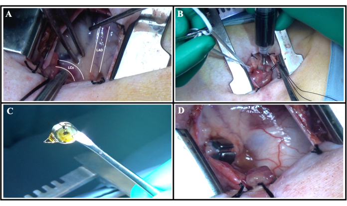
Figure 1: Surgical procedure. (A) Coronary anatomy once the left atrial appendage was retracted. The LCx and LAD are traced and labeled. (B) The injection of gold microspheres in the left atrial appendage while the LCx is occluded with the vessel loop. (C) The ameroid constrictor, lubricated, and positioned on the Allis clamp for placement. (D) Ameroid constrictor placed around the LCx with the keyhole positioned outward. Please click here to view a larger version of this figure.
| Section | Gold | Weight (g) | Gold/wt |
| 1A | 63037.6 | 0.658 | 95801.8237 |
| 2A | 38206.2 | 0.347 | 110104.323 |
| 3A | 48130.6 | 0.426 | 112982.629 |
| 4A | 11124.4 | 0.241 | 46159.3361 |
| 5A | 8742.4 | 0.623 | 14032.7448 |
| 6A | 33249.4 | 0.353 | 94190.9348 |
| 3B | 0 | 0.505 | 0 |
| 4B | 0 | 0.577 | 0 |
| 5B | 9198 | 1.049 | 8768.35081 |
| 6B | 75049.3 | 0.849 | 88397.2909 |
Table 1: Representative table of results from gold quantification. Highlighted in red are the left ventricular sections that are most ischemic, as illustrated by zero quantification of gold in the tissue, and represent the LCx territory. The highlighted green row demonstrates the non-ischemic left ventricular myocardial section determined by the largest quantification of gold in the tissue and depicts the territory supplied by the left anterior descending artery.
Discussion
The main advantage of using an ameroid constrictor is the simplicity of the device; however, this presents limitations in the inability to control closure rates or the degree of stenosis around the coronary artery. This can lead to variability in the induction of myocardial ischemia. Alternatives to the use of ameroid constrictors include the use of silk ties, hydraulic occluders, or serial placement of flow probes12,13,14,15,16. The use of silk ties and occluders is often employed to create a fixed stenosis model providing a precise percentage of occlusion of a given coronary artery but is often offset by higher mortality rates12,15. Occluders additionally have increased rates of arterial dissection due to the introduction of such devices14. The use of serial flow probes again offers standard reductions in coronary flow but requires sophisticated equipment, multiple procedures, and more technical expertise to reliably model17. In contrast, ameroid constrictor placement is technically simple, cost-effective, and produces a relatively low mortality rate of about 20%18. Additionally, we ensure that the placement of the ameroid is around the most proximal portion of the LCx at its takeoff from the left main coronary artery. This strategy plays a vital role in achieving a consistent ischemic burden across all subjects.
The main cause of mortality has been demonstrated to be ventricular arrhythmia around the time of complete closure of the ameroid constrictor: 2 weeks postoperatively. In a previously published protocol of ameroid placement, the group utilized prophylactic oral amiodarone postoperatively through the end of the study6. Two concerns are raised with this use: 1) there was no decrease in mortality rate compared to this protocol, and 2) the use of amiodarone negatively affects the inotropic capability of the heart with administration, so it may confound the results when investigating new therapies19. Additionally, the use of amiodarone has effects on the reduction of afterload and changes in coronary artery resistance, adding further evidence against postoperative use of this drug19. Alternative agents that may be used prophylactically could be class I antiarrhythmic medications, such as lidocaine; however, these medications additionally have dose-dependent negative effects on inotropy and chronotropy20. In regard to other causes of mortality, it has been reported that decompensated heart failure may occur with ameroid placement6. We rarely have observed heart failure leading to mortality within the cohorts of swine. This may be due to the effective and consistent placement of the ameroid on the proximal LCx, leading to ischemia persistently to about 20% of the myocardium. The ischemic area reflects a large enough territory to thoroughly investigate therapeutic agents and limits affecting a large enough area to induce left ventricular heart failure.
In the above-described protocol, control animals have had ameroid constrictors placed that subsequently do not receive experimental therapies. We typically do not include sham animals within the protocol as we have consistently shown the control animals have reduced overall cardiac function. For example, controls typically have a mean ejection fraction (EF) of around 30-35%, about half of normal swine21. Furthermore, histological analysis of the left ventricular myocardium within the ischemic territory reliably demonstrates fibrosis, oxidative stress, and apoptosis. With such long-standing evidence of reliable induction of myocardial ischemia, we feel the inclusion of sham swine would not add benefit to the investigations. Groups starting the following protocol may wish to include sham swine as well as control-operated swine to ensure reliable induction of meaningful myocardial ischemia. Furthermore, based on our prior standard deviation of myocardial perfusion studies (0.150 mL/mg/min) and using a β of 0.1, we have determined an ideal sample size is 8 swine per group. Given the mortality rate, researchers should plan on starting with about 10 swine per group to maintain power.
As described, a critical step in this protocol is the use of gold microspheres for the mapping of myocardial perfusion. Prior groups, including our laboratory, have utilized multiple strategies for measuring myocardial perfusion, such as cardiac MRI, coronary angiography, cardiac positron emission tomography (PET), or contrast echocardiography22,23. Cardiac PET scanning using radioactive tracers has been extensively validated and remains a gold standard for noninvasive measurement of myocardial perfusion; however, this technique has several limitations, including the high cost and technical expertise of imaging protocols, the need for specific tracers and possible motion artifacts of the images24. Coronary angiography is advantageous for its ability to rapidly acquire data on coronary anatomy, ventricular function, and perfusion within one study; however, it requires investigators to be exposed to radiation and has multiple limitations in regards to image quality which is inversely proportional to heart rate making characterization under stress extremely limited23. Finally, cardiac MRI has become more widespread, using gadolinium contrast agents, resulting in high-resolution imaging, especially under stress conditions. However, this modality can be challenging in its application to animals requiring sedation and costly equipment6,25. Furthermore, the use of an ameroid constrictor adds further susceptibility artifacts due to a metallic foreign body, which led to the development of plastic ameroid constrictors to limit this interference26.
Nonetheless, microspheres represent the gold standard for invasive measurement of myocardial perfusion and have several advantages over the noninvasive imaging modalities, including low cost, simplified and reproducible protocol, and the ability to simultaneously measure regional metabolism by measuring other stably labeled compounds. In this first step of using gold microspheres, the left ventricular regions are accurately mapped. This allows a reliable illustration of the LCx ischemic territory and the LAD non-ischemic territory, especially after sectioning of the left ventricular. Now that the myocardium is mapped, perfusion to these territories can be measured during the terminal harvest with different isotopes of microspheres. We have found using both an ameroid constrictor and gold microspheres to be reliable, affordable, and reproducible.
Disclosures
The authors have no disclosures or conflicts of interest.
Acknowledgements
The research was funded by the National Institutes of Health with the following grant numbers: T32HL16051703 (K.C.M., C. S., D.D.H, M.K.), R01HL46716 (F.W.S), and R01HL128831 (F.W.S). We would like to thank the veterinary staff at Rhode Island Hospital and Brown University for their exceptional work and support on our multitude of projects.
Materials
| Name | Company | Catalog Number | Comments |
|---|---|---|---|
| #10 Scalpel | Medline | MDS15310 | |
| 0.9% Saline | Dechra/Vetivex | 17033-492-50 | Rate: 5 mL/kg during case |
| 0-Silk Ties | Ethicon | SA76G | Tie off Left Atrial Appendage |
| 1-0 PDS II on a CT Blunt Tip | Ethicon | Z359T | Close rib space |
| 2% Lidocaine | Phoenix | Dosing: 2–5 mg/kg | |
| 2-0 Silk Suture on SH Taper | Ethicon | K833H | Tack pericardium |
| 28-guage needle | Patterson Veterinary | 78032728 | For microsphere injection |
| 3 18G Needles | Patterson Veterinary | 78032793 | For drawing medication |
| 3-0 Vicryl on an RB Taper | Ethicon | VCP215H | Close pericardium |
| 3-0 Vicryl Reverse Cutting | Ethicon | VCP442H | Close muscular and deep dermal layers |
| 4-0 Monocryl | Ethicon | MCP496G | Close skin |
| 6-0 Polypropylene Suture | SharePoint | JS617N | For emergency repair of atrium |
| All Surgical Instruments | Steris and Medline | Requires Sales Representative | All Commonly Found in Hospital Operating Rooms |
| Ameroid Constrictors | Research Instruments SW, Inc. | CS-2.00-TI | Type: titanium (2.0 cm to 3.0 cm) |
| Analgesia: Buprenorphine | Hospira | Brand Name: Bupredine | Dosing: 0.03 mg/kg |
| Analgesia: Fentanyl Patch | Covetrus | 77870 | Dosing: 4 mg/kg |
| Anesthesia: Telazol | Zoetis | (tiletamine and zolazepam) | Dosing: 4.4 mg/kg |
| Anesthesia: xylazine (Rompun) | Dechra | N/A | Dosing: 2.2 mg/kg |
| Chlorhexidine 4% | BD E-Z Scrub | 371073 | Surgical Prep |
| Cuffed Endotracheal Tube | RUSCH | 7-0 typically | May need different sizes depending on swine size and weight |
| ECG Monitoring System | MidMark | N/A | Multiparameter Monitor |
| Electrocautery | Bovie Specialist Pro | 1250S | |
| Excede (Ceftiofur) | Covetrus | 23814 | Single-dose IM injection |
| Gold STERIspheres | BioPAL, Inc. | C-15H20 | Amount: 5cc per surgery |
| Isoflurane, USP | Dechra | Dosing: 0.75%–3.0% | |
| Mini White Vessel Loops (X-ray Detectable) | DeRoyal | 30-711 | To isolate the LCx |
| Multiple 5cc Syringes | Medline | TMOSS05L | For multiple injections |
| Nitroglycerin | Baxter | Nitroglycerin in 5% dextrose injection (200mcg/mL) | Spray 2–3 cc topically |
| Red Rubber Catheter | Cardinal Health | 888766008 | Place into chest to extract air |
| Sterile Gloves | Encore | 820955 | |
| Suction System | GOMCO | 4042 | Better if wall suction of own facility |
| Suction tip: Yankauer | Medline | DYND50130 | |
| Surgical Drapes | Patterson Veterinary General Surgery Pack | 78929436 | 4 small adhesive drapes and one large laparotomy drape |
| Surgical Lubricant | SurgiLube | 0281-0205-43 | Must be sterile |
| Systemic Heparin | Covetrus | 82496 | Dosage: 80 IU/kg |
| Yorkshire Swine | CBSET, Inc. | N/A | Ideally around 11-weeks-old |
References
- Martin, S. S., Aday, A. W., Almarzooq, Z. I., et al. heart disease and stroke statistics: a report of US and global data from the American Heart Association. Circulation. 149 (8), e347-e913 (2024).
- Tsao, C. W., Aday, A. W., Almarzooq, Z. I., et al. heart disease and stroke statistics-2023 update: a report from the American Heart Association. Circulation. 147 (8), e93-e621 (2023).
- Pound, P., Ebrahim, S., Sandercock, P., Bracken, M. B., Roberts, I. Where is the evidence that animal research benefits humans. BMJ. 328 (7438), 514-517 (2004).
- Jia, H., Chang, Y., Song, J. The pig as an optimal animal model for cardiovascular research. Lab Anim (NY). 53 (6), 136-147 (2024).
- Litvak, J., Siderides, L. E., Vineberg, A. M. The experimental production of coronary artery insufficiency and occlusion. Am Heart J. 53 (4), 505-518 (1957).
- Keeran, K. J., et al. A chronic cardiac ischemia model in swine using an ameroid constrictor. J Vis Exp. (128), e56190(2017).
- Hughes, G. C., Post, M. J., Simons, M., Annex, B. H. Translational physiology: porcine models of human coronary artery disease: implications for preclinical trials of therapeutic angiogenesis. J Appl Physiol. 94 (5), 1689-1701 (2003).
- Reinhardt, C. P., Dalhberg, S., Tries, M. A., Marcel, R., Leppo, J. A. Stable labeled microspheres to measure perfusion: validation of a neutron activation assay technique. Am J Physiol Heart Circ Physiol. 280 (1), H108-H116 (2001).
- Harris, D. D., et al. Proteomic analysis and sex-specific changes in chronically ischemic swine myocardium treated with sodium-glucose cotransporter-2 inhibitor canagliflozin. J Am Coll Surg. 238 (6), 1045-1055 (2024).
- Sabe, S. A., et al. Sitagliptin therapy improves myocardial perfusion and arteriolar collateralization in chronically ischemic myocardium: a pilot study. Physiol Rep. 11 (11), e15744(2023).
- Stone, C. R., et al. Crafting a rigorous, clinically relevant large animal model of chronic myocardial ischemia: what have we learned in 20 years. Methods Protoc. 7 (1), 17(2024).
- Chen, C., et al. Functional and structural alterations with 24-hour myocardial hibernation and recovery after reperfusion: a pig model of myocardial hibernation. Circulation. 94 (3), 507-516 (1996).
- Chen, C., et al. Impact of delayed reperfusion of myocardial hibernation on myocardial ultrastructure and function and their recoveries after reperfusion in a pig model of myocardial hibernation. Cardiovasc Pathol. 9 (2), 67-84 (2000).
- Fallavollita, J. A., Perry, B. J., Canty, J. M. Jr 18F-2-deoxyglucose deposition and regional flow in pigs with chronically dysfunctional myocardium: evidence for transmural variations in chronic hibernating myocardium. Circulation. 95 (7), 1900-1909 (1997).
- Liedtke, A. J., Renstrom, B., Nellis, S. H., Hall, J. L., Stanley, W. C. Mechanical and metabolic functions in pig hearts after 4 days of chronic coronary stenosis. J Am Coll Cardiol. 26 (3), 815-825 (1995).
- Young, L. H., Renfu, Y., Russell, R., et al. Low-flow ischemia leads to translocation of canine heart GLUT-4 and GLUT-1 glucose transporters to the sarcolemma in vivo. Circulation. 95 (2), 415-422 (1997).
- Bolukoglu, H., et al. An animal model of chronic coronary stenosis resulting in hibernating myocardium. Am J Physiol. 263 (1 Pt 2), H20-H29 (1992).
- Radke, P. W., et al. Evaluation of the porcine ameroid constrictor model of myocardial ischemia for therapeutic angiogenesis studies. Endothelium. 13 (1), 25-33 (2006).
- Remme, W. J., et al. Acute hemodynamic and antiischemic effects of intravenous amiodarone. Am J Cardiol. 55 (6), 639-644 (1985).
- Nawada, T., et al. Evaluation of negative inotropic and antiarrhythmic effects of class 1 antiarrhythmic drugs. Int J Clin Pharmacol Ther. 32 (7), 347-355 (1994).
- Paslawska, U., et al. Normal electrocardiographic and echocardiographic (M-mode and two-dimensional) values in Polish Landrace pigs. Acta Vet Scand. 56 (1), 54(2014).
- Robich, M. P., et al. Anti-angiogenic effect of high-dose resveratrol in a swine model of metabolic syndrome. Surgery. 148 (2), 453-462 (2010).
- Salerno, M., Beller, G. A. Noninvasive assessment of myocardial perfusion. Circ Cardiovasc Imaging. 2 (5), 412-424 (2009).
- Qayyum, A. A., Kastrup, J. Measuring myocardial perfusion: the role of PET MRI and CT. Clin Radiol. 70 (6), 576-584 (2015).
- Sirajuddin, A., et al. Ischemic heart disease: noninvasive imaging techniques and findings. Radiographics. 41 (4), 990-1021 (2021).
- Hecht, S., Adams, W. H., Narak, J., Thomas, W. B. Magnetic resonance imaging susceptibility artifacts due to metallic foreign bodies. Vet Radiol Ultrasound. 52 (4), 409-414 (2011).
Reprints and Permissions
This article has been published
Video Coming Soon