Method Article

Establishment of a Rat Peri-Implantitis Model via Maxillary Titanium Implant Placement and Porphyromonas gingivalis-Ligature Induction

DOI:

10.3791/69682

March 6th, 2026

In This Article

Summary

Loading...
$$\rightleftharpoonup{xx}$$ $$\longleftharp{xx}$$, $$\longrightharp{xx}$$,

This study established a rat model of peri-implantitis using maxillary titanium implants, involving teeth extraction, surgical implantation of titanium fixtures, and subsequent induction of inflammatory bone loss via Porphyromonas gingivalis-soaked ligature placement around the implant neck after healing.

Abstract

Loading...
$$\rightleftharpoonup{xx}$$ $$\longleftharp{xx}$$, $$\longrightharp{xx}$$,

Peri-implantitis (PI) is a leading cause of dental implant failure, characterized by inflammatory soft tissue lesions and progressive bone loss, necessitating reliable animal models for mechanistic and therapeutic research. This study established a novel rat model that synergistically combines mechanical irritation with bacterial infection to better mimic the pathogenesis of human PI. Following the extraction of the maxillary first molars in 12 male Sprague-Dawley rats (4-week-old) and an 8-week healing period, titanium implants were placed. After 4 weeks of osseointegration, the PI group (n = 6) received ligatures soaked with Porphyromonas gingivalis (P. gingivalis) around the implant necks, while the negative controls (NC) group (n = 6) received no ligature treatment. Two weeks post-induction, analyses via Micro-computed tomography (micro-CT) and histology revealed significant alveolar bone loss and severe soft tissue inflammation, including marked inflammatory cell infiltration and tissue destruction, in the PI group compared to the NC group. This validated model effectively recapitulated key features of clinical PI. Its limitations include a simplified mono-pathogen challenge and an accelerated disease course compared to human PI. Nonetheless, it provides a robust and clinically relevant tool for investigative and therapeutic studies.

Introduction

Loading...
$$\rightleftharpoonup{xx}$$ $$\longleftharp{xx}$$, $$\longrightharp{xx}$$,

With advancements in prosthetic dentistry, dental implants see continuously expanding clinical application1. However, peri-implantitis (PI), as the most prevalent complication, significantly compromises the long-term survival of dental implants and leads to persistent detrimental effects on patients' oral health2. PI is a pathological inflammatory condition of the peri-implant tissues, primarily driven by dysbiosis of the plaque biofilm microbiome, and is characterized by progressive inflammation of the peri-implant soft tissues and subsequent loss of supporting bone3,4. With patient-level and implant-level prevalence rates estimated at 19.53% and 12.53% respectively5, and considering the complex and costly nature of its management, PI represents a substantial clinical and public health burden6.

The elucidation of PI's complex pathogenesis and the development of effective therapies fundamentally rely on robust animal models that faithfully replicate the human disease7,8. While large animal models like dogs and minipigs offer anatomical similarities, their use in deep mechanistic inquiry is limited by high costs, ethical and housing concerns, and a lack of species-specific molecular tools9. Rodent models, by contrast, present advantages of cost-effectiveness, easier handling, and abundant genetic manipulability, facilitating detailed investigation of molecular pathways10. The mouse model, however, is often limited by its small size and the technical challenges of surgical procedures11. These limitations are compounded by procedural issues in established methods, such as the focal nature and premature ligature loss common in murine periodontitis models12. The rat emerges as a pragmatic compromise, offering a larger operative field, established utility in bone healing research, and the availability of transgenic strains for probing specific genetic factors13, making it a highly suitable candidate for PI modeling. Despite its potential, the adoption of rat PI models is limited by technical intricacies in surgical procedures, including reliable tooth extraction and consistent implant placement.

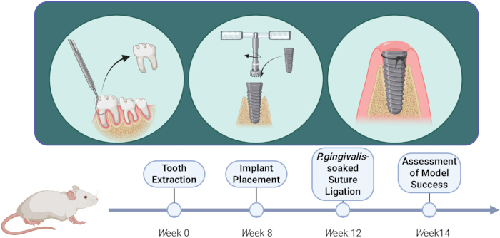
Existing rat model induction techniques include silk ligature placement14,15, local pathogen inoculation16, and lipopolysaccharide injection17. Previous infection models that relied on oral bacterial lavage often result in unstable biofilm formation and inconsistent inflammatory responses18. The silk ligature method is common for promoting rapid plaque accumulation. However, ligation alone provides a non-physiological mechanical insult and fails to incorporate a controlled pathogenic challenge, inadequately representing the infectious disease etiology of PI19. Monoinfection with pathogens like P. gingivalis, on the other hand, often struggles to establish a stable biofilm and may not consistently induce robust inflammation20. We hypothesize that the ligation procedure delivers dual insults, the silk suture acts as a mechanical irritant and a scaffold for complex biofilm formation, while the delivered keystone pathogen, P. gingivalis, instigates and perpetuates a dysbiotic inflammatory response21. To visually summarize this integrated dual-pathogenic approach and its temporal progression, a schematic overview of the experimental procedure and timeline for establishing this combined P. gingivalis-ligation rat model is provided (Figure 1). This article details the establishment and validation of this combined P. gingivalis-ligation model, designed to reliably reproduce the critical bone and soft tissue pathology of human PI, thereby enhancing the model's clinical relevance and utility for preclinical research.

Tooth implant process diagram; stages: extraction, placement, P.gingivalis ligation, success assessment.
Figure 1: Schematic Diagram of the experimental procedure timeline for establishing a rat peri-implantitis model induced by the bacteria-loaded suture ligation method. Please click here to view a larger version of this figure.

Access restricted. Please log in or start a trial to view this content.

Protocol

Loading...
$$\rightleftharpoonup{xx}$$ $$\longleftharp{xx}$$, $$\longrightharp{xx}$$,

The sample size was determined based on common practices in previously published rodent peri-implantitis (PI) models and the constraints of available resources22. Twelve 4-week-old male Sprague-Dawley rats (provided by the Experimental Animal Center of Southwest Medical University, License No.: SCXK (Chuan) 2024-0046) were used for tooth extraction, implant placement, and PI induction. All rats were maintained in a standard specific pathogen-free (SPF) barrier facility, with animals from different experimental groups housed separately. Cage locations within the rack were randomized on a weekly basis. Rats were assigned to either the negative control (NC) or PI group (n = 6 per group) using a random number generator. The order of surgical procedures for all rats is randomized. All procedures are approved by the Animal Research Ethics Committee of Southwest Medical University (Approval No. 20241111-012) and adhere to animal welfare guidelines. This study is conducted in accordance with the 3R principles (Replacement, Reduction, Refinement) for ethical animal research. The reagents and the equipment used are listed in the Table of Materials.

1. Pre-extraction preparation

  1. Sterilize all surgical instruments (#5 dental explorer, needle holder, ophthalmic forceps, medical cotton swabs, tissue forceps, etc.) by autoclaving. Perform all subsequent procedures within a Class II biosafety cabinet to maintain aseptic conditions.
  2. Administer intraperitoneal injection of tribromoethanol (250 mg/kg) to anesthetize the rats23 (following institutionally approved protocols). Confirm anesthetic depth by the absence of a withdrawal reflex in response to a hind paw pinch.
  3. Apply ophthalmic lubricant to prevent corneal desiccation during procedures.
  4. Thoroughly disinfect all work surfaces using a 75% ethanol solution.
  5. Position the rat in a supine position on a stable surgical platform. Retract the maxillary and mandibular incisors using rubber bands to achieve and maintain maximal mouth opening. Illuminate the oral cavity with a surgical headlight.
  6. Grasp the tongue gently with tissue forceps and retract it to one side. Ensure the tongue is held securely to maintain a clear and unobstructed view of the maxillary molars.

2. Tooth extraction procedure

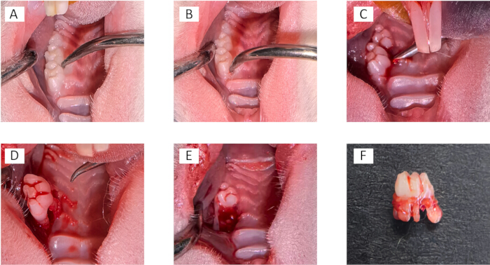
  1. Disinfect the surgical site with povidone-iodine. Position a #5 dental explorer at the distal surface of the maxillary first molar (interproximal space between the first and second molars), and apply mesiodistal force to achieve initial luxation (Figure 2A).
  2. Reposition the explorer to the mesial surface of the first molar, and reapply mesiodistal force to further mobilize the tooth (Figure 2B).
  3. Place the tip of the explorer into the palatal gingival sulcus. Apply a combination of gentle bucco-palatal rotational movement and sustained occlusal pressure along the long axis of the tooth. Continue until the tooth is fully dislocated from the socket (Figure 2C,D).
  4. Carefully inspect the extracted tooth under direct vision to confirm that all roots are intact and no fractures are present. Gently irrigate the empty socket with a stream of normal saline from a sterile syringe to remove any residual blood clots or debris (Figure 2E,F).
  5. Achieve hemostasis by applying sterile cotton swab pressure for 1 min. Return the tongue to anatomical position. Maintain the animal on the heating pad until full recovery from anesthesia.
  6. Provide a soft diet and antibiotic-supplemented drinking water (Amoxicillin 250 mg/kg) for one week.
    ​NOTE: Apply controlled, progressive force during luxation and avoid abrupt movements to prevent root fracture or alveolar bone damage.

Dental extraction sequence, oral surgery method, post-operative result, educational keyword.
Figure 2: Tooth extraction procedure. A dental probe was inserted into the distal and proximal aspects of the rat maxillary first molar to release the periodontal ligaments (A,B). Palatal application of rotational and occlusal forces to luxate the tooth (C,D). The socket was thoroughly examined for any residual root fragments (E). An intact rat maxillary first molar is shown (F). Please click here to view a larger version of this figure.

3. Implant placement surgery

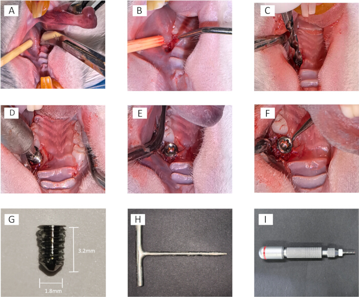
  1. Eight weeks post-extraction, create a 5 mm mesiodistal incision over the edentulous ridge using a sterile 12c blade, penetrating to the bone surface (Figure 3A).
  2. Utilize #5 explorer for blunt dissection to elevate full-thickness mucoperiosteal flaps, exposing the surgical site (Figure 3B).
  3. Connect a tungsten carbide drill (1.5 mm diameter) to a manual screwdriver adapter and perform osteotomy by clockwise manual rotation with intermittent saline irrigation for thermal control, preparing a 4 mm deep implant site within the healed extraction socket while maintaining continuous tactile feedback to prevent cortical perforation (Figure 3C).
  4. Insert custom-made titanium implant (1.8 mm diameter, 3.2 mm length, machined surface) using clockwise rotation. Verify stability by the absence of mobility upon the explorer application (Figure 3D-F).
  5. Allow a one-week healing period with continued soft diet and antibiotic regimen as previously described.
    ​NOTE: Employ gentle surgical technique and avoid excessive axial force to prevent iatrogenic perforation of the maxillary sinus floor.

Surgical procedure for dental implant placement, sequence of images showing step-by-step process.
Figure 3: Implant placement procedure. A mesiodistal incision was made using a No. 12 curved blade (A). The mucosal flap was reflected with a dental probe (B). An osteotomy was performed using a 1.5 mm tungsten carbide drill bit attached to a hand drill (C). The implant was placed into the jawbone via a customized implant carrier (D). After implantation, a probe was used to check for mobility (E,F). Custom-made titanium implant (smooth surface thread, diameter 1.8 mm, length 3.2 mm), corresponding implant carrier (G,H), and mini hand drill (I). Please click here to view a larger version of this figure.

4. P. gingivalis -ligated suture preparation

  1. Culture P. gingivalis anaerobically at 37 °C in 5 mL brain heart infusion (BHI) broth until reaching 109 CFU/mL (OD600 =1.0).
  2. Transfer suspension at 1:20 dilution to 10 mL fresh BHI medium.
  3. Immerse sterile 3-0 silk sutures in inoculated medium completely. Incubate anaerobically at 37 °C for 48-72 h to establish a mature biofilm.

5. PI induction

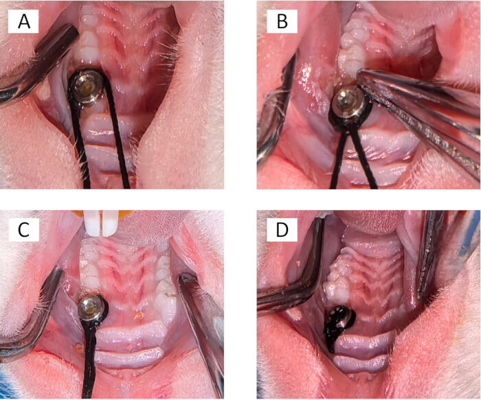
  1. Four weeks post-implantation, ligate a P. gingivalis-soaked suture around the implant neck using a sterile needle holder. Secure with a triple knot adjacent to the gingival tissue (Figure 4A-D).
  2. Maintain ligature for 2 weeks while monitoring inflammatory progression.
  3. Perform daily ligature inspection. Replace if loosened or lost using an aseptic technique.
    NOTE: When tying knots, gently separate the soft tissue to avoid tearing, and ensure the suture knot is positioned subgingivally.

Oral surgery procedure images, sequence A-D; dental tools; surgical technique demonstration.
Figure 4: Suture-induced PI procedure: The prepared bacteria-soaked suture thread (3-0) was placed at the subgingival position on the distal end of the implant (A). The first knot was tied and secured with a needle holder to prevent knot dislocation (B). Subsequently, tie a triple knot to prevent suture slippage (C,D). Please click here to view a larger version of this figure.

6. Euthanasia and sample collection

  1. After a 2-week induction period, euthanize the animals via cervical dislocation under deep isoflurane anesthesia (following institutionally approved protocols). Dissect out the intact maxillae with the implants using sharp instruments.
  2. Immediately immerse specimens in 4% paraformaldehyde for 24-h fixation. Remove residual soft tissue with PBS irrigation and store in 70% ethanol.
    NOTE: Experiment may be paused at this stage with long-term storage in 70% ethanol at 4 °C.

7. Micro-computed tomography (micro-CT) analysis

  1. Scan specimens using micro-computed tomography according to established methodology24.
  2. Power on the scanner and computer. Launch the acquisition software. Click on the radiation safety icon and allow a 15 min warm-up.
  3. Open scanning chamber. Mount the specimen in a 15 mL conical tube stabilized with low-density foam, then secure the tube on the rotary stage and position it within the scanning chamber.
  4. Initiate the X-ray source (90 kV, 80 µA). Click on the preview icon to confirm optimal positioning. Set the parameters: 12 µm resolution, 20 Hz exposure, 4000 total frames.
  5. Reconstruct images using Recon 1.7.4.0 software.
  6. Analyze the following bone parameters in Avatar 1.7.4.0 software by delineating an ROI in the maxillary first molar region: bone volume fraction (BV/TV), bone surface to bone volume ratio (BS/BV), trabecular thickness (Tb.Th), and trabecular number (Tb.N).

8. Histological processing

  1. Decalcify fixed specimens in 10% EDTA (pH 7.4) with solution changes every 72 h for 4 weeks. Verify a completion by a needle penetration test.
  2. Dehydrate specimens through an ethanol series: 75% ethanol for 4 h, 85% ethanol for 2 h, 90% ethanol for 2 h, 95% ethanol for 1 h, and two changes of 100% ethanol for 30 min each.
  3. Clear specimens in xylene with two changes for 10 min each. Infiltrate with paraffin using three changes for 1 h each.
  4. Embed tissues in molten paraffin and cool on a -20 °C freezing stage. Trim the blocks and store at 4 °C for 10 h before sectioning.
  5. Section paraffin blocks at 5 µm thickness. Float sections on a 45 °C water bath and mount on glass slides. Dry slides at 37 °C for 1 h.
  6. Deparaffinize sections through two changes of xylene for 5 min each. Rehydrate through a graded ethanol series: 100% ethanol for 5 min, 90% ethanol for 5 min, 80% ethanol for 5 min, and 70% ethanol for 5 min.
  7. Rinse sections in distilled water for 1 min. Stain with hematoxylin for 8 min, then rinse in running tap water.
  8. Differentiate in 1% acid alcohol for 3 s. Rinse in running tap water for bluing. Counterstain with eosin for 5 min.
  9. Dehydrate sections through graded ethanol series: 70% ethanol for 10 s, 80% ethanol for 10 s, 90% ethanol for 10 s, and 100% ethanol for 10 s. Clear in two changes of xylene for 5 min each.
  10. Mount coverslips with neutral balsam and examine under a light microscope.
    NOTE: Xylene is hazardous. Use in a fume hood with appropriate PPE.

Access restricted. Please log in or start a trial to view this content.

Results

Loading...
$$\rightleftharpoonup{xx}$$ $$\longleftharp{xx}$$, $$\longrightharp{xx}$$,

All twelve 4-week-old Sprague-Dawley rats enrolled in the study successfully completed all surgical procedures and exhibited stable osseointegration prior to ligation, meeting the pre-defined inclusion criterion of recovery without complications. No animals are excluded from the analysis. Rats are randomly assigned to the NC or PI group (n = 6 per group) using a random number generator. Researchers performing micro-CT quantification and histopathological evaluations remain blinded to group assignments throughout outcome ...

Access restricted. Please log in or start a trial to view this content.

Discussion

Loading...
$$\rightleftharpoonup{xx}$$ $$\longleftharp{xx}$$, $$\longrightharp{xx}$$,

The present study demonstrates the feasibility of inducing PI in a rat model using sutures soaked with P. gingivalis, and provides a detailed documentation of the associated surgical procedures. Rodent models, particularly in rats, are well-established in dental research for studying periodontitis and PI, owing to their accessibility, low maintenance cost, and high reproductive efficiency, as supported by existing literature15,25. Although most surgical ...

Access restricted. Please log in or start a trial to view this content.

Disclosures

Loading...
$$\rightleftharpoonup{xx}$$ $$\longleftharp{xx}$$, $$\longrightharp{xx}$$,

The authors affirm that there are no potential conflicts of interest that might have influenced the work reported in this paper.

Acknowledgements

Loading...
$$\rightleftharpoonup{xx}$$ $$\longleftharp{xx}$$, $$\longrightharp{xx}$$,

This work is supported by grants from the Sichuan Medical Association (Grant No. S2024052); the Luzhou Science and Technology Bureau (Grant No. 2024JYJ070), and the Affiliated Stomatology Hospital of Southwest Medical University (Grant No. 2025DS03). The authors also gratefully acknowledge the technical assistance provided by the Animal Experiment Center of Southwest Medical University. The scientific diagram is created with BioRender.

Access restricted. Please log in or start a trial to view this content.

Materials

List of materials used in this article
NameCompanyCatalog NumberComments
#5 dental explorerZhengzhou Kangdetai Medical Co., Ltd., Zhengzhou,CN6970327120856Tooth luxation and extraction
1% acid alcoholSinopharm Chemical Reagent Co., Ltd, Shanghai, CN10000018 (Ethanol) & 10000118 (Hcl)Hematoxylin differentiation
10% EDTA (pH 7.4)Servicebio Technology Co., Ltd, Wuhan, CNG1105Bone decalcification
100% ethanol solutionMacklin Biochemical Technology Co., Ltd., Shanghai, ChinaE708068Complete dehydration and solvent preparation
12c blade and surgical scalpelTianda Medical Co., Ltd., Huaian, CN6951933811154Mucoperiosteal flap creation
3-0 silk ligaturesJinhuan Medical, Shanghai, CNNC9201232Ligature
4% paraformaldehydeBiosharp, Hefei, CNBL539ATissue fixation
70% EthanolLaboratory preparation-Sample storage
75% ethanol solutionMacklin Biochemical Technology Co., Ltd., Shanghai, ChinaE885996Surface disinfection and Initial dehydration
Amoxicillin 50 μg/mLNorth China Pharmaceutical Co., Ltd, Shijiazhuang, CNH13020635Antibiotic supplementation in drinking water
Avatar softwareAvatar Integrated Systems Inc, Munich, DE-Bone parameters analysis
BHI mediumCrown Lab Supplies Inc, British Columbia, CA110052P. gingivalis culture
CT-VoxelBruker Corporation, Billerica, MA, USA-Micro-CT image visualization
EosinBeijing Solarbio Science & Technology Co., Ltd, Beijing, CN
G1100
Cytoplasmic staining
Ethanol series ( 80%, 90%, 95%)Laboratory preparationGradual dehydration/rehydrate of tissues
GraphPad PrismGraphPad Software, Inc., San Diego, CA, USAVersion 9.0.0Statistical analysis and graph generation
HeadlampShenzhen Supfire Lighting Co., Ltd, Shenzhen, CNHL23-XIntraoral illumination
HematoxylinBeijing Solarbio Science & Technology Co., Ltd, Beijing, CNH8070Nuclear staining
Implant carrierSichuan Weisida Medical Device Co., Ltd,Sichuan, CNUnique productManual implant placement
IsofluraneRWD Life Science Co., Ltd, Shenzhen, CNR510-22-10 Inhalational anesthesia maintenance
Manual screwdriver adapterDiqin Decorative Materials Co., Ltd, Hangzhou, CNDQ-80013Osteotomy
Medical cotton swabsKekang Pharmaceutical Co., Ltd, Suzhou, CNHN-02Haemostasis
Micro-CT scannerServicebio Technology Co., Ltd, Wuhan, CNVNC-102μCT scans
Needle holderShanghai Medical Instruments Co., Ltd, Shanghai, CNJ32110Suture ligation
Neutral balsamBeijing Solarbio Science & Technology Co., Ltd.
Beijing, CN
G8590Histological section mounting
Ophthalmic forcepsSolingmed Technology Co., Ltd, Shenzhen, CNSS-5002Ligature placement
ParaffinAmin Noori General Trading LLC, Dubai, AE8012-95-1Tissue embedding
PBSProcell Life Science & Technology Co., Ltd, Wuhan, CNPB180327Tissue irrigation
Porphyromonas gingivalisEquivalent-Inflammation induction
PovidoneiodineChengdu Yong'an Pharmaceutical Co., Ltd, Chengdu, CNH51023539Disinfection
Recon softwareRecon Technology Ltd, Cayman Islands,GBVersion 1.7.4.0Micro-CT image reconstruction
Rubber bandZhejiang Yueyi Rubber Products Co., Ltd, Taizhou, CNVYR43*1.4Jaw traction and fixation
SD RatsExperimental Animal Center of Southwest Medical University, Luzhou, CNSCXK(Chuan)2024-0046Surgical procedures
Sharp surgical scissorsHunan Cofoe Medical Technology Co., Ltd., Changsha, CN1.0806E+13Thread cutting
Tissue forcepsFine Science Tools, Foster City, CA, USA11000-12Tongue traction and fixation
Titanium implants (Ø1.8×3.2mm)Sichuan Weisida Medical Device Co., Ltd,Sichuan, CNUnique productTitanium implant
Tribromoethanol (Avertin)Henan Saihou Biotechnology Ltd., Zhengzhou, CN-Intraperitoneal anesthesia
XyleneSinopharm Chemical Reagent Co., Ltd, Shanghai, CN10023418Tissue clearing and dewaxing

References

Loading...
$$\rightleftharpoonup{xx}$$ $$\longleftharp{xx}$$, $$\longrightharp{xx}$$,
  1. Gupta, R., Gupta, N., Weber, D. K. Dental implants. , StatPearls Publishing LLC. Treasure Island (FL). Copyright 2025 (2025).
  2. Lee, C. T., Huang, Y. W., Zhu, L., Weltman, R. Prevalences of peri-implantitis and peri-implant mucositis: Systematic review andmeta-analysis. J Dent. 62, 1-12 (2017).
  3. Berglundh, T., et al. Peri-implant diseases and conditions: Consensus report of workgroup 4 of the 2017 world workshop on the classification of periodontal and peri-implant diseases and conditions. J Clin Periodontol. 45 (Suppl 20), S286-S291 (2018).
  4. Schwarz, F., Derks, J., Monje, A., Wang, H. L. Peri-implantitis. J Clin Periodontol. 45 (Suppl 20), S246-S266 (2018).
  5. Diaz, P., Gonzalo, E., Villagra, L. J. G., Miegimolle, B., Suarez, M. J. What is the prevalence of peri-implantitis? A systematic review and meta-analysis. BMC Oral Health. 22 (1), 449(2022).
  6. Herrera, D., et al. Prevention and treatment of peri-implant the EFP S3 level clinical practice guideline. J Clin Periodontol. 50 (Suppl 26), 4-76 (2023).
  7. Fu, J. H., Wang, H. L. Breaking the wave of peri-implantitis. Periodontol. 84 (1), 145-160 (2020).
  8. Sant'anna, H. R., et al. Peri-implant repair using a modified implant macrogeometry in diabetic rats: Biomechanical and molecular analyses of bone-related markers. Materials (Basel). 15 (6), 2317(2022).
  9. Ribitsch, I., et al. Large animal models in regenerative medicine and tissue engineering: To do or not to do. Front Bioeng Biotechnol. 8, 972(2020).
  10. He, Q., et al. Development of a rat model for type 2 diabetes mellitus peri-implantitis: A preliminary study. Oral Dis. 28 (7), 1936-1946 (2022).
  11. DeAraújo Silva, D. N., et al. Experimental model of ligature-induced peri-implantitis in mice. J Vis Exp. (207), e66316(2024).
  12. Chadwick, J. W., Glogauer, M. Robust ligature-induced model of murine periodontitis for the evaluation of oral neutrophils. J Vis Exp. (155), e59667(2020).
  13. Kantarci, A., Hasturk, H., Van Dyke, T. E. Animal models for periodontal regeneration and peri-implant responses. Periodontol. 68 (1), 66-82 (2000).
  14. Varon-Shahar, E., et al. Peri-implant alveolar bone resorption in an innovative peri-implantitis murine model: Effect of implant surface and onset of infection. Clin Implant Dent Relat Res. 21 (4), 723-733 (2019).
  15. Hiyari, S., et al. Ligature-induced peri-implantitis and periodontitis in mice. J Clin Periodontol. 45 (1), 89-99 (2018).
  16. Tzach-Nahman, R., Mizraji, G., Shapira, L., Nussbaum, G., Wilensky, A. Oral infection with Porphyromonas gingivalis induces peri-implantitis in a murine model: Evaluation of bone loss and the local inflammatory response. J Clin Periodontol. 44 (7), 739-748 (2017).
  17. Pirih, F. Q., et al. Ligature-induced peri-implantitis in mice. J Periodontal Res. 50 (4), 519-524 (2015).
  18. Pirih, F. Q., et al. A murine model of lipopolysaccharide-induced peri-implant mucositis and peri-implantitis. J Oral Implantol. 41 (5), e158-e164 (2015).
  19. Reinedahl, D., Chrcanovic, B., Albrektsson, T., Tengvall, P., Wennerberg, A. Ligature-induced experimental peri-implantitis-a systematic review. J Clin Med. 7 (12), 492(2018).
  20. Gao, L., Yu, X. Q., Cai, Y. Effect of molar ligation and local Porphyromonas gingivalis inoculation on alveolar bone loss in the mouse. Beijing Da Xue Xue Bao Yi Xue Ban. 49 (1), 31-35 (2017).
  21. Wongtim, K., et al. Overexpression of PD-L1 in gingival basal keratinocytes reduces periodontal inflammation in a ligature-induced periodontitis model. J Periodontol. 93 (1), 146-155 (2022).
  22. Koutouzis, T., Eastman, C., Chukkapalli, S., Larjava, H., Kesavalu, L. A novel rat model of polymicrobial peri-implantitis: A preliminary study. J Periodontol. 88 (2), e32-e41 (2017).
  23. Veríssimo, L. F., et al. Cardiovascular effects of early maternal separation and escitalopram treatment in rats with depressive-like behaviour. Auton Neurosci. 256, 103223(2024).
  24. Kim, Y., Brodt, M. D., Tang, S. Y., Silva, M. J. MicroCT for scanning and analysis of mouse bones. Methods Mol Biol. 2230, 169-198 (2021).
  25. Kuraji, R., Hashimoto, S., Ito, H., Sunada, K., Numabe, Y. Development and use of a mouth gag for oral experiments in rats. Arch Oral Biol. 98, 68-74 (2019).
  26. Lu, S., et al. Effect of ovariectomy on tissue-level changes in rat maxilla. Int J Oral Maxillofac Implants. 34 (4), 865-872 (2019).
  27. Heitzer, M., et al. Establishing a new periodontitis-like intrabony maxillary defect in rats for investigation on bone regeneration. Sci Rep. 15 (1), 39358(2025).
  28. Moiseev, D., et al. A new way to model periodontitis in laboratory animals. Dent J (Basel). 11 (9), 219(2023).
  29. Marchesan, J., et al. An experimental murine model to study periodontitis. Nat Protoc. 13 (10), 2247-2267 (2018).
  30. Nasution, D. L. I., Tjahajawati, S., Indriyanti, R., Amaliya, Anti-inflammatory effectiveness of Peperomia pellucida (l.) kunth in rats induced with periodontitis. Biochem Biophys Rep. 40, 101856(2024).
  31. Meng, L. W., et al. Comparison of three methods for establishing rat peri-implantitis model. Beijing Da Xue Xue Bao Yi Xue Ban. 55 (1), 22-29 (2023).
  32. Zhang, H., et al. Application of immediate implant placement techniques in peri-implantitis modeling. J Craniofac Surg. 34 (8), 2544-2550 (2023).
  33. Ortiz-Sánchez, B. J., Juárez Avelar, I., Rodriguez-Sosa, M. Murine model of advanced periodontitis induced by nylon ligature in the second upper molar. J Vis Exp. (219), e67848(2025).
  34. Lafaurie, G. I., et al. Microbiome and microbial biofilm profiles of peri-implantitis: A systematic review. J Periodontol. 88 (10), 1066-1089 (2017).
  35. Zhang, Q., Liu, J., Ma, L., Bai, N., Xu, H. Lox-1 is involved in TLR2-induced RANKL regulation in peri-implantitis. Int Immunopharmacol. 77, 105956(2019).
  36. Qiao, S., et al. Oral microbial profile variation during canine ligature-induced peri-implantitis development. BMC Microbiol. 20 (1), 293(2020).
  37. De Avila, E. D., Van Oirschot, B. A., Van Den Beucken, J. Biomaterial-based possibilities for managing peri-implantitis. J Periodontal Res. 55 (2), 165-173 (2020).
  38. Iizumi, T., et al. Effect of antibiotic pre-treatment and pathogen challenge on the intestinal microbiota in mice. Gut Pathog. 8, 60(2016).
  39. Heitz-Mayfield, L. J. A., Salvi, G. E. Peri-implant mucositis. J Clin Periodontol. 45 ((Suppl 20)), S237-S245 (2018).
  40. Javed, F., Romanos, G. E. Efficacy of photobiomodulation in the management of experimentally-induced peri-implant osseous defects: An evidence-based review of studies on animal models. J Dent. 159, 105833(2025).
  41. Yu, S., et al. Adjunctive diode laser therapy and probiotic lactobacillus therapy in the treatment of periodontitis and peri-implant disease. J Vis Exp. (183), e63893(2022).

Access restricted. Please log in or start a trial to view this content.

Reprints and Permissions

Request permission to reuse the text or figures of this JoVE article

Request Permission

Tags

Peri Implantitis ModelRat Implant ModelTitanium Implant PlacementPorphyromonas GingivalisLigature InductionAlveolar Bone LossSoft Tissue InflammationMicro CT AnalysisHistological AnalysisDental Implant Failure
Video Coming Soon

Related Articles