This study established and optimized a partial renal artery ligation model in Syrian hamsters (2K1C), which stably induces RAS-related secondary hypertension in a background of hyperlipidemia, providing a highly reproducible platform for cardiovascular research.
Method Article
Establishing a Standardized Renal Artery Ligation Procedure for Hypertension in Syrian Golden Hamsters
* These authors contributed equally
In This Article
Summary
Abstract
Hypertension is considered a major global health threat, which can lead to severe complications, including vascular remodeling, heart failure, myocardial infarction, and end-stage renal disease. Syrian golden hamsters, a model with naturally expressing cholesteryl ester transfer protein (CETP), are susceptible to diet-induced hyperlipidemia. To establish a reliable and reproducible hamster model for studying the relationship between renal artery stenosis (RAS), secondary hypertension, and cardiovascular disease, the two-kidney, one-clip (2K1C) Goldblatt model was employed. This study details a refined surgical procedure for inducing RAS in Syrian golden hamsters via partial renal artery ligation, emphasizing critical factors such as surgical isolation technique and ligature material selection, which significantly impact outcomes. Implementation of this standardized surgical protocol resulted in approximately 90% of treated hamsters exhibiting a consistent systolic blood pressure elevation of 25–30 mmHg. This optimized renal artery ligation procedure provides a robust method for generating a hypertensive hamster model with high consistency, enhanced reproducibility, and utility for cardiovascular disease research.
Introduction
Renal artery stenosis (RAS) is a leading cause of secondary hypertension and constitutes a significant risk factor for cardiovascular morbidity1. A primary challenge in clinical research is the difficulty in delineating the causal pathway connecting dyslipidemia, hypertension, and end-organ damage, as comorbidities and concurrent medications frequently confound patient studies. This underscores the critical need for an animal model that accurately recapitulates this complex pathophysiology in a controlled, reproducible experimental setting.
The two-kidney, one-clip (2K1C) Goldblatt model is a well-established method for inducing renovascular hypertension in rodents. However, conventional species such as mice and rats present considerable limitations. Notably, these rodents lack cholesteryl ester transfer protein (CETP), a key enzyme in human high-density lipoprotein metabolism2. As a result, they are resistant to diet-induced hypercholesterolemia and atherosclerosis, failing to mimic the metabolic profile commonly observed in patients with RAS. Furthermore, the hypertensive response to renal artery clipping in these models is often attenuated and highly variable; for instance, C57BL/6 mice exhibit a minimal blood pressure increase, while Sprague-Dawley rats show considerable inter-individual variability, which impedes reproducible mechanistic and therapeutic investigations3,4.
In contrast, the Syrian golden hamster (Mesocricetus auratus) possesses unique physiological advantages for such research. Its natural expression of CETP enables the development of a human-like lipoprotein profile, including elevated LDL-C and slightly increased HDL-C, upon feeding a high-fat and high-cholesterol diet, leading to the spontaneous formation of atherosclerotic lesions in the aorta and coronary arteries5. This provides a highly relevant metabolic and vascular background for studying cardiovascular disease. Moreover, recent evidence indicates that hamsters mount a significantly more robust blood pressure response to renin-angiotensin-aldosterone system (RAAS) activation compared to mice, and chymase, making them particularly suitable for hypertension research6.
Despite these advantages, the utility of the hamster 2K1C model has been limited by the lack of a standardized and survivable surgical protocol. Existing techniques, which range from complete ligation with high mortality rates to operator-dependent partial ligation, yield inconsistent results and poor reproducibility7. Therefore, to fully exploit the hamster’s dual susceptibility to metabolic and hypertensive disease, we developed a refined and highly reproducible surgical approach for the 2K1C model. This optimized protocol ensures consistent induction of hypertension against a background of diet-induced dyslipidemia, thereby offering a robust and integrated experimental platform to investigate the synergistic effects of RAS, hyperlipidemia, and cardiovascular injury.
Protocol
The animal protocols were approved by the laboratory animal ethics committee of Jiangmen International Health Innovation Institute (N2023008) and Peking University (DLASBE0730). Male Syrian golden hamsters, 10–12 weeks of age, were used for the experiment. The details of the reagents and equipment used were listed in the Table of Materials.
1. Preoperative preparation
- Feed the animals a normal chow diet and provide clear water under standard conditions (temperature 21–24 °C, humidity 40%–60%, 12-h light-dark cycle).
- Sterilize all surgical instruments and materials before use. Disinfect the surgical platform with 75% ethanol. Preheat a heating pad to approximately 38 °C before surgery.
- Place the hamster in an anesthesia induction chamber supplied with 3% isoflurane (following institutionally approved protocols).
- Confirm deep anesthesia by the absence of a pedal withdrawal reflex. Maintain anesthesia via a nose cone with 1.5%–2% isoflurane.
- Remove the hair from the mid-left lumbar region using electric clippers.
- Disinfect the exposed skin with povidone-iodine.
2. Surgical procedure
- Make an approximately 1.5–2 cm longitudinal incision in the skin along the midline of the left lumbar region using a scalpel. Bluntly dissect the underlying muscles to separate the latissimus dorsi from the iliocostalis layer. Create an approximately 1 cm access channel along the direction of the muscle fibers.
- Gently retract the retroperitoneal fat using blunt forceps to expose the kidney. Exteriorize the left kidney and identify the renal pedicle. Separate the renal artery and vein using blunt forceps.
- Place a 0.27 mm diameter needle adjacent to the renal artery. Loop a 6-0 polypropylene suture around both the artery and the needle. Tie a single overhand knot loosely.
- Carefully withdraw the needle, leaving a circumferential constriction that reduces the luminal diameter to 0.27 mm. Confirm successful stenosis by observing visible narrowing, but not complete cessation, of blood flow accompanied by a mild change in renal color.
- Return the kidney to the renal fossa. Close the muscle layer and skin separately using 4-0 and 3-0 nonabsorbable polypropylene sutures, respectively. Disinfect the surgical area with povidone-iodine followed by ethanol.
- Place the animal on a heating pad and monitor respiratory status until consciousness returns. Administer carprofen (5 mg/kg, subcutaneously) for up to 3 days after surgery. House the hamster individually for at least 1 week.
3. Blood pressure measurement
NOTE: Measure blood pressure non-invasively at 2 and 4 weeks post-operation using a Tail-cuff blood pressure monitoring system.
- Anesthetize the hamsters in a chamber supplied with 3% isoflurane.
- Place the animal in a prone position on an operating board and fit a mask to maintain anesthesia.
- Attach a cuff to the forelimb and record blood pressure for at least 2 min for each animal.
4. Ultrasound examination
NOTE: Assess renal hemodynamics and structural adaptation 4 weeks after surgery using color Doppler and three-dimensional volume ultrasound systems.
- Anesthetize the hamsters in a chamber supplied with 3% isoflurane.
- Place the animal in a prone position on an operating board and fit a mask to maintain anesthesia.
- Remove abdominal hair using a depilatory cream to ensure optimal acoustic contact. Apply coupling gel before placing the ultrasound probe.
- Position the probe over the left kidney and obtain a clear longitudinal B-mode image.
- Measure renal length and cortical thickness using the caliper function.
- Activate color Doppler mode and visualize renal blood flow.
- Record peak systolic velocity, end-diastolic velocity, and calculate the resistive index.
- Perform three-dimensional scanning to reconstruct renal volume.
- Save all images and measurement data.
- Remove the probe and monitor the animal until recovery.
Results
In this study, these hamsters were randomly divided into two groups: 8 in the sham surgery group and 11 in the surgical group. All data are presented as means ± standard deviations unless otherwise stated. The approximate operative time was 20–25 min per animal. There were no intraoperative deaths, and the survival rate within 9 weeks of surgery was 100%. 2 weeks after surgery, noninvasive blood pressure was measured. The surgery, with or without ligation, had a lesser effect on appetite, and body weight in both groups showed similar changes (Figure 1A). The systolic and diastolic blood pressures in the ligation group were 125.5 ± 16.97 and 85.33 ± 13.06 (mmHg), respectively, and in the sham group were 79.50 ± 13.92 and 53.50 ± 18.79 (mmHg), respectively. And the mean blood pressure was significantly increased 1.5-fold in the ligation group compared with the sham group. The heart rate in both groups was similar (Figure 1B–E).
Blood biochemistry was performed after 2 weeks of renal artery ligation. Plasma samples were collected, and blood glucose, triglycerides, total cholesterol, and HDL-cholesterol levels were measured. Strikingly, total cholesterol and HDL cholesterol levels were significantly lower in the ligation group than in the sham group (Table 1). Meanwhile, the levels of uric acid, urea, and creatinine in the blood were measured 4 weeks after surgery. The results showed that the level of uric acid in the ligation group was 7.5-fold higher than in the sham group. And the plasma urea level was also significantly increased in the ligation group compared with the control (Table 1). To test renal function in vivo, color Doppler ultrasound was performed, and the results showed that after ligation, kidney size and blood flow were significantly decreased (Figure 1F). The compensatory effect was observed and compared with the control; the right kidney in the ligation animals was considerably larger than the kidney in the sham group (Figure 1G). And the renin level was also significantly increased in the ligation group, when compared with the control (Figure 1H).
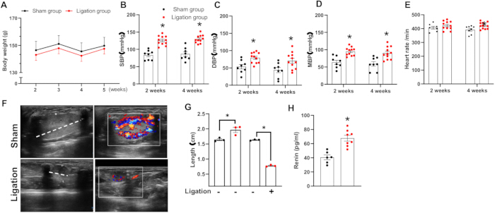
Figure 1: Biochemical parameters, blood pressure, and renal function. Body weight was measured 2–5 weeks after surgery (A). Systolic blood pressure, diastolic blood pressure, and heart rate were measured at 2 and 4 weeks after surgery (B–E). Renal function was evaluated in vivo using color Doppler imaging; representative images (F) and quantitative data (G) are shown. Plasma renin levels were measured 4 weeks after surgery, and quantitative data are presented (H). Data are presented as mean ± SD. n = 8 for the sham group and n = 11 for the ligation group. Normality was assessed prior to statistical analysis. A two-tailed Student’s t-test was performed, and P ≤ 0.05 was considered statistically significant. Please click here to view a larger version of this figure.
| Sham group (n=8) | Ligation group (n=11) | |
| Glucose (mg/dl) | 87.3±13.6 | 117.5±15.2* |
| Triglyceride (mg/dl) | 211.7 ±104.1 | 210.4 ±94.6 |
| Cholesterol (mg/dl) | 262.9 ±41.3 | 191.4 ±61* |
| HDL-C (mg/dl) | 171.95±24.67 | 116.28±18.11* |
| Uric acid (mmol/l) | 0.04±0.01 | 0.30±0.01* |
| Urea (mmol/l) | 5.42 ±0.78 | 6.99 ±0.70* |
| Creatinine (mg/dl) | 0.56 ±0.08 | 0.59 ±0.07 |
Table 1: Blood biochemical and renal function parameters. Values are presented as mean ± SD; n indicates the number of animals. A two-tailed Student’s t-test was performed, and P ≤ 0.05 was considered statistically significant.
| Problem | Possible reason | Solution |
| Body weight loss exceeding 20% in the first week after surgery. | Surgical trauma | 1. Employ meticulous surgical technique to minimize operative injury. |
| 2. Ensure the procedure is performed by an experienced surgeon. | ||
| Surgical bleeding | Ineffective ligation (knot tied too loosely). | Use blunt-tipped scissors and surgical forceps for careful dissection to avoid vascular injury. |
| No apparent color change in the ligated kidney compared to the sham group. | Ineffective ligation (knot tied too loosely). | Ensure ligatures are secure. The knot must be tight enough to completely occlude the target vessel/ureter. |
| The experimental animal sustains an external injury/trauma. | Inadequate postoperative housing or animal care. | House animals individually (single-cage housing) for at least 2 weeks postoperatively to prevent aggression and cross-contamination. |
| The blood pressure cuff does not fit the hamster properly. | Equipment incompatibility. | Standard mouse/rat tail-cuff systems are not suitable. Use specialized equipment designed for forelimb blood pressure measurement in hamsters. |
Table 2: Troubleshooting guide.
Discussion
The two-kidney, one-clip (2K1C) model is a classic animal model in renal vascular hypertension research. With historical roots dating back to the last century, it effectively simulates the pathogenesis of human renovascular hypertension. The underlying mechanism involves renal artery stenosis-induced ischemia, which activates the renin-angiotensin system (RAS), ultimately leading to systemic hypertension. This model not only closely mirrors the pathological progression of human hypertension but also offers significant advantages, including operational simplicity and high reproducibility.
In comparison, the one-kidney, one clip (1K1C) model induces more pronounced hypertension, but lacks compensatory mechanisms present in the 2K1C model. Transgenic models enable precise exploration of specific gene functions but involve complex procedures and higher costs. Drug-induced methods allow for a comprehensive analysis of the complex interactions within the RAS8. Integrating multiple models allows for a comprehensive analysis of the complex interactions within the RAS. Therefore, the 2K1C model plays an irreplaceable role in simulating chronic renovascular hypertension and investigating RAS pathophysiological mechanisms, particularly in drug screening and systemic impact assessment. Furthermore, combining the 2K1C approach with genetically modified models or drug-induced methods to target specific RAS effectors can further elucidate the complexity of the RAS system and its multifaceted roles in various diseases.
In this study, a unilateral partial renal artery ligation model was established in Syrian golden hamsters. The combination of renal artery stenosis and hypertension enabled the successful construction of a repeatable and quantifiable "metabolic-renal vascular" dual stress model. Postoperative blood pressure remained persistently elevated, which is fully consistent with the classic hypertensive characteristics of the 2K1C model. A significant reduction in renal blood flow with increased renin level accurately replicated the core pathophysiological change of the 2K1C model9, confirming the successful induction of essential renovascular hypertension features. Renal artery stenosis caused ischemic atrophy in the clipped kidney, whereas the contralateral, untouched kidney exhibited typical compensatory hypertrophy and functional enhancement, as observed in our study. Previously, a high-renin genetic hypertension hamster model was bred, and the systolic blood pressure was increased 1.7-fold compared with wild-type hamsters. However, the high blood pressure was inconsistent throughout the lives of these hamsters10. In mice, angiotensin II infusion via osmotic minipumps is widely used as the hypertension model11.
Recently, a report showed that a chronic model of Ang II-dependent hypertension in the hamster was established, and female animals showed heightened vulnerability to Ang II-induced hypertension12. The existing literature indicates that the use of hamsters as hypertension models remains relatively limited, yet they demonstrate unique advantages for investigating specific diseases, such as metabolic syndrome13. Syrian golden hamsters are widely recognized as ideal models for studying diet-induced metabolic syndrome, due to their high sensitivity to diets rich in lipids, sucrose, and cholesterol14. These models systematically develop phenotypes including obesity, insulin resistance, and dyslipidemia. As a primary risk factor for cardiovascular diseases (CVD), hypertension can lead to severe consequences such as myocardial hypertrophy, heart failure, and atherosclerosis. As a primary risk factor for CVD, hypertension can lead to severe consequences such as myocardial hypertrophy, heart failure, and atherosclerosis. The persistent hypertension induced by the 2K1C model provides an ideal platform for studying cardiovascular remodeling and dysfunction. Using the 2K1C hamster model, researchers can systematically evaluate structural and functional changes in the heart (particularly the right ventricle), including parameters of myocardial fibrosis and ventricular wall thickening15,16. However, missing details about performing 2K1C in hamsters limited its use. It is known that the circulating renin-angiotensin II system generates angiotensin II. However, recent studies indicate that chymase, a non-ACE angiotensin II-forming enzyme, contributes to angiotensin II formation in human vascular tissues, making it a therapeutic target for hypertension17,18. Unlike mouse and rat chymases, the hamster vascular tissue chymase, like human chymase, catalyzes angiotensin I to angiotensin II19,20.
Given the frequent co-occurrence of hypertension with hyperlipidemia, diabetes, and other metabolic disorders in metabolic syndrome, the 2K1C hypertension model in hamsters is particularly valuable for revealing interactions between renovascular hypertension, lipid metabolism abnormalities, and insulin resistance. For instance, researchers can investigate whether 2K1C-induced hypertension exacerbates lipid metabolism disorders or how high-fat diets modulate the severity of hypertension and target organ damage.
One limitation of this protocol is the size of the cap for ligation. A preliminary experiment using a 0.31 mm clip gap resulted in a suboptimal increase in blood pressure (71.0 ± 8.72 mmHg vs. 63.34 ± 12.22 mmHg in the sham control group), indicating that clip gap size is a critical determinant for successful blood pressure induction. In the current study, young, normal-weight hamsters were used. Whether it's still working for an aged or overweight hamster is unknown. Aging and obesity can also impair vasoconstriction and vasodilation. It would be better to test the size of the gap in these studies in the future.
Another limitation is the sex difference. In the current study, male animals were used. To minimize estrogen interference with experimental results, male animals are typically used in research. Whether it elicits a consistent response across both genders remains unknown. Also, a troubleshooting table was provided for researchers concerned about this model (Table 2).
In summary, a hypertensive hamster model was established using the 2K1C protocol in this study. The consistency and reproducibility of hypertension in hamsters will provide significant new insights into the complex interactions between renovascular hypertension, hyperlipidemia, and cardiovascular diseases.
Acknowledgements
This research was funded by the Innovation Team Program supported by the National Key Research and Development Program of China (2022YFA1105403), the Application Foundation Project supported by Jiangmen City (2220002000296), and the Innovation Team Program supported by Guangdong Province (2020KCXTD038).
Materials
| Name | Company | Catalog Number | Comments |
|---|---|---|---|
| 0.37 wire diameter suture | Johnson & Johnson MedTech, China | JH-18-5100 | |
| 6-0 Polypropylene suture | Yangzhou Huanyu Medical Equipment Co., Ltd., China | N/A | |
| Animal anesthesia machine | Midmark Corporation, USA | V2330299 | |
| Carprofen | Macklin, China | C830557-5g | |
| Cholesterol Assay Kit | Zhongsheng Beikong, China | 100000180 | |
| Color Doppler ultrasound system | Mindray Medical, China | Resona8 | |
| Glucose Assay Kit | Zhongsheng Beikong, China | 100000240 | |
| High-density lipoprotein cholesterol assay kit | Zhongsheng Beikong, China | 100020235 | |
| LDLR KO Syrian golden hamster | Peking University, China | N/A | |
| Muromachi MK-2000ST Non-Invasive Blood Pressure Monitor for Mice & Rats | Muromachi Kikai Co., Ltd., Japan | 140305MU | |
| Pet heating pad | Meitai, China | DXT30*35-1X | |
| Rat Renin ELISA kit | Covide, China | KWD-E7398R-B | |
| Suture needle | Johnson & Johnson MedTech, China | JH-18-5037 | |
| Triglyceride Assay Kit | Zhongsheng Beikong, China | 100000220 | |
| Vertical pressure steam sterilizer | Yamato Scientific, Japan | J3170372 |
References
- Schwarz, K., et al. Prevalence and risk factors of renal artery stenosis in patients undergoing simultaneous coronary and renal artery angiography: A systematic review and meta-analysis of 31,689 patients from 31 studies. Diseases. 12 (9), 208(2024).
- Hogarth, C. A., Roy, A., Ebert, D. L. Genomic evidence for the absence of a functional cholesteryl ester transfer protein gene in mice and rats. Comp Biochem Physiol B Biochem Mol Biol. 135 (2), 219-229 (2003).
- Ong, J., et al. Renal sensory nerves increase sympathetic nerve activity and blood pressure in 2-kidney 1-clip hypertensive mice. J Neurophysiol. 122 (1), 358-367 (2019).
- Chelko, S. P., Schmiedt, C. W., Lewis, T. H., Lewis, S. J., Robertson, T. P. A novel vascular clip design for the reliable induction of 2-kidney, 1-clip hypertension in the rat. J Appl Physiol. 112 (3), 362-366 (2012).
- Guo, X., et al. LDL receptor gene-ablated hamsters: A rodent model of familial hypercholesterolemia with dominant inheritance and diet-induced coronary atherosclerosis. EBioMedicine. 27, 214-224 (2018).
- Li, P., Chen, P. M., Wang, S. W., Chen, L. Y. Time-dependent expression of chymase and angiotensin converting enzyme in the hamster heart under pressure overload. Hypertens Res. 25 (5), 757-762 (2002).
- Hall, S. K. Acute renal vascular occlusion: An uncommon mimic. J Emerg Med. 11 (6), 691-700 (1993).
- Cui, Y., et al. An antihypertensive drug-AT1 inhibitor attenuated BRCA development promoted by chronic psychological stress via Ang II/PARP1/FN1 pathway. Biochim Biophys Acta Mol Basis Dis. 1870 (3), 167031(2024).
- Salguero, G., et al. Renovascular hypertension by two-kidney one-clip enhances endothelial progenitor cell mobilization in a p47phox-dependent manner. J Hypertens. 26 (2), 257-268 (2008).
- Suzuki, H., Ikezaki, H., Hong, D., Rubinstein, I. PGH2-TxA2-receptor blockade restores vasoreactivity in a new rodent model of genetic hypertension. J Appl Physiol. 88 (6), 1983-1988 (2000).
- Lu, H., et al. Subcutaneous angiotensin II infusion using osmotic pumps induces aortic aneurysms in mice. J Vis Exp. (103), e53191(2015).
- Ji, H., et al. Paradoxical sex differences in a hamster model of angiotensin II-dependent hypertension and associated renal injury. Biol Sex Differ. 16 (1), 86(2025).
- Jordania da Silva, V., et al. Obesity induction in hamster that mimics the human clinical condition. Exp Anim. 66 (3), 235-244 (2017).
- Bhathena, J., et al. Diet-induced metabolic hamster model of nonalcoholic fatty liver disease. Diabetes Metab Syndr Obes. 4, 195-203 (2011).
- Shiota, N., et al. Chymase is activated in the hamster heart following ventricular fibrosis during the chronic stage of hypertension. FEBS Lett. 406 (3), 301-304 (1997).
- Joyner, W. L., Mohama, R. E., Gilmore, J. P. Specificity of arginine vasopressin and angiotensin II for microvessels in the hamster cheek pouch after the induction of renovascular hypertension. Microvasc Res. 35 (1), 8-20 (1988).
- Ansary, T. M., et al. Effects of the selective chymase inhibitor TEI-F00806 on the intrarenal renin-angiotensin system in salt-treated angiotensin I-infused hypertensive mice. Exp Physiol. 103 (11), 1524-1531 (2018).
- Li, M., et al. Involvement of chymase-mediated angiotensin II generation in blood pressure regulation. J Clin Invest. 114 (1), 112-120 (2004).
- Kinoshita, I., et al. Increase in chymase-positive mast cells in recurrent pleomorphic adenoma and carcinoma ex pleomorphic adenoma of the parotid gland. Int J Mol Sci. 22 (23), 12613(2021).
- Dell'Italia, L. J., Collawn, J. F., Ferrario, C. M. Multifunctional role of chymase in acute and chronic tissue injury and remodeling. Circ Res. 122 (2), 319-336 (2018).