This protocol aims to enable precise, selective transection of intestine-innervating sympathetic and parasympathetic nerves in mice using the superior mesenteric artery as a consistent anatomical landmark, providing a focused and reproducible model to study autonomic regulation of intestinal physiology and disease.
Method Article
Murine Model for Selective Transection of Intestinal Autonomic Nerves
Opens in a new tab
In This Article
Summary
Loading...
$$\rightleftharpoonup{xx}$$
$$\longleftharp{xx}$$,
$$\longrightharp{xx}$$,
Abstract
Loading...
$$\rightleftharpoonup{xx}$$
$$\longleftharp{xx}$$,
$$\longrightharp{xx}$$,
Autonomic nerves comprise the sympathetic and parasympathetic systems, which act in a largely antagonistic manner to regulate diverse physiological functions. With growing interest in the roles of autonomic innervation in intestinal physiology and pathology, there is an increasing need for experimental models that enable precise manipulation of gut-directed sympathetic and parasympathetic inputs. Here, we present a surgical protocol to selectively transect the sympathetic and parasympathetic nerves innervating the intestine. Using the superior mesenteric artery (SMA) as a reproducible anatomical landmark, this approach allows reliable identification of intestinal autonomic nerve bundles and subsequent targeted transection. In contrast to broader denervation approaches such as ganglionectomy or subdiaphragmatic vagotomy, this approach is intended to achieve a more anatomically localized transection of intestine-directed autonomic bundles, which may help reduce extra-intestinal interference. The injury is controllable and is typically associated with minimal bleeding, supporting procedural consistency and postoperative recovery. Although the surgery requires appropriate microsurgical training, the protocol is adaptable, enabling users to tailor key parameters to specific experimental aims. Importantly, this model provides a practical platform to investigate the roles of sympathetic and parasympathetic nerves in regulating intestinal function under both physiological and disease conditions. This article aims to describe the procedure in detail to facilitate its adoption by new users in their research.
Introduction
Loading...
$$\rightleftharpoonup{xx}$$
$$\longleftharp{xx}$$,
$$\longrightharp{xx}$$,
The gastrointestinal tract is under continuous control of the autonomic nervous system (ANS), with sympathetic and parasympathetic pathways coordinating fundamental aspects of intestinal physiology, including motility, secretion, epithelial barrier function, and immune homeostasis1,2. In the colon, extrinsic sympathetic inputs can exert region-dependent effects across multiple cellular compartments, underscoring that broad ANS perturbations may obscure site-specific intestinal phenotypes1. Conversely, parasympathetic circuits, classically mediated by vagal signaling and downstream cholinergic mechanisms, are increasingly recognized as integral to maintaining intestinal immune balance and shaping mucosal responses, further motivating experimental strategies that can disentangle sympathetic versus parasympathetic contributions with anatomic precision2.
Beyond physiological regulation, autonomic activity has emerged as a relevant modifier of intestinal disease biology, including colorectal cancer (CRC). Recent work in CRC demonstrates a bidirectional sympathetic nerve–mesenchymal program, in which β2-adrenergic signaling and NGF-expressing cancer-associated fibroblasts reinforce neural–stromal interactions and promote tumor progression, providing direct evidence that sympathetic circuits can be functionally coupled to CRC pathogenesis3. In parallel, cholinergic signaling has been linked to CRC growth and tumor-associated immune features, as shown by pharmacologic inhibition of muscarinic receptor 3 in an orthotopic mouse model and by connecting cholinergic signaling with immune checkpoint expression and other malignant hallmarks4,5. Importantly, autonomic influences on intestinal disease may also converge with other modulators, such as psychological stress and the gut microbiota. The enteric nervous system can relay stress signals to intestinal inflammation6, and microbial signals can engage gut–brain circuits that tune gut-extrinsic sympathetic neurons, collectively supporting a multi-factorial framework in which neural regulation intersects with immune and microbial ecology6,7.
These converging lines of evidence highlight the need for experimental approaches that can isolate gut-directed autonomic effects with sufficient anatomical precision to support causal inference across both physiological and pathological settings. Despite rapid progress in circuit mapping and neuromodulation, many commonly used interventions remain limited in organ specificity. Systemic pharmacologic manipulation, chemical sympathectomy, and subdiaphragmatic vagotomy can influence broad autonomic outputs and thereby alter immune and metabolic set points beyond the intestine, complicating causal attribution to colon-specific neural inputs8. For example, vagal circuits can suppress inflammatory cytokine production9, and vagal stimulation can modulate pancreatic endocrine function and β cell dynamics, illustrating that upstream or non-selective interventions may introduce extra-intestinal effects that confound interpretation of intestinal phenotypes10. These limitations are particularly consequential for studies aiming to isolate colon-restricted autonomic mechanisms in physiology and pathology. The overall goal of the method is therefore to enable anatomically guided, selective transection of gut-directed sympathetic and parasympathetic fibers in mice, providing a practical model for interrogating intestinal autonomic regulation with reduced off-target impact.
In summary, this protocol reports a more precise murine intestinal autonomic denervation approach with minimal disruption to the autonomic innervation of other abdominal organs, thereby enhancing interpretability by reducing potential bias from extra-intestinal organ effects.
Protocol
Loading...
$$\rightleftharpoonup{xx}$$
$$\longleftharp{xx}$$,
$$\longrightharp{xx}$$,
All animal procedures were approved by the Institutional Animal Care and Use Committee (IACUC) of Beijing Friendship Hospital, Capital Medical University (NO. 24-2026). Adult wild-type male C57BL/6J mice were used in this study (8–10 weeks old, 20–30 g). Detailed information on reagents and equipment is provided in the Table of Materials.
Inclusion criteria: Mice of the same strain, sex, and age range were used within each experiment to minimize biological variability. Only healthy animals demonstrating normal activity, grooming behavior, food and water intake, and stool output during the acclimatization period were included. In addition, mice were acclimated to the animal facility for at least 7 days before surgery.
Exclusion criteria: Mice were excluded in cases of uncontrolled bleeding or major vascular injury during surgery, intestinal perforation, or significant tissue damage incompatible with survival. Animals that failed to recover from anesthesia, defined as not becoming ambulatory within 2 h after surgery, were also excluded. Additional exclusion criteria included postoperative complications such as persistent wound dehiscence, severe infection, or weight loss exceeding 15% within 48 h or 20% over any 72 h period. Mice with surgical misidentification of the nerve branch, confirmed by post-hoc histological evaluation, were also excluded from the study.
1. Preoperative preparation
- Sterilize all surgical instruments.
- Administer preventive analgesia to all mice 30 min prior to surgery to avoid pain sensitization. Administer meloxicam (5 mg/kg body weight) via subcutaneous injection.
- Prewarm the heating pad to 37 °C and place it underneath the surgical platform.
- Spray the surgical area (including the anesthesia induction chamber) with 75% ethanol, and wipe thoroughly with paper towels. Place sufficient paper towels at the bottom of the induction chamber.
- Inspect the anesthesia induction chamber and tubing to confirm there are no leaks. Refill and ensure an adequate supply of isoflurane. Induce anesthesia with 3%–3.5% isoflurane delivered in 1 L/min oxygen. Monitor the mouse continuously; a surgical plane is typically reached within 1–2 min.
- Place the mouse in the supine position with all four limbs fully extended, and secure with hypoallergenic tape. Fit and secure the nose cone. Reduce isoflurane to 1.5%–2.5% for maintenance. Assess anesthetic depth by the absence of a pedal withdrawal response (no reaction to toe pinch) and regular respiration (55–65 breaths/min).
NOTE: During prolonged microsurgical procedures, anesthetic depth is continuously monitored beyond the initial surgical plane. In addition to assessing the pedal withdrawal reflex and respiratory rate every 5–10 min, we monitor respiratory pattern (e.g., regularity and depth) and heart rate via visual observation or contact with the thoracic wall. Mucous membrane color (assessed from the paws or lips) is also checked periodically to ensure adequate oxygenation. If the mouse shows any signs of lightening anesthesia, such as increased respiratory rate (>80 breaths/min), spontaneous movements, or a return of the pedal reflex, the isoflurane concentration is increased in increments of 0.25%–0.5% until an adequate surgical plane is restored. Conversely, if respiration becomes shallow or irregular, or if the heart rate drops markedly, isoflurane is reduced by 0.25%–0.5% until stable cardiorespiratory function returns. This dynamic adjustment ensures that the mouse remains at an appropriate anesthetic depth throughout the procedure while minimizing cardiorespiratory depression. - Apply ophthalmic ointment to both eyes to prevent corneal drying and injury under anesthesia.
- Shave the abdominal hair from the level of the pubic symphysis to the area below the costal margin using an electric hair shaver. Then, move the surgical platform under a stereomicroscope.
- Disinfect the surgical site using povidone-iodine swabs in an outward, concentric pattern (from the center to the periphery), covering the area from below the xiphoid to the pubic symphysis and laterally to both flanks. Repeat 3 times. Then wipe with 70% ethanol in the same manner. Ensure the field is free of any loose hair.
- Don a surgical mask and disposable cap, wear a sterile surgical gown, and put on sterile gloves.
- Cover the mouse with a sterile fenestrated drape. Adjust the fenestration to approximately 2 × 3 cm and align it with the abdominal midline to fully expose the operative field. Secure the four corners of the drape.
2. Laparotomy and exposure
- Using sterile forceps, gently lift a small fold of skin just cranial to the pubic symphysis. With sterile sharp-tipped scissors, make a small initial incision at the apex (~0.3 cm), then extend the skin incision cranially along the midline for 1.5–2.0 cm.
NOTE: After inserting the sharp scissor tips into the initial opening, angle the tips upward and perform a controlled “push-cut” along the undersurface of the skin to avoid cutting too deeply and injuring the abdominal musculature or viscera. Use forceps and scissors to bluntly dissect the loose subcutaneous connective tissue from the underlying abdominal wall musculature. Dissect slightly wider than the skin incision to facilitate subsequent exposure. - Approximately 0.5 cm caudal to the xiphoid process, carefully insert the tips of sterile fine forceps into the linea alba and lift a small tent of tissue.
- Using sterile, sharp-tipped scissors, create a small opening (2–3 mm) at the lifted site. At this point, the smooth peritoneum and/or underlying organs (e.g., a liver lobe) should be visible.
- Insert one blade of sterile blunt-tipped scissors into the peritoneal cavity, keeping it closely opposed to the abdominal wall. Under direct visualization, extend the incision along the linea alba toward the cranial direction until the total length matches the skin incision.
NOTE: Keep the scissor tip angled upward throughout and proceed only under direct vision to ensure that no bowel loops or blood vessels are beneath the scissors.
3. Identification of the superior mesenteric artery and nerve transection
- Gently retract the abdominal wall laterally using sterile tape strips to expose the peritoneal cavity.
- Apply moderate pressure with a cotton swab to the abdominal wall to allow the intestines to exteriorize. Gently move the small intestine, colon, and cecum to the left side of the mouse.
NOTE: Cover the exposed bowel with a sterile cotton pad or gauze moistened with warm (≈37 °C) sterile saline to prevent tissue drying and heat loss. - Using the right hand, gently displace the stomach, duodenum, and colon en bloc toward the left upper quadrant with a sterile, moistened cotton swab (or blunt-tipped micro forceps). The abdominal aorta, anterior to the spine, should be clearly visible at this stage, sometimes partially covered by a small amount of retroperitoneal fat.
- Identify the left renal vein and the celiac trunk. The superior mesenteric artery (SMA) arises as a single arterial branch from the anterior wall of the abdominal aorta, typically ~2–4 mm caudal to the celiac trunk and at or slightly above the level of the left renal vein.
NOTE: The left renal vein is a dark, thick vein coursing horizontally across the anterior aspect of the abdominal aorta before draining into the inferior vena cava. The celiac trunk is usually the first major arterial branch arising from the anterior wall of the abdominal aorta, located just cranial (superior) to the left renal vein, and commonly trifurcates into the hepatic, splenic, and left gastric arteries. In contrast, the SMA is a single trunk without hepatic/splenic branches. It enters the root of the mesentery and supplies the jejunum, ileum, and proximal colon; tracing its course into the mesentery can help confirm correct identification. - Identify the milky-white, cord-like bundles running parallel to the SMA as the target nerve fibers. The nerve bundle located superior to the SMA corresponds to the parasympathetic fibers, whereas the bundle located inferior to the SMA corresponds to the sympathetic fibers (Figure 1A).
- Using micro forceps and micro scissors, very gently separate the fat and lymphatic tissue surrounding the SMA to fully expose the nerve fibers. Gently lift the sympathetic or parasympathetic nerve bundle with micro forceps to separate it from the SMA, then transect it quickly and carefully (Figure 1).
NOTE: During dissection, keep the tips of the forceps and scissors oriented toward the lateral wall of the artery and avoid directing the instruments vertically into deeper planes. If major bleeding occurs (e.g., hemorrhage from the SMA or abdominal aorta that cannot be controlled within 30 s by applying pressure with a sterile cotton swab), the procedure is immediately terminated. The animal is humanely euthanized while still under anesthesia and is excluded from all subsequent analyses. This endpoint is predefined in accordance with IACUC-approved humane endpoints to prevent unnecessary suffering. Additional troubleshooting guidance for common intraoperative complications is provided in Supplementary File 1.
4. Closure of the abdominal cavity
- Remove all intra-abdominal gauze/cotton pads. Carefully inspect the abdominal cavity for any active bleeding, with particular attention to the SMA dissection field, the nerve transection site, and the abdominal wall cut edges.
- Gently irrigate the peritoneal cavity with a small volume of warm sterile saline (0.5–1.0 mL), then carefully wick away excess fluid using a sterile dry cotton swab.
- Perform an instrument and gauze/cotton count to ensure that no foreign material is retained within the abdominal cavity.
- Using 6-0 absorbable suture, close the peritoneum and muscle layer with a continuous running stitch starting from one end of the incision. Each bite must incorporate the peritoneum and the abdominal wall musculature on both sides.
NOTE: Lift the wound edges with forceps and place each stitch under direct visualization to avoid catching underlying bowel or omentum. If needed, gently displace the intestines away from the incision using a moistened cotton swab. - Close the skin with interrupted sutures using 5-0 silk braided non-absorbable suture. Place sutures at ~2 mm intervals and tie knots with moderate tension, approximating (but not strangulating) the skin edges to avoid ischemia.
NOTE: Poor wound apposition may result in an incisional hernia. Ensure the muscle layer is tightly sealed, and the skin edges are well aligned. - Gently clean the closed skin incision once more with a povidone-iodine swab.
- Mark the mouse (ear or tail) with the date of surgery.
- Remove the nose cone and transfer the mouse to a clean, soft recovery cage placed on a 37 °C warming pad (single housing). Maintain the animal in lateral recumbency to reduce the risk of aspiration.
NOTE: Confirm that the warming pad remains stable at 37 °C to prevent hypothermia. If necessary, administer 1 mL of warm sterile saline subcutaneously for volume support. - Continuously monitor the mouse until it has fully regained consciousness and can maintain a prone position independently.
NOTE: Postoperative analgesia was continued with meloxicam (5 mg/kg, subcutaneous, once every 24 h for up to 48 h after surgery), with additional veterinary assessment if signs of persistent pain or distress were observed.
5. Postoperative monitoring parameters
NOTE: Postoperative monitoring was performed twice daily for the first 72 h and then daily for the duration of the study. Parameters assessed included:
- Monitor general appearance, including grooming, coat condition, posture, and activity level.
- Measure body weight daily; define a loss of more than 15% of preoperative body weight within 48 h or 20% over any 72 h period as a humane endpoint.
- Inspect the surgical site for signs of infection, dehiscence, or discharge.
- Monitor behavior, including spontaneous movement, response to handling, and signs of distress (e.g., vocalization and restlessness).
- Evaluate any animal showing persistent signs of pain or distress despite analgesia by veterinary staff and, if necessary, euthanize the animal.
Results
Loading...
$$\rightleftharpoonup{xx}$$
$$\longleftharp{xx}$$,
$$\longrightharp{xx}$$,
Using the SMA as a consistent anatomical landmark, intestinal autonomic nerve bundles can be identified as thin, pearly-white structures running parallel to the vessel. Based on their anatomical position relative to the SMA, the superior bundle was designated as the presumed parasympathetic bundle and the inferior bundle as the presumed sympathetic bundle (Figure 1A,D). A positive intraoperative result is achieved when the target bundle is clearly visualized and completely interrupted, with no residual fibers bridging the transection plane. Any milky or whitish fluid occasionally observed at the transection site was not considered confirmatory evidence of successful nerve transection, because a similar appearance may arise from disruption of surrounding lymphatic or connective tissue. Successful denervation was therefore evaluated by combined anatomical observation and downstream validation (Figure 1B,C,E). Suboptimal intraoperative outcomes include difficulty distinguishing the bundle from surrounding connective tissue, incomplete transection with residual fibers bridging the cut ends, or bleeding that obscures the surgical field, each of which may lead to partial denervation and variable downstream validation.
To confirm functional disruption of gut-directed autonomic projections, retrograde tracing was performed using fluorescent cholera toxin B (CTB). Mice were anesthetized, and abdominal surgery was performed as described above. Then, 0.5 µL of 1% CTB-555 in PBS containing 0.1% Fast Green was administered as a single focal submucosal injection into the proximal colon adjacent to the cecum using a microsyringe. The injection site was washed several times with PBS to minimize spillover of tracer to adjacent tissues, and the abdominal wall was closed with sutures. Successful injection was defined by localized retention of Fast Green within the bowel wall without visible leakage onto the serosal surface or adjacent tissues; representative successful and failed injections are shown in Figure 2. Two days post-injection, the relevant ganglia were dissected (NG for parasympathetic projections; CG for sympathetic projections) and fixed overnight in 4% PFA, embedded in OCT, cryosectioned at 10 µm, counterstained with DAPI, and imaged by confocal microscopy11.
In this paradigm, successful selective transection is indicated by the absence of CTB-555 labeling in the upstream ganglion (Figure 3A,C,E), consistent with interruption of autonomic projections. In contrast, sham-operated mice are expected to exhibit positive CTB-555 labeling in the corresponding ganglion, because the nerve branch is exposed and separated but not transected, preserving intact autonomic projections. (Figure 3B,D,F). Therefore, weak or absent labeling in sham-operated mice typically indicates a tracer-related technical issue (e.g., insufficient injection into the bowel wall, leakage, spillover, or suboptimal tracer uptake or transport), whereas residual labeling in transected animals suggests incomplete transection or sparing of collateral fibers. These scenarios can be distinguished from true biological variability by verifying injection quality, standardizing the post-injection interval, and maintaining consistent sectioning and imaging settings12,13.
To further support pathway-specific denervation, colon tissues were collected after transection and subjected to immunohistochemical analysis. Following transection of the presumed sympathetic bundle, TH immunoreactivity was markedly reduced in both the proximal and distal colon, indicating decreased sympathetic innervation (Figure 4). Following transection of the presumed parasympathetic bundle, ChAT immunoreactivity was markedly reduced in both the proximal and distal colon, indicating decreased cholinergic parasympathetic input (Figure 5). These findings provide additional histological support for the effectiveness of the denervation procedure.
Together, anatomical confirmation of nerve interruption at the SMA and loss of CTB-555 retrograde labeling in the pathway-specific ganglion provide a practical range of outcomes for interpreting denervation efficacy, spanning complete, partial, and failed denervation.
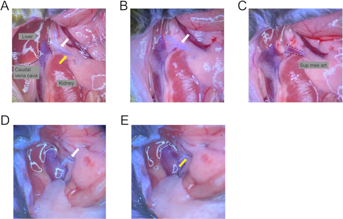
Figure 1: Selective transection of intestine-innervating autonomic nerves in a murine model. (A) Representative operative view after exposure of the superior mesenteric artery (SMA), showing two white, translucent nerve bundles running parallel to the vessel. The white arrow indicates the parasympathetic branch on the superior aspect of the SMA, and the yellow arrow indicates the sympathetic branch on the inferior aspect of the SMA. (B) After transection of the inferior sympathetic branch, only the superior parasympathetic branch remains, as indicated by the white arrow. (C) After subsequent transection of the superior parasympathetic branch, only the SMA remains visible, as indicated by the arrow. (D) Representative operative view from another mouse, with the white arrow indicating the parasympathetic branch. (E) After transection of the superior parasympathetic branch, only the inferior sympathetic branch remains, as indicated by the yellow arrow. Please click here to view a larger version of this figure.
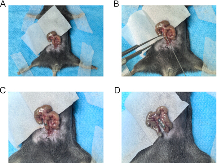
Figure 2: Intramural injection into the proximal colon for retrograde tracing. (A) The cecum is gently exteriorized from the abdominal cavity to expose the proximal colon. (B) The proximal colon is lifted carefully with microforceps, and the tracer solution is injected into the intestinal wall using a microsyringe. (C) Representative image of a successful injection, showing that the Fast Green dye remains localized within the submucosal layer without obvious leakage. (D) Representative image of an unsuccessful injection, showing leakage of the Fast Green dye into the intestinal lumen. Please click here to view a larger version of this figure.
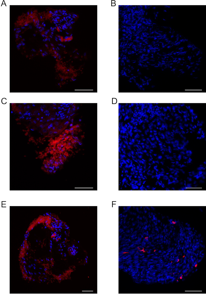
Figure 3: CTB-555 retrograde tracing validates selective intestinal autonomic denervation. Mice received a focal submucosal colonic injection of CTB-555 (red) for retrograde tracing; nuclei were counterstained with DAPI (blue). (A) Selective parasympathetic transection group; retrograde labeling was evaluated in the left NG. (B) Sham-operated control corresponding to (A), retrograde labeling was evaluated in the NG. (C) Selective parasympathetic transection group; retrograde labeling was evaluated in the right NG. (D) Sham-operated control corresponding to (C), retrograde labeling was evaluated in the NG. (E) Selective sympathetic transection group; retrograde labeling was evaluated in the CG. (F) Sham-operated control corresponding to (E); retrograde labeling was evaluated in the CG. Scale bars = 50 µm. Please click here to view a larger version of this figure.
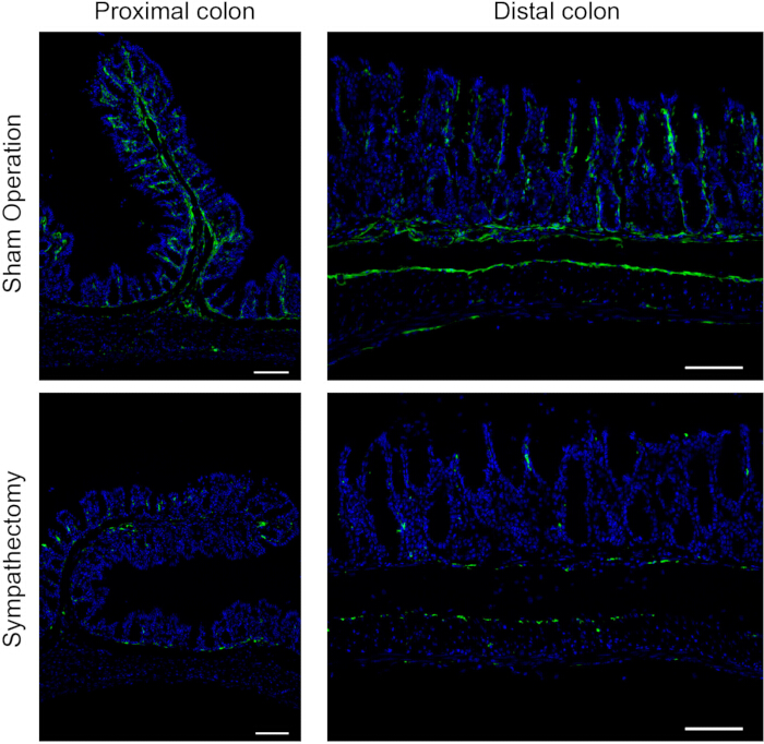
Figure 4: Tyrosine hydroxylase (TH) immunofluorescence staining in the proximal and distal colon after sympathectomy. Representative immunofluorescence images of the proximal colon and distal colon from the sympathectomy group and the sham operation group. TH is shown in green, and nuclei are counterstained with DAPI (blue). The upper panels show the sham operation group, and the lower panels show the sympathectomy group. The left panels represent the proximal colon, and the right panels represent the distal colon. Scale bars = 100 µm. Please click here to view a larger version of this figure.
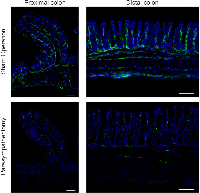
Figure 5: ChAT immunofluorescence staining in the proximal and distal colon after parasympathectomy. Representative immunofluorescence images of the proximal colon and distal colon from the parasympathectomy group and the sham operation group. ChAT is shown in green, and nuclei are counterstained with DAPI (blue). The upper panels show the sham operation group, and the lower panels show the parasympathectomy group. The left panels represent the proximal colon, and the right panels represent the distal colon. Scale bars = 100 µm. Please click here to view a larger version of this figure.
Supplementary File 1: Troubleshooting guide for common intraoperative complications.Please click here to download this file.
Discussion
Loading...
$$\rightleftharpoonup{xx}$$
$$\longleftharp{xx}$$,
$$\longrightharp{xx}$$,
The murine SMA arises from the anterior wall of the abdominal aorta, inferior to the celiac trunk, and provides arterial inflow to major abdominal viscera, including the intestine14. Anterior to the SMA lie the colon and its mesentery, whereas posteriorly it is closely related to the left renal vein and the abdominal aorta; the superior mesenteric vein typically courses along its right side. In mice, the SMA is extremely delicate and is surrounded by critical organs and major vessels. Importantly, equally fine sympathetic and parasympathetic nerve fibers adhere tightly to and run alongside the SMA. Therefore, transection of either the sympathetic or parasympathetic trunk requires a high degree of precision.
Two major challenges are encountered during this procedure. The first is reliable intraoperative identification of the SMA and the sympathetic and parasympathetic fibers traveling with it. The second is that, because the nerves are intimately opposed to the vessel wall, nerve transection can easily result in SMA injury and bleeding. To minimize this risk, a glass probe can be used to very carefully separate the nerve bundle from the SMA and adjacent connective tissue over a distance of ~3–5 mm before transection. All maneuvers should be performed gently to avoid traction injury to the nerve and to prevent tearing of the accompanying vessels.
Injury to abdominal organs may occur during laparotomy. If this happens, immediately apply gentle pressure with a sterile cotton swab moistened with warm saline to control bleeding. Small superficial lacerations of a liver lobe can often be managed with compression. However, an intestinal perforation should be repaired immediately with 8-0 sutures. Major injuries should prompt consideration of humane endpoints. If a small vessel ruptures intraoperatively, apply pressure with sterile gauze until hemostasis is complete. To reduce the likelihood of intestinal injury, gently and efficiently exteriorize and reposition the bowel using cotton swabs, and carefully elevate the liver to improve visualization when needed.
In cachectic or aged mice, the linea alba may be less distinct. In such cases, starting directly inferior to the xiphoid process is helpful, as this region often provides the clearest midline convergence of the abdominal musculature. In older or obese mice, the SMA and its accompanying nerve fibers may be obscured by fat; careful dissection of adipose tissue with forceps and microscissors may be required to achieve adequate exposure. If excessive adiposity, inadvertent injury to a major vessel, or any other cause results in profuse hemorrhage, the procedure must be terminated and the animal euthanized. During abdominal closure, if marked resistance is felt during needle passage or if the bowel is seen to be lifted with the suture, withdraw the needle and re-suture the layer. An unrecognized suture through the intestine can lead to fatal bowel obstruction and peritonitis.
The celiac ganglia are located along the anterior surface of the abdominal aorta around the origins of the celiac trunk and the SMA and are a key component of the sympathetic nervous system, providing sympathetic input to organs such as the gastrointestinal tract, liver, pancreas, and spleen11. The superior mesenteric ganglion lies posterior to the origin of the SMA and consists predominantly of sympathetic fibers that innervate the jejunum, ileum, cecum, and colon15. The present approach is designed to achieve targeted transection of a major subset of intestine-directed sympathetic fibers traveling along the SMA, while minimizing dissection around the abdominal aorta and ganglia. This method should therefore be interpreted as a regionally targeted denervation model rather than complete elimination of all sympathetic inputs to the colon, because additional sympathetic fibers may reach distal colonic regions through alternative pathways. This anatomical strategy may help reduce the risk of inadvertent injury to the abdominal aorta or the SMA compared with conventional ganglionectomy techniques.
Subdiaphragmatic vagotomy refers to the transection of the vagal trunks below the diaphragm to block vagal input to the stomach, liver, pancreas, and intestine16,17,18. In comparison, the present approach may carry a lower risk of injury to the left hepatic lobe or the esophagus and can provide a simpler, clearer operative field, potentially reducing technical difficulty and shortening operative and anesthetic time. In addition, this strategy may help reduce the risk of respiratory and heart rate disturbances that can occur following bilateral vagal transection.
Although this method allows direct visualization of the target perivascular bundles during surgery, it remains possible that additional anatomically inconspicuous nerve branches contribute to intestinal innervation. For this reason, successful denervation should be interpreted on the basis of combined anatomical observation, retrograde tracing, and immunohistochemical validation, rather than gross appearance alone. Future studies may further subdivide and selectively transect these fibers to clarify their specific roles. Given the challenges of distinguishing sympathetic from parasympathetic fibers and the potential for bleeding complications, it is recommended to first practice the dissection and transection on cadaveric mice to become familiar with the relevant anatomy before performing survival surgery.
Murine models offer advantages, including low cost and ease of handling, and they are widely used to build experimental systems tailored to diverse research questions. The nerve-transection strategy described here provides flexible, investigator-controlled selectivity for disrupting either sympathetic or parasympathetic input to the intestine. This approach may facilitate mechanistic studies of autonomic regulation of intestinal physiology and tumor biology and provides an experimental framework for investigating the roles of sympathetic and parasympathetic pathways in gut-related oncologic and physiologic settings.
Acknowledgements
Loading...
$$\rightleftharpoonup{xx}$$
$$\longleftharp{xx}$$,
$$\longrightharp{xx}$$,
We sincerely thank the Laboratory Animal Center, the Beijing Clinical Research Institute, and Beijing Friendship Hospital for their excellent animal care and support. This work was supported by the Natural Science Foundation of China(82300646); Beijing Municipal Administration of Hospitals Incubating Program (PX2020001, PX20240103); Capital’s Funds for Health Improvement and Research (2024-2-2028)
Materials
| Name | Company | Catalog Number | Comments |
|---|---|---|---|
| 5-0 silk braided non-absorbable suture | Ethicon | W500 | |
| 6-0 PGA Suture | Jinhuan | CR631 | |
| Anesthesia Induction Chambers | RWD Life Science | V100 | |
| Animal Anesthesia Machine | RWD Life Science | R500IP | |
| Anti-Choline Acetyltransferase antibody | Abcam | ab181023 | |
| Artificial Tears Lubricant Ophthalmic Ointment | Akorn | 59399-162-35 | |
| C57BL/6J Mice | Shanghai Model Organisms CenterTechnology | SM-001 | |
| Cholera Toxin Subunit B (CTB), Alexa FluorTM 555 Conjugate | Thermo Scientific | C34776 | |
| Compound Microscope | Yuyan Instruments | 512 | |
| DAPI and Hoechst Nucleic Acid Stains | Invitrogen | D3571 | |
| Fast Green | Thermo Scientific | F7252 | |
| Fine Hemostatic Forceps | Jinzhong surgical instrument | JCG020 | |
| Hypoallergenic Surgical Tape | 3M Blenderm | 70200419342 | |
| Isoflurane | RWD Life Science | R510-22-10 | |
| Micro Forceps(Curved) | Jinzhong surgical instrument | WCC200 | |
| Micro Forceps(Straight) | Jinzhong surgical instrument | WCC190 | |
| Micro Needle holders | Jinzhong surgical instrument | WBA050 | |
| Micro scissors | Jinzhong surgical instrument | Y3F010 | |
| Micro syringe | Hamilton | 7632-01 | |
| Ophthalmic Scissors(pointed tip) | Jinzhong surgical instrument | JC2303 | |
| Ophthalmic Scissors(rounded tip) | Jinzhong surgical instrument | JC2303 | |
| Povidone-Iodine Swabstick | Winner Medical | 206 | |
| Tissue and Cell Fixative (4% Paraformaldehyde, PFA) | MedChemExpress | HY-DY3003 | |
| Tissue OCT-Freeze Medium | Sakura | C2077 | |
| Tyrosine Hydroxylase (E2L6M) Rabbit Monoclonal Antibody | Cell Signaling Technology | 58844S |
Reprints and Permissions
Request permission to reuse the text or figures of this JoVE article
Request Permission