This review synthesizes recent literature on venous pulsatile tinnitus associated with internal jugular venous outflow disturbance, summarizing current evidence on clinical presentation, imaging evaluation, mechanisms, management strategies, and future research directions.
Review Article
Tinnitus and Tinnitus-Like Symptoms in Cerebral Venous Outflow Disorders: A Critical Review
In This Article
Summary
Abstract
Venous pulsatile tinnitus and tinnitus-like symptoms associated with internal jugular venous outflow disturbance represent an emerging clinical topic with specific diagnostic and management challenges. A narrative review was conducted using English-language literature identified through searches of PubMed, Web of Science, and Google Scholar. The review focused on studies published through February 2026 that addressed venous pulsatile tinnitus, internal jugular vein stenosis or compression, cerebral venous outflow disturbance, imaging and hemodynamic assessment, and management strategies. This review summarizes current evidence on vascular anatomy, clinical presentation, pathophysiological mechanisms, imaging evaluation, and treatment. Structural venous abnormalities and external compression may produce disturbed flow and local vascular sound, particularly in classical venous pulsatile tinnitus, whereas venous congestion, intracranial pressure dysregulation, altered cerebral perfusion, and central auditory plasticity remain proposed mechanisms for other tinnitus-like symptoms. MRI/MRV, CTV/CTA, and Doppler ultrasonography provide complementary anatomical and hemodynamic information. Invasive angiography is reserved for selected patients, with venous manometry added when pressure-gradient assessment is clinically relevant. Management options include conservative care, intracranial pressure management, endovascular treatment, surgical decompression, and supportive tinnitus therapy. However, current evidence is derived mainly from selected retrospective cohorts and small case series and does not establish the prevalence or causal contribution of IJVOD in unselected patients with venous pulsatile tinnitus. Standardized terminology, validated diagnostic criteria, improved patient selection, and prospective multicenter studies are required to enhance diagnostic confidence and enable robust assessment of long-term outcomes.
Introduction
Tinnitus, the perception of sound without an external source, can manifest as ringing, humming, buzzing, or rhythmic noises. Its causes are diverse. Pulsatile tinnitus is characterized by rhythmic sound perception that is often synchronized with the heartbeat. It has particular clinical importance because an underlying vascular or structural abnormality may be identifiable in selected patients1,2,3,4. While causes can be arterial or venous, venous mechanisms have garnered more attention in recent years. This is largely due to the fact that many venous causes are visible on imaging and may be amenable to specific treatments. Pulsatile tinnitus accounts for a minority of tinnitus presentations, but it carries disproportionate clinical importance because vascular or structural causes are identified in a substantial proportion of affected patients. Venous causes are increasingly evaluated in patients with pulse-synchronous symptoms, particularly when symptoms vary with head position or ipsilateral jugular compression2,3,5. However, the prevalence and causal contribution of extracranial internal jugular venous outflow disturbance in unselected patients with venous pulsatile tinnitus remain uncertain.
This review distinguishes venous pulsatile tinnitus from internal jugular venous outflow disturbance (IJVOD)-associated tinnitus-like symptoms. Classical venous pulsatile tinnitus is most often associated with a local vascular sound source near the temporal bone, including the sigmoid sinus, jugular bulb, emissary veins, or dural arteriovenous shunts2,5. By contrast, pulse-synchronous tinnitus directly attributable to extracranial internal jugular vein compression remains insufficiently characterized and validated2,6. Reported IJVOD-associated symptoms may include nonspecific tinnitus-like sensations without established pulse synchrony1,7. These symptoms should not be considered equivalent to classical venous pulsatile tinnitus. However, the prevalence and causal contribution of IJVOD in unselected patients with venous pulsatile tinnitus remain uncertain. When these pathways become narrowed, compressed, or anatomically variant, normal venous flow can be disrupted. Conditions such as internal jugular vein stenosis, extrinsic compression from nearby structures, jugular bulb anomalies, venous sinus stenosis, or dural sinus wall defects can cause turbulent blood flow. When located near the temporal bone, disturbed venous flow may generate or transmit sound to adjacent auditory structures and may contribute to pulse-synchronous tinnitus5,8.
The relevance of this topic goes beyond the auditory symptom itself. For many individuals, venous pulsatile tinnitus is a source of significant distress and disruption, and its explanation often requires a thorough vascular assessment. Emerging research indicates that abnormal venous hemodynamics might do more than just create a local sound source; it could also affect central auditory pathways, potentially playing a role in why symptoms persist and cause considerable distress9,10. This interplay between venous flow, intracranial pressure, and auditory processing has broadened the conceptual framework for venous pulsatile tinnitus. Venous pulsatile tinnitus is therefore increasingly viewed not merely as a mechanical phenomenon but as a condition that may involve both vascular and neural components.
Despite these insights, the field faces several ongoing challenges. Terminology remains inconsistent across studies, diagnostic approaches vary between institutions, and establishing a clear causal link between internal jugular venous abnormalities and tinnitus symptoms is often complex. Some imaging findings may be incidental, while others are clinically significant and treatable. This diagnostic uncertainty can lead to delays, complicate patient counseling, and hinder the comparison of treatment outcomes. Therefore, there is a pressing need for a clearer framework that better connects patient symptoms, imaging results, hemodynamic changes, and therapeutic decisions.
This review critically examines current evidence on venous pulsatile tinnitus and IJVOD-associated tinnitus-like symptoms, with particular attention to the limited and heterogeneous evidence concerning extracranial internal jugular venous abnormalities. The review focuses on terminology, clinical presentation, anatomical and pathophysiological mechanisms, imaging-based diagnostic strategies, management options, and emerging research directions. The synthesis of recent clinical and mechanistic findings is intended to clarify the possible role of venous outflow disturbance in tinnitus, support more standardized evaluation protocols, and identify priorities for individualized management and future research.
Review and Perspective
Evidence base and methodological limitations
The current evidence base for venous pulsatile tinnitus and tinnitus-like symptoms associated with internal jugular venous outflow disturbance remains heterogeneous. Most available studies are retrospective, single-center, or based on small case series, and many use different definitions of tinnitus phenotype, venous stenosis severity, hemodynamic significance, and treatment response. These limitations restrict direct comparison across studies and make it difficult to establish causality between an imaging abnormality and the patient’s symptoms.
A major methodological challenge is selection bias. Patients referred for advanced imaging, venous manometry, stenting, or surgical decompression often represent highly selected populations with severe or refractory symptoms. As a result, reported treatment success rates may not be generalizable to broader clinical populations. In addition, many studies lack standardized baseline symptom scores, validated tinnitus-specific outcome measures, blinded imaging review, control groups, or long-term follow-up.
Therefore, current conclusions should be interpreted as suggestive rather than definitive. Available evidence generally favors a cautious diagnostic approach that integrates careful symptom phenotyping, multimodal imaging, and hemodynamic correlation before invasive treatment is considered. However, specific diagnostic thresholds, optimal patient selection criteria, and comparative effectiveness among conservative, endovascular, and surgical strategies remain insufficiently validated. Current evidence is derived mainly from selected retrospective cohorts, small case series, and case reports and does not establish the prevalence or causal contribution of IJVOD in unselected patients with venous pulsatile tinnitus.
Vascular anatomy and hemodynamics
The internal jugular veins are the main extracranial drainage routes for blood leaving the brain. Compromising their outflow can significantly impact intracranial venous hemodynamics11,12,13. Narrowing of these veins can arise from intrinsic issues, such as malformed valves, septa, or intraluminal webs, or from extrinsic compression by neighboring structures like an elongated styloid process, tumors, or anatomical variants at the C1 transverse process level. These abnormalities may reduce the venous lumen and may be associated with locally increased velocity or disturbed flow5,7. Venous stenosis and related morphological abnormalities may produce disturbed flow patterns capable of generating or transmitting vascular sound14. Other venous contributors include enlarged emissary or condylar veins, jugular bulb abnormalities, and sigmoid sinus diverticula15. The vertebral venous plexus provides an important collateral pathway, particularly in the upright position, when cerebral venous outflow shifts partly away from the internal jugular veins. This positional redistribution may influence cerebrospinal fluid and intracranial pressure dynamics, although its role in IJVOD remains uncertain16. IJVOD may be considered within the broader spectrum of cerebral venous congestion. A multi-institutional framework emphasizes that venous pathology should be interpreted according to its anatomical location and its interactions with cerebrospinal fluid dynamics and intracranial pressure17. More recently, an imaging-based classification of cerebral venous outflow insufficiency has been proposed, although its diagnostic and prognostic value requires external validation18. Venous dominance may also influence jugular bulb morphology. A high jugular bulb, particularly with bony dehiscence, may facilitate local venous sound transmission, although its presence alone does not establish symptom causality. In a retrospective study of 228 patients, dominant-side internal jugular vein stenosis was independently associated with a dominant-side high jugular bulb, suggesting a possible relationship among venous dominance, altered outflow, and jugular bulb morphology19. However, these cross-sectional findings do not establish causality or demonstrate that a high jugular bulb directly causes tinnitus.
Not all venous abnormalities cause symptoms. Whether disturbed venous flow translates into perceptible tinnitus likely depends on individual hemodynamic factors. Variables such as flow velocity, vessel wall compliance, pressure gradients, and the degree of local turbulence all influence whether abnormal flow becomes an audible signal8. This individual variability helps explain why similar imaging findings may be silent in one patient yet profoundly symptomatic in another. Currently, there is no universally agreed-upon threshold for when venous turbulence becomes clinically significant, and methods for assessing these parameters vary widely. Consequently, comparing results across different studies is challenging, and well-defined imaging or hemodynamic criteria for clinically important disease are still lacking.
Altered venous drainage can also disrupt intracranial pressure dynamics. Elevated cerebral venous pressure has been associated with rhythmic auditory symptoms in selected cohorts, but the direction and clinical significance of this relationship remain uncertain20. However, the relationship is not straightforward because some reports describe persistent tinnitus despite apparent hemodynamic correction. One theory posits that a combination of high-velocity jet flow and venous wall mobility is necessary to convert normal physiological flow into a pathological auditory signal21. While this model is mechanistically appealing, the precise thresholds for wall motion, velocity, and pressure transmission needed to generate tinnitus remain poorly defined. Taken together, the evidence supports a multifactorial model where structural venous anomalies and aberrant flow dynamics interact to produce clinically perceptible venous sounds.
The venous system is also integral to regulating intracranial pressure. Hemodynamically significant internal jugular vein stenosis may impair venous drainage and may be associated with venous congestion or altered intracranial pressure, particularly when collateral pathways are insufficient. The coexistence of jugular narrowing and intracranial hypertension suggests an association, but does not establish that fixed IJV stenosis causes elevated intracranial pressure or tinnitus20. Whether internal jugular vein stenosis contributes to increased intracranial pressure may depend on its hemodynamic significance and collateral venous drainage. Elevated intracranial pressure is unlikely to aggravate fixed jugular stenosis; therefore, evidence linking cerebral venous sinus stenosis to intracranial pressure should not be extrapolated to internal jugular venous outflow disturbance. Persistent venous hypertension may also contribute to progressive symptoms like headache, visual disturbances, and papilledema22. Given that most evidence comes from retrospective series and small observational studies, prospective, mechanism-focused research is needed to clarify these temporal and causal relationships.
Clinical interpretation becomes even more complex when venous narrowing stems from external compression23. The C1 transverse process has been increasingly recognized as a potential site of extracranial internal jugular vein (IJV) compression in selected symptomatic cohorts, although its prevalence and clinical significance in unselected populations remain uncertain24,25,26. The so-called jugular “nutcracker” phenomenon involves extrinsic compression of the internal jugular vein between adjacent bony and vascular structures18. In Eagle syndrome, an elongated styloid process may compress the vein, leading to symptoms such as neck discomfort, pain on swallowing, headache, or tinnitus27,28. A persistent diagnostic challenge lies in differentiating clinically significant venous compression from incidental anatomical variation1. Although modern imaging can precisely define the location and severity of compression, imaging findings alone cannot establish whether the abnormality is responsible for the reported symptoms. This ambiguity highlights the need for an individualized assessment that synthesizes anatomical data, hemodynamic significance, the patient's symptom profile, and their response to clinical maneuvers5.
Clinical manifestations
Venous pulsatile tinnitus is typically described as a pulse-synchronous whooshing, rushing, or humming sound. Modulation with head position, the Valsalva maneuver, or gentle ipsilateral jugular compression may support a venous source but is not diagnostic2,3,5. IJVOD-associated tinnitus-like symptoms may include nonspecific brain noise or intracranial buzzing without established pulse synchrony; however, the frequency and specificity of these symptoms remain uncertain6,18,29.
Patients with impaired jugular venous outflow frequently present with complaints beyond isolated auditory symptoms. Associated issues may include chronic headache, dizziness, transient visual obscurations, neck pain, sleep disturbance, and a sensation of cranial pressure or fullness. Impaired jugular venous outflow has also been associated with several other neurological conditions, such as normal pressure hydrocephalus, transient global amnesia, white matter lesions, leukoaraiosis, and cognitive impairment30,31,32,33. In some patients, these broader manifestations may reflect underlying intracranial pressure abnormalities or a more generalized impairment of venous drainage. Internal jugular vein compression is often underdiagnosed because its symptoms can be subtle, nonspecific, and variable over time29. Reported prevalence and severity of these associated symptoms vary considerably across studies, reflecting both clinical heterogeneity and a lack of validated diagnostic criteria.
The functional and psychological burden of venous pulsatile tinnitus is substantial. The persistent, pulse-synchronous noise can interfere with sleep, concentration, emotional regulation, and daily activities. Patients with refractory forms of this condition have shown higher rates of anxiety, depression, and social withdrawal34,35. Symptoms tend to be particularly intrusive at night when background noise is minimal, exacerbating sleep disruption and psychological distress36. These consequences underscore why delayed or incorrect diagnosis is clinically significant, as it can lead to ineffective treatments, prolonged patient uncertainty, and further deterioration in quality of life. Therefore, a comprehensive clinical evaluation should address not only the potential vascular mechanisms but also the broader psychosocial impact of the condition3.
Imaging modalities and clinical applications
Imaging is central to the evaluation of pulsatile tinnitus because the appropriate modality depends on the suspected vascular mechanism and anatomical location1,3,4,37. MRI/MRV, CTV/CTA, and Doppler ultrasonography provide complementary information rather than interchangeable diagnostic results.
MRI and MRV offer excellent soft-tissue contrast for non-invasive assessment of intracranial venous sinuses, soft-tissue lesions, and external compression. However, contrast-enhanced brain MRI may overestimate internal jugular vein stenosis compared with contrast-enhanced CT38. Therefore, suspected IJV stenosis should be confirmed with complementary imaging before invasive treatment is considered38. These techniques are especially useful for identifying subtle structural lesions like venous valves, septa, or diverticula that might be missed on routine imaging. Their diagnostic performance for venous malformations is favorable when dedicated venous protocols are used38. Advanced MRI techniques have further expanded the utility of MRI-based evaluation. Quantitative flow analysis can help characterize disturbed venous hemodynamics and pinpoint high-risk flow patterns39. Time-of-flight angiographic sequences and time-resolved contrast-enhanced methods improve the delineation of venous anatomy and dynamic flow abnormalities linked to venous sinus stenosis37. Four-dimensional (4D) flow MRI can estimate trans-stenotic velocity differences, which sometimes correlate with cerebral venous pressure gradients in conditions like transverse sinus stenosis40. Arterial spin labeling (ASL) has also proven valuable for detecting subtle shunting lesions, including small dural arteriovenous fistulas that conventional post-contrast or time-of-flight sequences might overlook37,41. Although ASL does not directly measure venous outflow impairment, it offers indirect yet useful insights into the perfusion consequences of cerebral venous compression. Impaired venous drainage can disturb cerebral blood flow distribution, disrupt autoregulation, and lead to detectable reductions in perfusion42,43. Considering these advances together, MRI-based evaluation has become a first-line, non-invasive imaging strategy for many patients with venous pulsatile tinnitus4. Limitations of MRI include longer acquisition times, suboptimal visualization of bony anatomy, and contraindications in certain patients.
CT-based techniques complement MRI by providing superior visualization of bone, skull base relationships, and calcified or compressive lesions. CTV and CTA are particularly helpful when extrinsic compression, from an elongated styloid process, a hypertrophic C1 transverse process, or another bony variant, is suspected1,27. Jugular foramen-IJV caliber mismatch and abnormal collateral veins may help distinguish acquired stenosis from physiological IJV slenderness, but require clinical and hemodynamic correlation44. These CT-based techniques also provide high spatial resolution and rapid acquisition, which are advantageous in routine practice. CTV also enables detailed assessment of high jugular bulb and jugular bulb dehiscence, which may be relevant to local venous sound transmission. However, a recent systematic review and meta-analysis showed that both variants are relatively common and that prevalence estimates vary substantially with anatomical definitions, imaging protocols, and measurement planes. Clinical relevance should therefore be determined by symptom laterality, adjacent bony dehiscence, and hemodynamic concordance rather than by the presence of these variants alone45. Dynamic CT approaches can reveal how venous caliber and adjacent anatomy change with posture, making them especially valuable for evaluating patients with position-dependent symptoms1,46. The downsides of CT include exposure to ionizing radiation, the frequent need for iodinated contrast, and limited ability to assess real-time venous hemodynamics.
Ultrasonography is convenient, non-invasive, and easily repeatable, enabling real-time evaluation of venous flow. Doppler ultrasound can detect alterations in flow velocity, segmental narrowing, collateral redistribution, and positional variation, making it well-suited for initial screening and follow-up13. Jugular venous flow measurements may vary with respiratory phase and examination conditions, highlighting the need for standardized ultrasound acquisition and interpretation47. The diagnostic utility of ultrasonography is limited for lesions near the jugular bulb or distal transverse sinus due to acoustic window constraints. In such cases, cross-sectional imaging remains necessary if clinical suspicion is high. Recent work suggests that a flow velocity of 27.5 cm/s or less in the upper internal jugular vein (J3 segment) may serve as a practical screening marker for J3 stenosis, with reasonable sensitivity and specificity48. Increased combined bilateral flow volume at the J3 level has also been proposed as a non-invasive indicator of intracranial hypertension. However, these Doppler-derived thresholds have not yet undergone broad external validation across independent cohorts, imaging laboratories, or standardized acquisition protocols. These Doppler-derived thresholds should therefore be regarded as investigational screening markers rather than standalone diagnostic criteria. Interpretation should be integrated with symptoms, cross-sectional imaging, collateral drainage patterns, and hemodynamic assessment.
No single imaging modality is perfect for every case, and invasive angiographic evaluation may still be justified in carefully selected patients. MRI/MRV is generally best for soft-tissue and intracranial assessment, CT-based studies excel at defining bony anatomy and extrinsic compression, and ultrasonography is valuable for screening and dynamic bedside evaluation. Although Digital subtraction angiography (DSA) is invasive and is not routinely required, it can define vascular anatomy, collateral pathways, and dynamic flow. Combined venous manometry allows direct measurement of trans-stenotic pressure gradients, although the clinical thresholds may differ between venous sinus stenosis and extracranial IJV stenosis5. DSA should therefore be reserved for selected patients with suspected high-risk vascular lesions, inconclusive non-invasive imaging, or planned intervention49. The highest diagnostic yield is typically achieved through a rational, multimodal approach tailored to the suspected disease mechanism. Future efforts should focus on standardizing imaging pathways, refining diagnostic thresholds for each modality, and improving the correlation between radiographic abnormalities and symptom generation. A proposed diagnostic workflow integrating clinical phenotyping, multimodal imaging, hemodynamic assessment, and mechanism-based management is summarized in Figure 1.
Pathophysiological mechanisms
Several interconnected mechanisms may explain how venous outflow disturbance contributes to venous pulsatile tinnitus and related tinnitus-like symptoms. One widely discussed mechanism involves the generation of turbulent or vortex-like flow within narrowed or externally compressed venous segments5. This disturbed flow can create vibrations or pressure oscillations that transmit through adjacent bone and soft tissue to the cochlea or other auditory structures7,50. This explanation is anatomically plausible, especially when the abnormal venous segment lies close to the temporal bone or skull base.
A second proposed mechanism centers on progressive vascular remodeling. Chronic turbulence has been proposed to promote endothelial stress and vascular remodeling, although direct longitudinal evidence remains limited51,52. This process could help explain why venous pulsatile tinnitus becomes chronic or progressively more severe in some individuals. Furthermore, altered venous drainage might influence pressure transmission within the subarachnoid space, contributing to subtle or overt intracranial pressure abnormalities. These pressure changes, in turn, could facilitate the transmission of vascular sounds to the auditory system.
A third, more hypothetical mechanism involves central auditory plasticity. Repetitive pulse-synchronous input may alter central auditory processing and help explain persistent symptoms after partial correction of a venous lesion, although longitudinal evidence is limited. Perfusion studies have also reported altered cerebral blood flow or neurovascular activity in some patients with venous pulsatile tinnitus or venous outflow abnormalities42. These findings suggest a possible relationship among venous drainage, cerebral perfusion, and sensory processing, but do not establish that perfusion changes cause tinnitus. Local vascular sound generation remains better supported, whereas central auditory and perfusion-related mechanisms remain provisional. This multifactorial model carries important clinical implications and suggests that persistent symptoms may not always indicate ongoing structural obstruction alone. Successful management, therefore, might require attention to both the vascular lesion and its downstream auditory or psychological consequences. The model also highlights the need for future studies that integrate hemodynamic, neuroimaging, and patient-reported symptom data within the same cohorts.
Management approaches
Management of venous pulsatile tinnitus and tinnitus-like symptoms reported in association with venous outflow disturbance should be individualized. Decisions should be based on anatomical findings, hemodynamic severity, symptom burden, and the presence of associated conditions like intracranial hypertension. Conservative management is generally appropriate for patients with mild symptoms, equivocal imaging findings, or no clearly correctable structural lesion. For selected patients where elevated intracranial pressure is part of the clinical picture, medical therapy aimed at reducing pressure, including diuretics, may provide symptom relief53,54. Pharmacologic treatment for associated anxiety, insomnia, or mood disturbances can also help reduce tinnitus-related distress, although such measures typically do not address the primary vascular issue.
Recent studies have increasingly identified the transverse process of the first cervical vertebra (C1) as a site of extracranial internal jugular vein compression in selected symptomatic cohort24,25,26. However, its prevalence in unselected populations and its causal relationship with venous pulsatile tinnitus or IJVOD-associated tinnitus-like symptoms remain uncertain7,27. Anatomical narrowing alone should not determine treatment. Clinical relevance should be assessed by integrating symptom phenotype and laterality, collateral venous drainage, dynamic imaging findings, and, when clinically appropriate, hemodynamic measurements7,27.
Potential management options include observation and symptom-directed care, selected cervical rehabilitation or stabilization approaches, surgical decompression with partial resection of the C1 transverse process and release of adjacent soft tissues, and adjunctive venoplasty or stenting in carefully selected patients25,26. Evidence supporting cervical curve correction or stabilization remains preliminary55,56. Stenting alone may also be insufficient when fixed osseous compression remains uncorrected26. Treatment selection should therefore be individualized through multidisciplinary assessment, and current options should not be interpreted as validated treatment standards7.
Endovascular intervention, particularly stenting of the venous sinus or internal jugular vein, has become an important option for severe or refractory cases with demonstrable stenosis and supportive hemodynamic evidence57,58,59. The goal is to restore venous patency, reduce pressure gradients across the stenosis, and diminish pathological turbulence60. Selected observational studies have reported rapid improvement in pulsatile tinnitus after venous sinus or IJV stenting, although response rates may not be generalizable to unselected patients49,53,58. Long-term outcomes may depend on lesion type, procedural factors, and identification of all relevant sources of obstruction49. Persistent extrinsic compression, suboptimal stent placement, or untreated adjacent abnormalities can lead to restenosis or treatment failure60. Published observational series have reported short-term symptom improvement in selected patients, but recurrence, thrombosis, restenosis, and local neurovascular complications remain concerns53,58,59.
Surgical decompression may be considered when clinically relevant venous obstruction results from fixed osseous compression7,26,60, including styloid- or C1-related compression. Procedures may include styloidectomy, partial C1 transverse process resection, and release of adjacent soft tissues, with venoplasty or venous reconstruction considered in selected cases24,25,26. Evidence is derived mainly from retrospective studies and small case series, and direct comparisons with conservative or endovascular treatment are lacking7.
Supportive therapies also play a crucial role. Sound therapy, counseling, and cognitive-behavioral interventions can lessen distress and improve coping mechanisms, especially for patients with chronic symptoms, an incomplete response to vascular treatment, or no clear interventional target. While not curative, these approaches can significantly enhance quality of life. Overall, current clinical practice favors a stepwise, multidisciplinary strategy. This begins with careful diagnostic confirmation and then escalates treatment based on symptom severity and anatomical evidence. A major unmet need in the field remains the development of clearer criteria for selecting treatments and more robust long-term outcome data.
Clinical outcomes in venous pulsatile tinnitus largely depend on the type of lesion, its anatomical complexity, hemodynamic significance, and how completely the abnormality can be corrected. Observational reports suggest that patients with focal stenosis may have better outcomes after endovascular treatment than patients with diffuse, multifocal, or predominantly extrinsic lesions; however, this association has not been consistently validated. Coexisting intracranial hypertension can worsen symptom severity and influence treatment response, making its recognition and management particularly important. Long-term clinical follow-up may be appropriate after intervention because restenosis, thrombosis, or symptom recurrence has been reported53,58,59. Follow-up imaging should be guided by the procedure, symptoms, and suspected complications rather than applied uniformly to all patients. A concise comparison of conservative, pressure-directed, endovascular, surgical, and supportive approaches is provided in Table 1.
The principal clinical challenge is determining whether an anatomical venous abnormality is incidental or symptomatically relevant. No validated framework currently integrates symptom phenotype, laterality, collateral drainage, imaging findings, and hemodynamic measurements. Treatment comparisons are further limited by heterogeneous selection criteria and outcome measures. These uncertainties should be considered before invasive evaluation or intervention.
Future perspectives
The possible association between IJVOD and venous pulsatile tinnitus or tinnitus-like symptoms has received increasing research attention, but its prevalence, causal relevance, and treatment implications remain uncertain. Recent work has expanded the conceptual framework beyond a purely structural or mechanical model to a more complex framework that incorporates venous anatomy, abnormal flow dynamics, intracranial pressure regulation, and central auditory processing. This broader perspective helps explain the considerable variability in symptom severity, associated neurological complaints, and treatment response among patients with similar imaging findings. This broader perspective also supports multidisciplinary evaluation integrating neurovascular anatomy, advanced imaging, otologic assessment, and patient-reported outcomes1.
Several key priorities should guide future progress. First, there is a clear need for more consistent terminology. Terms like pulsatile tinnitus, venous pulsatile tinnitus, and cerebral tinnitus are often used interchangeably or inconsistently, which hinders communication across specialties and complicates the comparison of study results. Adopting a more standardized nomenclature would improve study design, patient stratification, and the interpretation of outcomes. Future studies should distinguish classical venous pulsatile tinnitus from other tinnitus-like symptoms associated with IJVOD by reporting pulse synchrony, symptom phenotype, anatomical findings, and hemodynamic relevance61.
Second, diagnostic pathways require greater standardization. Although multimodal imaging is now central to evaluation, there is no universally accepted algorithm for determining when an internal jugular venous abnormality is incidental versus clinically causative. Future research should aim to establish more robust imaging-based and hemodynamic criteria to better differentiate symptomatic lesions from benign anatomical variants5.
A third priority is the development of more comprehensive outcome measures. Current reports often focus on anatomical success or short-term symptom relief, while long-term functional outcomes, quality-of-life metrics, recurrence rates, and patient-reported symptom severity are less consistently assessed. The creation and adoption of standardized, disease-specific outcome tools would allow for more meaningful comparisons across different treatment strategies, be they conservative, endovascular, or surgical. Given the relative heterogeneity of venous pulsatile tinnitus and variations in local practice, prospective multicenter registries and collaborative studies will likely be essential to generate the necessary sample sizes.
Several emerging research avenues hold promise for improving both understanding and management. One major direction is personalized hemodynamic modeling. Advances in three-dimensional reconstruction, computational fluid dynamics, and image-based simulation now allow for a detailed study of how factors like vessel curvature, stenosis geometry, wall compliance, and valvular abnormalities influence local flow disturbances. These methods could help identify which venous lesions are most likely to be symptomatic, optimize planning for stenting or surgical decompression, and even predict the hemodynamic consequences of an intervention before it is performed14,62.
Future research should explore biomarkers of endothelial dysfunction, venous remodeling, and microvascular inflammation while defining diagnostic and patient selection criteria through multicenter studies. Treatment should be tailored to the dominant mechanism: patients with focal stenosis and concordant hemodynamic findings may benefit from targeted intervention, whereas those with mixed structural, pressure-related, and auditory factors may require multimodal therapy. Standardized outcomes integrating anatomical, hemodynamic, and patient-reported data are also needed to compare treatment strategies and support more precise care.
Conclusions
Venous pulsatile tinnitus and IJVOD-associated tinnitus-like symptoms are distinct clinical phenotypes that may overlap in selected patients but should not be considered equivalent. Available evidence suggests that structural venous abnormalities, altered hemodynamics, intracranial pressure disturbances, and central auditory mechanisms may contribute differently across these phenotypes. Advances in imaging and individualized interventions have improved the identification of potentially treatable lesions and supported management tailored to patient-specific anatomy and physiology.
Despite this progress, important challenges remain, including inconsistent terminology, limited diagnostic standardization, uncertain lesion-symptom causality, and a lack of long-term comparative outcome data. The most urgent research priorities are to establish validated diagnostic criteria, define hemodynamic and imaging thresholds that predict symptom relevance, identify patients most likely to benefit from conservative, endovascular, surgical, or combined treatment, and develop standardized outcome measures that include symptom severity, recurrence, complications, and quality of life. Addressing these priorities will be essential for translating emerging mechanistic insights into more reliable diagnosis, safer patient selection, and more durable clinical benefit.
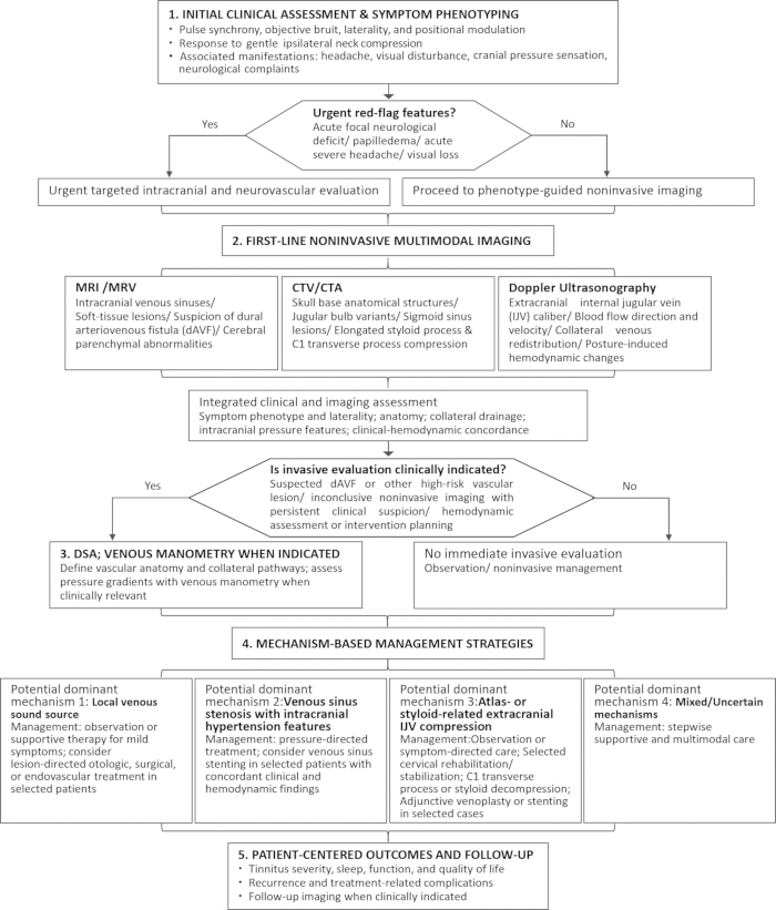
Figure 1: Proposed diagnostic workflow for venous pulsatile tinnitus and tinnitus-like symptoms associated with internal jugular venous outflow disturbance. Clinical evaluation begins with symptom phenotyping and assessment for red-flag features. MRI/MRV and CTV/CTA are used to evaluate intracranial venous abnormalities, local venous sound sources, skull-base variants, and cervical structural compression, while Doppler ultrasonography provides dynamic assessment of extracranial jugular venous flow. DSA is reserved for selected patients with suspected dural arteriovenous fistula, inconclusive non-invasive imaging, disabling symptoms, or planned intervention; venous manometry may be added when assessment of a pressure gradient is clinically relevant. Management is guided by the dominant mechanism, clinical and hemodynamic concordance, and individualized risk-benefit assessment. This proposed workflow is a mechanism-oriented clinical framework and does not replace individualized multidisciplinary evaluation. Abbreviations: CTA, computed tomography angiography; CTV, computed tomography venography; DSA, digital subtraction angiography; MRI, magnetic resonance imaging; MRV, magnetic resonance venography. Please click here to view a larger version of this figure.
| Management Strategy | Potential Indications | Advantages | Limitations and Risks | Evidence Gaps |
| Conservative management | Mild or intermittent symptoms; equivocal imaging findings; absence of clearly correctable structural lesions; patients with high procedural risks | Noninvasive; avoids procedural risks; permits serial assessment of symptoms and imaging findings | Fails to resolve structural venous obstruction; incomplete symptom relief; continued follow-up may be needed if symptoms or imaging findings change | Few prospective studies investigating natural disease history; no validated predictors for spontaneous improvement or deterioration |
| Intracranial pressure management | Clinical manifestations of intracranial hypertension (headache, papilledema, visual disturbances, elevated lumbar puncture opening pressure); venous sinus stenosis with pressure-related tinnitus | Targets pressure-induced exacerbation of tinnitus; may reduce pressure-related symptoms and tinnitus burden in selected patients | Not suitable for isolated mechanical venous compression; highly variable therapeutic response; long-term use may be limited by medication side effects | No unified criteria to identify pressure-mediated tinnitus; lack of standardized outcome assessment tools |
| Endovascular treatment | Severe, refractory symptoms with concordant clinical, anatomical, and hemodynamic findings; selected venous sinus stenosis or IJV stenosis after multidisciplinary evaluation | Directly restores venous patency; reduces trans-stenotic pressure gradients and blood flow turbulence; may provide rapid symptom relief in selected patients | Risks include thrombosis, restenosis, in-stent stenosis and vascular injury; requires periprocedural antithrombotic therapy; the regimen and duration vary by procedure and center; unstable long-term efficacy in patients with extrinsic venous compression | Absence of randomized controlled trials; inconsistent patient inclusion criteria across studies; limited long-term head-to-head comparative data |
| Surgical decompression | Styloidectomy, partial C1 transverse process resection, and release of adjacent compressive soft tissues in selected patients with concordant clinical, anatomical, and hemodynamic findings | Directly addresses fixed osseous or soft-tissue compression | Technically demanding; risks include neurovascular injury, incomplete decompression, postoperative scarring, and recurrent compression; comparative evidence is limited | Insufficient comparative trials versus endovascular monotherapy or combined treatment regimens; scarce standardized long-term follow-up datasets |
| Supportive tinnitus therapy | Persistent distress after vascular-targeted therapy; no clear interventional indications; mixed or undefined pathogenic mechanisms; severe insomnia, anxiety and markedly impaired quality of life | Noninvasive; may improve coping, sleep, and psychological well-being; can be combined with lesion-directed treatment | Does not treat underlying venous pathology; benefits vary and depend on adherence and access to appropriate services | Insufficient disease-specific research evidence focusing exclusively on populations with venous pulsatile tinnitus |
Table 1: Management strategies for venous pulsatile tinnitus or tinnitus-like symptoms associated with internal jugular venous outflow disturbance. Management should be individualized according to symptom phenotype, lesion laterality, anatomical findings, collateral drainage, intracranial pressure features, and hemodynamic concordance. The proposed indications are based mainly on observational evidence and should not be interpreted as validated treatment criteria. Abbreviations: C1, first cervical vertebra; IJV, internal jugular vein.
Disclosures
The authors have no conflicts of interest to disclose.
Acknowledgements
This work was supported by the 2023 Tianjin Municipal Health Commission Traditional Chinese Medicine and Integrated Chinese-Western Medicine Research Project (Grant No. 2023116).
References
- Alkhatib SG, et al. Imaging of pulsatile tinnitus. Radiographics. 2024;44:10.
- Narsinh KH, et al. Diagnostic approach to pulsatile tinnitus. JAMA Otolaryngol Head Neck Surg. 2022;148:476-483.
- Pacheco-López S, et al. Pulsatile tinnitus: a comprehensive clinical approach to diagnosis and management. J Clin Med. 2025;14:4428.
- Duvvuri M, Ali H, Amans MR. Non-invasive imaging modalities for diagnosing pulsatile tinnitus: a comprehensive review and recommended imaging algorithm. J Neurointerv Surg. 2025;13:916-924.
- Abdalkader M, et al. State of the art: venous causes of pulsatile tinnitus and diagnostic considerations guiding endovascular therapy. Radiology. 2021;300:2-16.
- Zhou D, et al. Clinical characteristics and neuroimaging findings in internal jugular venous outflow disturbance. Thromb Haemost. 2019;119:308-318.
- Zhou D, et al. Understanding jugular venous outflow disturbance. CNS Neurosci Ther. 2018;24:473-482.
- Valluru K, et al. Sound measurement in patient-specific 3D printed bench models of venous pulsatile tinnitus. Otol Neurotol. 2020;41:e7-e14.
- Haider HF, et al. Pathophysiology of subjective tinnitus: triggers and maintenance. Front Neurosci. 2018;12:866.
- Song J, et al. Objectification and differential diagnosis of vascular pulsatile tinnitus by transcanal sound recording and spectrotemporal analysis: a preliminary study. Otol Neurotol. 2016;37:613-620.
- Fulop GA, et al. Role of age-related alterations of the cerebral venous circulation in the pathogenesis of vascular cognitive impairment. Am J Physiol Heart Circ Physiol. 2019;316(5): H1124-H1140.
- Chung C, et al. Jugular venous hemodynamic changes with aging. Ultrasound Med Biol. 2010;36:1776-1782.
- Yin J, et al. Ultrasound evaluation of internal jugular venous insufficiency and its association with cognitive decline. Diagnostics (Basel). 2025;15:1427.
- Lv K, et al. Hemodynamic mechanisms in venous pulsatile tinnitus: a 4D flow MRI analysis of transverse-sigmoid sinus abnormalities. J Magn Reson Imaging. 2026;64:359-373.
- Amans MR, et al. MR venous flow in sigmoid sinus diverticulum. AJNR Am J Neuroradiol. 2018;39:2108-2113.
- Johnson JN, et al. The human craniospinal venous system and its influence on postural intracranial pressure: a review. J Neurosurg. 2024;141:1484-1493.
- Arun A, et al. A proposed framework for cerebral venous congestion. Neuroradiol J. 2022;35:94-111.
- Li H, et al. Cerebral venous outflow insufficiency: a study on symptoms and venous stenosis classification. MedComm (2020). 2026;7(3):e70609.
- Wang Z, et al. High jugular bulb in patients with non-thrombotic internal jugular venous and transverse sinus stenosis: clues to pathogenesis. CNS Neurosci Ther. 2024;30(3):e14424.
- Narsinh KH, et al. Management of vascular causes of pulsatile tinnitus. J Neurointerv Surg. 2022;14:1151-1157.
- Abdalkader M, et al. Cerebral venous disorders: diagnosis and endovascular management. J Neuroradiol. 2023;50:581-592.
- Succop BJ, et al. Non-invasive treatment of venous pulsatile tinnitus with an internal jugular vein compression collar. Laryngoscope. 2024;134:3342-3348.
- Greene PF. Internal jugular vein compression: a benign entity or an underappreciated phenomenon? eNeurologicalSci. 2025;40:100577.
- Peng X, et al. Surgical treatment for cognitive impairment caused by internal jugular vein stenosis: a clinical study of atlas transverse process resection. Front Neurol. 2026;17:1776658.
- Lu S, et al. Effectiveness of atlas transverse process resection combined with release and perfusion techniques in treating clinical symptoms caused by internal jugular vein stenosis secondary to external compression. World Neurosurg. 2025;202:124397.
- Ma H, et al. Case report: isolated surgical decompression for compressive internal jugular vein stenosis: case series and literature review. Front Surg. 2025;12:1639108.
- Mantovani G, et al. Styloid jugular nutcracker: the possible role of the styloid process spatial orientation—a preliminary morphometric computed study. Diagnostics (Basel). 2023;13:298.
- Mejia-Vergara AJ, et al. Styloidogenic jugular venous compression syndrome with papilloedema: case report and review of the literature. Neuroophthalmology. 2021;46:54-58.
- Scerrati A, et al. Styloidogenic-cervical spondylotic internal jugular venous compression, a vascular disease related to several clinical neurological manifestations: diagnosis and treatment—a comprehensive literature review. Ann Transl Med. 2021;9:718.
- Kuriyama N, et al. Retrograde jugular flow associated with idiopathic normal pressure hydrocephalus. Ann Neurol. 2008;64:217-221.
- Han K, et al. Transient global amnesia linked to impairment of brain venous drainage: an ultrasound investigation. Front Neurol. 2019;10:67.
- Kapadia A, Dmytriw AA. Venous dysfunction plays a critical role in “normal” white matter disease of aging. Med Hypotheses. 2021;146:110457.
- Adachi U, et al. Association of age-related spontaneous internal jugular vein reflux with cognitive impairment and incident dementia. J Alzheimers Dis. 2023;96:1221-1230.
- Amans M, et al. More than just noise: association of pulsatile tinnitus with anxiety, depression, and reduction of quality of life. Interv Neuroradiol. 2023;31:381-385.
- Pandey A, et al. Challenges in the use of venous sinus stenting in the treatment of idiopathic intracranial hypertension and pulsatile tinnitus. World Neurosurg. 2024;184:372-386.
- Su H, et al. Long-term outcomes of stenting for pulsatile tinnitus caused by cerebral venous sinus stenosis. Otol Neurotol. 2025;46:112-117.
- Cummins DD, et al. MRI and MR angiography evaluation of pulsatile tinnitus: a focused, physiology-based protocol. J Neuroimaging. 2021;32:253-263.
- Abdalkader M, et al. Differential assessment of internal jugular vein stenosis in patients undergoing CT and MRI with contrast. Tomography. 2024;10:266-276.
- Li X, et al. Effects of different morphologic abnormalities on hemodynamics in patients with venous pulsatile tinnitus: a four-dimensional flow magnetic resonance imaging study. J Magn Reson Imaging. 2021;53:1744-1751.
- Ding H, et al. Correlation between trans-stenotic blood flow velocity differences and the cerebral venous pressure gradient in transverse sinus stenosis: a prospective 4-dimensional flow magnetic resonance imaging study. Neurosurgery. 2021;89:549-556.
- Amukotuwa SA, et al. Arterial spin-labeling improves detection of intracranial dural arteriovenous fistulas with MRI. AJNR Am J Neuroradiol. 2018;39:669-677.
- Liu L, et al. Cerebral blood flow alterations in non-auditory tinnitus: implications for cerebral venous congestion pathophysiology. Brain Imaging Behav. 2026;20:72.
- Li X, et al. Altered cerebral blood flow in patients with unilateral venous pulsatile tinnitus: an arterial spin labeling study. Br J Radiol. 2021;94:20200990.
- Wang M, et al. Differentiation between anatomical slenderness and acquired stenosis of the internal jugular veins. CNS Neurosci Ther. 2022;28:1849-1860.
- Tudose RC, Rusu MC. Global prevalence of high jugular bulb and jugular bulb dehiscence: a systematic review and meta-analysis. Anat Sci Int. 2026;10:1007.
- Jain V, Markovic M, Agarwal M. Imaging of pulsatile tinnitus. Semin Roentgenol. 2024;59:203-214.
- Gao X, et al. Intracranial pressure, lateral sinus patency, and jugular ultrasound hemodynamics in patients with venous pulsatile tinnitus. Front Neurol. 2022;13:992416.
- Li M, et al. A hemodynamic screening tool for symptomatic internal jugular vein stenosis in J3 segment. Phlebology. 2025;40:581-589.
- Patsalides A, et al. Venous sinus stenting for the treatment of isolated pulsatile tinnitus: results of a prospective trial. Interv Neuroradiol. 2020;27:266-274.
- Midtlien JP, et al. Styloidectomy for jugular decompression in venous outflow disorders. World Neurosurg. 2025:124717.
- Abouelleil M, et al. Commentary: venous causes of pulsatile tinnitus: clinical presentation, clinical and radiographic evaluation, pathogenesis, and endovascular treatments: a literature review. Neurosurgery. 2022;90(3):e65-e66.
- Essibayi MA, Oushy SH, Lanzino G, Brinjikji W. Venous causes of pulsatile tinnitus: clinical presentation, clinical and radiographic evaluation, pathogenesis, and endovascular treatments: a literature review. Neurosurgery. 2021;89:760-768.
- Bai C, et al. Long-term safety and efficacy of stenting on correcting internal jugular vein and cerebral venous sinus stenosis. Ann Clin Transl Neurol. 2023;10:1305-1313.
- Bai C, et al. Pathogenesis and management in cerebrovenous outflow disorders. Aging Dis. 2021;12:203-222.
- Hauser RA, Matias D, Rawlings B. The ligamentous cervical instability etiology of human disease from the forward head-facedown lifestyle: emphasis on obstruction of fluid flow into and out of the brain. Front Neurol. 2024;15:1430390.
- Hauser RA, et al. Brain-based symptoms reported in young adults evaluated for cervical spine etiology: a retrospective, cross-sectional clinical study with preliminary data on over 200 patients. Front Neurol. 2026;17:1749266.
- Fargen KM, et al. Internal jugular vein stenting for symptomatic severe rotational stenosis. J Neurointerv Surg. 2026;2025:24659.
- Fargen KM, et al. The promise, mystery, and perils of stenting for symptomatic internal jugular vein stenosis: a case series. Neurosurgery. 2024;95:400-407.
- Ognard J, et al. Future is ven(o)us: a 5-year narrative update on the venous route for therapeutics in neurointervention. AJNR Am J Neuroradiol. 2026;47:313-321.
- Petersingham G, et al. Invasive surgical management of cervical internal jugular venous compression: a literature review. J Clin Neurosci. 2025;137:111304.
- Reddy A, et al. Flow analysis of central venous outflow tract: a new approach to understanding pulse-synchronous tinnitus. Otolaryngol Head Neck Surg. 2024;171:1485-1496.
- Hu L, Chen L. Editorial for “Hemodynamic mechanisms in venous pulsatile tinnitus: a 4D flow MRI analysis of transverse-sigmoid sinus abnormalities”. J Magn Reson Imaging. 2026;64:374-375.