This protocol describes a standardized artificial groove model for comparing five irrigant activation techniques, including a multisonic system and two erbium lasers, for removing a set of calcium silicate-based sealers during endodontic retreatment.
Research Article
Removal of Two Ready-to-Use Calcium Silicate-Based Sealers from Artificial Grooves Using a Multisonic System and Erbium Lasers: An In Vitro Study
In This Article
Summary
Abstract
Calcium silicate-based sealers bond to dentin and are difficult to remove during retreatment. This study compared five techniques for removing two ready-to-use calcium silicate-based sealers from artificial grooves. Grooves were cut in the apical and coronal thirds of 200 extracted maxillary central incisors and filled with CeraSeal (n = 100) or BioRoot Flow (n = 100). After 30 days, the sealer was removed using syringe needle irrigation, passive ultrasonic irrigation, a multisonic system (GentleWave G4), Er:YAG laser activation in R-SWEEPS mode, or Er,Cr:YSGG laser activation (n = 20/each group). Residual sealer was scored from 0 to 3 by two blinded observers. Scores were compared with the Kruskal-Wallis and Dunn tests, and the sealer and its interaction with the technique with the Scheirer-Ray-Hare test. The technique affected the residual score in both regions (p < 0.001), whereas no significant effect of the sealer or of the interaction was detected. GentleWave G4 produced the lowest scores and cleared the groove in up to 75% (15 of 20) of specimens, while syringe needle irrigation removed no sealer from any groove. The lasers and passive ultrasonic irrigation were intermediate, and passive ultrasonic irrigation did not differ from the needle for BioRoot Flow. Under the tested conditions, the removal of calcium silicate-based sealer from artificial grooves depended on the technique, whereas no statistically significant effect of the sealer was detected. No technique achieved complete removal.
Introduction
Calcium silicate-based sealers are widely used with the single-cone technique because they are biocompatible, stimulate biomineralization, and bond chemically to dentin1,2. The same chemistry complicates their removal. These materials react with dentinal tubule fluid and precipitate hydroxyapatite, forming a mineral infiltration zone and cement plugs within the tubules3,4, and, after setting, their hardness resists cutting instruments3. Retrievability reflects the physical strength gained upon setting, which depends on composition5,6,7. Despite these clinical advantages, complete removal during retreatment has not been achieved8,9.
Nickel-titanium retreatment systems reach the working length in most canals but leave measurable residue, whatever the file system or kinematics10,11,12, and retreatment without a solvent has produced the highest patency rates for these sealers13,14. Patency is not cleanliness. Bardini et al.9 regained the working length in every specimen, yet removed none of the sealer completely. Residue persists where instruments cannot reach because files cut a circular path and leave the eccentric extensions of the canal untouched5,15. Artificial grooves isolate this instrument-inaccessible compartment and allow irrigation methods to be compared directly8,16,17. Supplementary activation improves removal18, yet passive ultrasonic irrigation loses efficacy apically, where tip-wall contact dampens the oscillation, entrapped gas impedes cavitation, and the tip cannot reach the apical zone8,19,20.
Two technologies address these limits differently. The multisonic system (GentleWave G4) delivers broadband acoustic energy through a fluid platform that fills the entire canal system and has been tested during retreatment21,22. Erbium lasers act through laser-activated irrigation, in which the energy absorbed by the irrigant generates vapor bubbles whose growth and collapse produce photoacoustic streaming and shock waves23,24. Wavelength governs this mechanism. A 980 nm diode improved sealer removal over manual irrigation but produced no surfactant-enhanced cavitation, which the authors attributed to the need for a mid-infrared wavelength23. Er:YAG (2940 nm) and Er,Cr:YSGG (2780 nm) meet that requirement. Both wavelengths are strongly absorbed by water, so the irrigant itself converts the emitted light into the mechanical energy that drives the streaming, and the pulse energy, pulse duration, and tip geometry determine the resulting cavitation24. The two lasers were used at the low pulse energies intended for irrigant activation rather than for tissue ablation, but in different modes. In resonant shock wave enhanced emission photoacoustic streaming (R-SWEEPS), the Er:YAG laser delivers pairs of synchronized pulses timed to the oscillation of the first bubble, so that the second pulse accelerates its collapse and amplifies the emitted shock wave25, with the tip held at the canal orifice; the Er,Cr:YSGG laser activates the irrigant with single pulses delivered through a radial firing tip placed inside the canal.
Evidence for these energy-based methods in the groove model is thin. The closest study kept passive ultrasonic irrigation constant across all specimens, with neither a non-activated control nor a laser or multisonic arm26. Our group previously compared the same five techniques for the debridement of necrotic tissue and debris from minimally instrumented oval canals, where the two erbium lasers approached the multisonic system27; that study addressed loosely attached soft tissue in an instrumented canal, not a set sealer bonded to the dentin of a recess that no instrument reaches, and it did not include a material variable. BioRoot Flow, a ready-to-use formulation, has not been examined during retreatment, and published data cover only its powder-liquid predecessor12,15. Whether the most effective technique depends on the sealer also remains open, because the one study reporting such a difference never tested the interaction formally5.
The purpose of the present study was therefore to determine whether two energy-based activation approaches that bypass the apical limitations of ultrasonic activation, GentleWave G4 and erbium laser activation, remove set calcium silicate-based sealer from instrument-inaccessible sites, and whether their effect depends on the sealer formulation. To this end, syringe needle irrigation (SNI, the non-activated control), passive ultrasonic irrigation (PUI), GentleWave G4, R-SWEEPS, and Er,Cr:YSGG were compared for removing two ready-to-use calcium silicate-based sealers from artificial grooves in the apical and coronal thirds. The null hypotheses were that the technique would not affect the residual sealer, that the two sealers would not differ, and that the technique's effect would not depend on the sealer. This protocol is designed as a laboratory tool to isolate the energy transmitted through the irrigant from the cutting action of instruments; it is suited for comparing activation systems under standardized conditions and is not intended to reproduce the full canal anatomy or the material composition of a clinical retreatment case.
Protocol
This laboratory study was approved by the institutional ethics committee of Kocaeli Health and Technology University (approval number 31/22) and was conducted in accordance with the ethical principles outlined in the Declaration of Helsinki and the PRILE 2021 guidelines28 (Supplementary File 1 and Supplementary File 2). Informed consent for the use of extracted teeth was obtained from the donors. The specimen model, groove preparation, and scoring followed a described protocol8. All materials, instruments, and software used in this protocol are listed in the Table of Materials.
Sample size calculation
The sample size was based on a previous investigation that used the same artificial groove model and scoring system26. In that study, an a priori power analysis (effect size = 0.6, α = 0.05, power = 0.95) indicated n = 12/each group, which the authors increased to 15 to compensate for possible losses. To increase precision, the present study used n = 20/each group, giving 400 grooves in total. This calculation addressed the main effect of the removal technique; no separate a priori calculation was made for the technique × sealer interaction. A post hoc sensitivity analysis based on the chi-square distribution of the Scheirer-Ray-Hare statistic (statsmodels 0.14.6) showed that the 200 grooves analyzed in each region gave 80% power (α = 0.05) to detect an interaction of effect size w = 0.24 (4 degrees of freedom) and a sealer main effect of w = 0.20 (1 degree of freedom), so that effects smaller than these, below Cohen's medium effect size of w = 0.3, may have escaped detection.
Sample selection criteria
Two hundred extracted human maxillary central incisors were used. After extraction, the teeth were disinfected in 2% NaOCl, rinsed with distilled water, and stored in thymol solution until use. Teeth with a straight single root canal and an intact apex were included, and a single canal was confirmed radiographically. Teeth with curvature, calcification, previous treatment, resorption, or an initial apical size of #30 or larger were excluded. The crowns were sectioned to a standard length, with a working length of 18 mm. Randomization was performed in two stages with a web-based random sequence generator (random.org): the 200 teeth were first allocated to the two sealers (n = 100 each), and the teeth of each sealer were then allocated to the five removal techniques (n = 20/each group), so that allocation was stratified by sealer. The random sequences were generated and applied by an investigator who did not perform scoring, and the specimens were labeled with coded identifiers to keep observers blinded to both the sealer and the removal technique.
CAUTION: Extracted human teeth represent a potential biohazard. Handle all specimens with appropriate personal protective equipment, including gloves, a mask, and protective eyewear.
Sample preparation
After patency was confirmed with an ISO size 10 K-file, the canals were enlarged to size 40/0.04 at the working length using a rotary endodontic instrumentation system under irrigation with 3% sodium hypochlorite (NaOCl), followed by a rinse of 5 mL of 3% NaOCl and 5 mL of 17% ethylenediaminetetraacetic acid (EDTA). All NaOCl solutions were prepared from a 5.25% stock solution by dilution with distilled water: 3% NaOCl by mixing 5.25% NaOCl and distilled water at a volume ratio of 4:3 (for example, 100 mL of 5.25% NaOCl and 75 mL of distilled water), and 5% NaOCl at a volume ratio of 20:1 (for example, 100 mL of 5.25% NaOCl and 5 mL of distilled water).
CAUTION: Sodium hypochlorite is caustic to the skin, eyes, and mucous membranes. Prepare and handle all solutions with gloves and protective eyewear, and avoid extrusion beyond the apical foramen.
The roots were embedded in impression material within centrifuge tubes and split longitudinally with a diamond disc, without penetrating the canal space. Any root in which the canal space was penetrated during splitting was to be excluded and replaced by a tooth prepared in the same way; no root met this criterion in the present series. Two artificial grooves, 4 mm long, 0.2 mm wide, and 0.5 mm deep, were cut into the canal wall with a prophylaxis scaling tip mounted on a piezoelectric ultrasonic endodontic unit at 40% power, under a dental operating microscope at ×20 magnification. The grooves were prepared on the opposite canal walls, at 2 to 6 mm and 10 to 14 mm from the apex (Figure 1). After groove preparation, the smear layer was removed using 2 mL of 17% EDTA.
The grooves of 100 teeth were filled with CeraSeal, and those of the remaining 100 teeth with BioRoot Flow using gutta-percha points. Complete filling of the grooves was confirmed under the dental operating microscope at ×20 magnification. The root halves were reassembled within the Eppendorf tubes, the access cavity was sealed with a temporary filling material, the apical foramen and the root surfaces were covered with wax, and the specimens were kept at 37 °C and 100% humidity for 30 days. Setting of the sealer was then verified under the dental operating microscope at ×20 magnification.
Removal procedures
Specimens for each sealer were assigned to five removal techniques (n = 20 per group), with SNI serving as the non-activated control. The SNI, PUI, R-SWEEPS, and Er,Cr:YSGG groups shared one irrigation regimen of six delivery-activation cycles, three with 5% NaOCl followed by three with 17% EDTA. In each cycle, 5 mL of irrigant was delivered over 150 s at 2 mL/min with an irrigation pump, and the irrigant filling the canal was then activated for 20 s with the technique under test. The three NaOCl cycles were completed before the three EDTA cycles, so that each specimen received 15 mL of 5% NaOCl and 15 mL of 17% EDTA (30 mL in total) and six 20 s activations (three per irrigant, 120 s in total). The treatment time in the three activated groups was therefore approximately 17 min (6 × 150 s of delivery plus 6 × 20 s of activation).
In the SNI group, the irrigants were delivered through a 30-G needle placed 2 mm short of the working length without activation; this group received the same volumes, flow rate, and irrigant sequence as the activated groups, but no waiting interval was interposed between deliveries in place of the activation periods, so that its total exposure time was approximately 15 min (6 × 150 s).
In the PUI group, the irrigant was activated using an IrriSafe size 20 tip mounted on the piezoelectric ultrasonic endodontic unit at 40% power, with the tip positioned 2 mm short of the working length, for three 20 s cycles per irrigant.
In the R-SWEEPS group, the irrigant was activated with a 2940 nm Er:YAG laser in R-SWEEPS mode using a radial tip (400 µm diameter, 14 mm length) held at the canal orifice without entering the canal, for three 20 s cycles per irrigant with the air and water spray switched off. In this mode, each 15 mJ pulse is delivered as one of a synchronized pair at a pulse-pair repetition rate of 15 Hz (30 mJ per pair), giving an average power of 0.45 W, 9 J per 20 s activation, and 54 J per specimen; the pulse energy corresponds to an energy density of 11.9 J/cm2 at the 400 µm tip end face.
In the Er,Cr:YSGG group, the irrigant was activated with a 2780 nm Er,Cr:YSGG laser in H mode (25 mJ per pulse, 50 Hz, 1.25 W; 25 J per 20 s activation and 150 J per specimen) using an RFT2 radial firing tip held 2 mm short of the working length, for three 20 s cycles per irrigant with the air and water spray switched off. In both laser groups, the canal was filled with the irrigant delivered by the pump before the laser was activated, and the parameters were the manufacturers' preset settings for endodontic irrigant activation, applied without modification. Table 1 gives the complete irradiation parameters, including the energy per pulse, the energy density, and the total energy delivered.
In the GentleWave G4 group, the reassembled specimen was kept in the centrifuge tube, a platform was built over the coronal end of the root with the manufacturer's SoundSeal resin to create an airtight seal between the endodontic irrigation instrument and the canal opening, and the automated procedure cycle of the GentleWave G4 system with ProControl software was run with the manufacturer's procedure fluids in the following sequence: 3% NaOCl for 5 min, distilled water for 30 s, 8% EDTA for 2 min, and a final distilled water rinse for 30 s (approximately 8 min in total). The procedure instrument was held in place by the platform for the entire cycle. The irrigant concentrations, flow rates, degassing, and cycle times are preset by the manufacturer and cannot be modified by the operator, so this group differed from the other four groups in irrigant concentration, volume, and treatment time (see Discussion section).
Assessment of residual sealer
The root halves were separated and examined under the dental operating microscope at ×20 magnification. Residual sealer in each groove was scored on a four-point scale29: 0, no sealer in the groove; 1, sealer in less than half; 2, sealer in more than half; and 3, the groove entirely filled. Two calibrated observers, blinded to the sealer and the removal technique, independently scored the grooves; interobserver agreement was κ = 0.92 (Cohen's kappa). Disagreements were resolved by consensus.
Statistical analysis
Data were analyzed using Python 3.9.6 with the SciPy 1.13.1, statsmodels 0.14.6, and scikit-posthocs 0.14.0 libraries. Apical and coronal grooves were analyzed separately. Residual sealer scores across the 10 groups were compared using the Kruskal-Wallis test, followed by the Dunn test with Bonferroni correction. Epsilon-squared was calculated as the effect size. The effects of sealer and removal technique, together with their interaction, were examined using the Scheirer-Ray-Hare test. Within each removal technique, the two sealers were compared using the Mann-Whitney U test with Bonferroni correction, and Cliff's delta with bootstrapped 95% confidence intervals was reported as the effect size. The significance level was set at 5%.
Results
Assessment of residual sealer
A total of 400 grooves were scored, 200 apical and 200 coronal, and no specimens were lost during the procedures.
Residual sealer scores differed among the 10 groups in both regions (p < 0.001; Table 2). SNI left every groove completely filled. Its median score was 3.0 for both sealers in both regions, and none of the 80 grooves irrigated with a needle alone reached a score of 0. GentleWave G4 produced the lowest medians, 0.5 for CeraSeal and 1.0 for BioRoot Flow in the apical region, and 0.0 for both sealers in the coronal region. Complete removal after GentleWave G4 was recorded in 50% (10 of 20) of apical and 75% (15 of 20) of coronal CeraSeal grooves, and in 45% (9 of 20) and 60% (12 of 20) of the corresponding BioRoot Flow grooves. PUI, R-SWEEPS, and Er,Cr:YSGG gave medians between 1.0 and 3.0 and complete-removal rates between 10% (2 of 20) and 35% (7 of 20).
Comparisons among removal techniques
GentleWave G4 differed from SNI for both sealers in both regions (p < 0.001) and also yielded lower scores than PUI with BioRoot Flow in both regions (p < 0.001 apically, p = 0.004 coronally; Table 2). R-SWEEPS and Er,Cr:YSGG differed from SNI with CeraSeal in both regions (p = 0.020 and p = 0.004 apically, p = 0.001 and p = 0.011 coronally), whereas with BioRoot Flow, only the apical Er,Cr:YSGG comparison reached significance (p = 0.028). PUI did not differ from SNI with BioRoot Flow in either region, or from SNI with CeraSeal in the coronal region (Table 2). Bonferroni-adjusted p-values for all 45 pairwise comparisons in each region are listed in Supplementary File 3.
Effect of sealer type and technique-sealer interaction
No statistically significant effect of the sealer was detected in either region, although the coronal comparison approached the threshold (p = 0.065), and the interaction between sealer and removal technique was not significant (Table 2). Within each technique, the two sealers did not differ significantly after correction for multiple comparisons (p > 0.05; Supplementary File 3).
Residual sealer persisted in at least one groove in every group. The highest proportion of grooves free of sealer was 75% (15 of 20), recorded for CeraSeal treated with GentleWave G4 in the coronal region, and the lowest was zero for both sealers irrigated with a needle alone (Figure 2).
These findings indicate that, under the tested conditions, the amount of sealer left in an instrument-inaccessible groove depended on the removal technique; no statistically significant effect of the sealer formulation was detected; and none of the five techniques emptied every groove. Because the GentleWave G4 group also differed from the other groups in irrigant concentration, treatment duration, and regimen, the between-technique differences cannot be attributed solely to activation energy.
DATA AVAILABILITY:
The raw data supporting the findings of this study are available in Supplementary File 4.
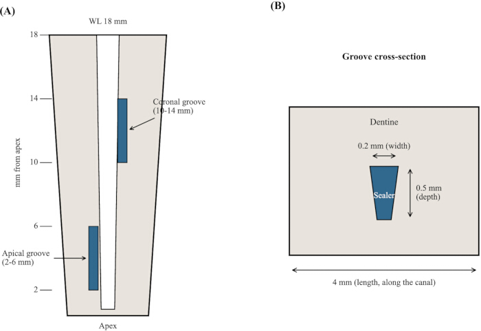
Figure 1: Schematic of the artificial groove model. (A) One apical groove (2–6 mm from the apex) and one coronal groove (10–14 mm from the apex) were cut on opposite canal walls of a root prepared to a working length of 18 mm. (B) Cross-section of a groove measuring 4 mm long, 0.2 mm wide, and 0.5 mm deep, filled with sealer. Please click here to view a larger version of this figure.
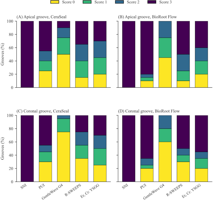
Figure 2: Distribution of residual sealer scores in the apical (A,B) and coronal (C,D) grooves for CeraSeal (A,C) and BioRoot Flow (B,D) after removal by five techniques. Scores range from 0 (no sealer in the groove) to 3 (groove entirely filled). n = 20 per group. Abbreviations: PUI = passive ultrasonic irrigation; SNI = syringe needle irrigation. Please click here to view a larger version of this figure.
| Parameter | R-SWEEPS | Er,Cr:YSGG |
| Laser type | Er:YAG | Er,Cr:YSGG |
| Wavelength (nm) | 2940 | 2780 |
| Device | LightWalker AT-S | Waterlase iPlus |
| Mode | R-SWEEPS (pulsed, synchronized paired pulses) | H mode (pulsed, single pulses) |
| Energy per pulse (mJ) | 15 | 25 |
| Energy per pulse pair (mJ) | 30 | Not applicable (single pulses) |
| Repetition rate (Hz) | 15 (pulse pairs) | 50 |
| Average power (W) | 0.45 | 1.25 |
| Tip | Radial, 400 µm diameter, 14 mm length | RFT2 radial firing tip |
| Energy density at the tip end face (J/cm²) | 11.9 | Not defined (lateral emission through the radial firing tip) |
| Tip position | Canal orifice, not entering the canal | 2 mm short of the working length |
| Air/water spray | Off/off | Off/off |
| Activation | 3 × 20 s per irrigant (6 × 20 s per specimen) | 3 × 20 s per irrigant (6 × 20 s per specimen) |
| Energy per 20 s activation (J) | 9 | 25 |
| Total energy per specimen (J) | 54 | 150 |
Table 1: Laser irradiation parameters used in the R-SWEEPS and Er,Cr:YSGG groups. Values are the manufacturers' preset parameters for irrigant activation and were applied without modification. In R-SWEEPS mode, each 15 mJ pulse is delivered as one of a synchronized pair, so that the 15 Hz repetition rate of pulse pairs gives an average power of 0.45 W (2 × 15 mJ × 15 Hz). Energy density is the pulse energy divided by the cross-sectional area of the tip end face; it is given for the 400 µm tip only, because the radial firing tip emits laterally and its end-face value is not defined. Total energy is the average power multiplied by the total activation time (6 × 20 s per specimen). The pulse duration is fixed by the selected mode of each device and was not modified. In both laser groups, the irrigant was delivered separately with the irrigation pump, and the laser was applied with the air and water spray switched off. Abbreviations: R-SWEEPS = resonant shock wave enhanced emission photoacoustic streaming.
| Technique | Sealer | Apical median (IQR) | Apical score 0, n (%) | Coronal median (IQR) | Coronal score 0, n (%) |
| SNI | CeraSeal | 3.0 (3.0-3.0)c | 0 (0) | 3.0 (3.0-3.0)d | 0 (0) |
| BioRoot Flow | 3.0 (3.0-3.0)c | 0 (0) | 3.0 (3.0-3.0)d | 0 (0) | |
| PUI | CeraSeal | 2.0 (0.8-3.0)ab | 5 (25) | 2.0 (0.0-3.0)bcd | 6 (30) |
| BioRoot Flow | 3.0 (3.0-3.0)bc | 2 (10) | 3.0 (1.8-3.0)cd | 4 (20) | |
| GentleWave G4 | CeraSeal | 0.5 (0.0-1.2)a | 10 (50) | 0.0 (0.0-0.2)a | 15 (75) |
| BioRoot Flow | 1.0 (0.0-1.2)a | 9 (45) | 0.0 (0.0-1.0)ab | 12 (60) | |
| R-SWEEPS | CeraSeal | 2.0 (1.0-3.0)ab | 3 (15) | 1.0 (0.0-2.2)abc | 7 (35) |
| BioRoot Flow | 2.5 (1.8-3.0)bc | 2 (10) | 2.5 (0.0-3.0)bcd | 6 (30) | |
| Er,Cr:YSGG | CeraSeal | 2.0 (1.0-3.0)ab | 4 (20) | 1.5 (0.8-3.0)abc | 5 (25) |
| BioRoot Flow | 2.0 (1.0-3.0)ab | 4 (20) | 3.0 (1.0-3.0)cd | 4 (20) |
Table 2: Residual sealer scores in the apical and coronal grooves after removal with five techniques. Scores range from 0 (no sealer in the groove) to 3 (groove entirely filled); score 0 denotes complete removal. n = 20/each group. Scores were compared with the Kruskal-Wallis test followed by the Dunn test with Bonferroni correction, separately for each region. Apical: H = 80.66, p < 0.001, epsilon-squared = 0.377. Coronal: H = 84.80, p < 0.001, epsilon-squared = 0.399. The sealer effect and the interaction between sealer and technique were tested by the Scheirer-Ray-Hare extension of the Kruskal-Wallis test and were not significant in either region (sealer: p = 0.147 apical, p = 0.065 coronal; interaction: p = 0.478 apical, p = 0.890 coronal). Identical superscript letters indicate no significant difference among groups within a region; different letters indicate a significant difference. Exact adjusted p-values for all pairwise comparisons are given in Supplementary File 3. Abbreviations: IQR = interquartile range; PUI = passive ultrasonic irrigation; SNI = syringe needle irrigation.
Supplementary File 1: PRILE 2021 checklist for the present study. Please click here to download this file.
Supplementary File 2: PRILE 2021 flowchart summarizing the experimental workflow. Please click here to download this file.
Supplementary File 3: Bonferroni-adjusted p-values. Bonferroni-adjusted p-values of the Dunn test for all 45 pairwise comparisons among the 10 groups in each region, and Mann-Whitney U comparisons between the two sealers within each technique with Cliff's delta and bootstrapped 95% confidence intervals. Please click here to download this file.
Supplementary File 4: Raw residual sealer scores for all 400 grooves. Please click here to download this file.
Discussion
The removal technique determined how much sealer remained, whereas no statistically significant effect of the sealer or of its interaction with the technique was detected, so the first null hypothesis was rejected and the other two were retained. GentleWave G4 left the least residual sealer and cleared the groove completely in up to 75% of specimens, whereas SNI removed none. The two lasers and PUI were intermediate, and their advantage over the needle varied by region and sealer.
The artificial groove was chosen because it isolates the variable of interest. A groove of fixed dimensions holds a reproducible volume of set sealer in a site that no instrument contacts, so the score reflects the energy transmitted through the irrigant rather than any cutting action8,16. Several steps in this protocol are critical for that isolation to hold, and each has a corresponding corrective action. First, the groove dimensions must be reproduced exactly; a groove cut deeper or wider than specified holds a different volume of sealer, and the scores are no longer comparable across specimens. The groove was prepared under a dental operating microscope at ×20 magnification, and the exclusion of any root in which the canal space was penetrated during splitting was specified in advance in the protocol. Second, the grooves must be completely filled before setting; a void left at placement is scored as removed material, which is why complete filling was verified under the dental operating microscope before the halves were reassembled. Third, the sealer must be fully set before removal begins, since a partially set sealer is displaced more easily; therefore, setting was confirmed under the dental operating microscope after the 30-day storage period. Fourth, the tip position and spray setting govern the energy that actually reaches the groove, so the laser and ultrasonic tips were placed at the depths specified above, and the air and water sprays were switched off, with the irrigant supplied separately by the pump. Lastly, scoring is the step most susceptible to observer drift; two observers were calibrated before scoring, remained blinded to the groups, and any disagreements were resolved by consensus, with an interobserver agreement of κ = 0.92. This agreement addresses the subjectivity that limits split-tooth assessment3. The sealers were placed without a master cone, so that the residual material was sealer alone and not gutta-percha26.
That no technique empties every groove aligns with the literature. Complete removal of these sealers has not been achieved with needle irrigation, ultrasonic activation, or rotary retreatment1,8,9, and residue concentrates apically, where access and fluid exchange are poorest1,20. The behavior of SNI was the clearest result. Delivered without activation, it left every groove full, confirming that irrigant volume and contact time alone (30 mL over 15 min) cannot displace set sealer from an uninstrumented recess and that some form of energy is required8,18.
GentleWave G4 separated from SNI for both sealers and both regions, and it also scored below PUI for BioRoot Flow. Its fluid platform circulates degassed irrigant through the entire canal and generates broadband acoustic energy that tip-to-wall contact does not dampen, the mechanism that weakens PUI apically19,21,22. The lasers acted via photoacoustic streaming and reduced the scores relative to the needle, mainly for CeraSeal; the effect on BioRoot Flow reached significance only apically with Er,Cr:YSGG. PUI did not differ from SNI for BioRoot Flow in either region, consistent with reports that ultrasonic activation is not universally effective against these sealers5. In our previous histological study of necrotic oval canals, the same two lasers, including GentleWave G4, were used to remove tissue remnants27. The ranking of the techniques was the same in the present study, with GentleWave G4 first, the needle last, and the lasers between them, but the gap between the lasers and GentleWave G4 was wider. The substrate explains the difference: necrotic tissue and debris are loosely attached and lie in an instrumented canal, whereas a set calcium silicate-based sealer is bonded to the dentin of a 0.5 mm deep recess that the streaming must enter, so the sustained, whole-canal fluid exchange of GentleWave G4 gains an advantage over the pulsed, tip-centered streaming of the lasers. The outcome measures also differ: a histomorphometric percentage in the previous study and an ordinal score here, so the two studies cannot be compared numerically.
No statistically significant difference between the two sealers was detected within any technique, and the technique that yielded the lowest residual scores did not vary with sealer. This contrasts with Peña-Bengoa et al.5, who observed that the most effective supplementary technique differed between two sealers, although they never tested the interaction formally. CeraSeal and BioRoot Flow are both tricalcium silicate-based2,7, and retrievability has been related to the physical strength a material gains on setting, which follows its composition5,6. Set strength was not measured in the present study, so the comparable scores cannot be attributed to a similar set strength; they indicate only that, under the tested conditions, any difference in retrievability between the two formulations was too small to be detected with 20 grooves per group, and the coronal comparison (p = 0.065) suggests that BioRoot Flow may leave slightly more residue. CeraSeal has ranked poorly elsewhere2, and a larger sample or a direct measurement of the set material would be needed to settle the question.
This study was conducted in vitro on single-rooted teeth, and its scores describe an artificial groove rather than a full canal, so the findings cannot be transferred directly to clinical retreatment. A groove is a flat-walled recess of fixed geometry with direct fluid access from the main canal, whereas the isthmuses, fins, and lateral canals of a natural root are narrower, longer, and less predictably connected to the main canal; the groove therefore, probably overestimates what any technique achieves clinically, and the ranking of the techniques, not the absolute removal rate, is the transferable result. Clinically, sealer retained in such recesses may shelter residual microorganisms from disinfection and limit the contact of the new irrigant and sealer with dentin, so a technique that empties the groove more often, as GentleWave G4 did here, may improve the cleanliness of the canal system during retreatment, although the effect of these differences on periapical healing remains to be tested. The grooves held sealer alone, without gutta-percha, and were assessed in two dimensions with an ordinal score; this score is the established outcome for the model8,16,26,29 and its reliability was supported by the observer agreement, but scanning electron microscopy would resolve the sealer-dentin interface at higher magnification, and micro-computed tomography would quantify the residual volume in three dimensions. The laser parameters were the manufacturers' presets, and the pulse duration, pulse energy, and repetition rate were not varied, although these settings, together with the tip design and position, influence cavitation and the outcome of laser-activated irrigation24,30. The main constraint concerns the GentleWave G4 arm. Its protocol is fixed by the manufacturer and uses 3% NaOCl and 8% EDTA for about 8 min, whereas the other groups receive 5% NaOCl and 17% EDTA for about 17 min (15 min in the SNI group, which had no activation intervals). GentleWave G4 achieved the lowest scores despite the lower concentration and shorter duration, so the direction of the confounder is conservative; however, the device and its irrigant regimen cannot be separated in this design, and the between-technique differences cannot be attributed to the activation energy alone. Future studies should equalize the irrigant and exposure time across systems and confirm these findings in fully obturated canals.
The protocol can be modified within limits. The sealer, the storage period before removal, the irrigant and its concentration, the activation device and its parameters, can be changed to address other questions, provided that they are held constant across the groups being compared. The GentleWave G4 arm illustrates the constraint: its irrigant regimen is fixed by the manufacturer, so any comparison with other techniques introduces differences in irrigant and duration that must be declared. Three elements should not be modified, because they define the model: the groove dimensions (4 × 0.2 × 0.5 mm) and positions, which fix the volume of sealer and its distance from the tip8,16; the verification of complete filling and of setting before removal; and the blinded scoring of the groove by two calibrated observers with the four-point scale29. If the grooves are filled using a master cone or a solvent is added, the score no longer isolates the sealer from other variables, and the experiment should be reported as a retreatment model rather than under this protocol. Troubleshooting mainly concerns the assembly: the impression material in the tube and the wax seal prevent leakage at the reassembled split during activation, and a specimen that leaks should be resealed before treatment rather than excluded. Beyond the two directions named above, the artificial-groove protocol can be used to screen new sealer formulations and solvents or chelating agents before full retreatment studies, to compare laser settings (pulse energy, pulse duration, repetition rate, tip design, and tip position) or ultrasonic tips under identical irrigant conditions, to test the removal of intracanal medicaments such as calcium hydroxide16, to combine the ordinal score with micro-computed tomographic or scanning electron microscopic assessment of the same grooves, and to model other instrument-inaccessible sites, such as isthmuses and lateral canals, by changing the groove geometry.
Conclusion
Within the limitations of this in vitro study, the removal of calcium silicate-based sealer from artificial grooves depended on the technique, whereas no statistically significant effect of the sealer was detected under the tested conditions. GentleWave G4 removed the most sealer, SNI removed the least, and the lasers and PUI fell between them. Because the GentleWave G4 regimen also differed in irrigant and duration, its advantage cannot be attributed to activation energy alone. No technique achieved complete sealer removal in all specimens, so retreatment of these materials remains incomplete regardless of the method chosen.
Disclosures
The authors deny any conflicts of interest related to this study.
AUTHOR CONTRIBUTIONS:
Taha Özyürek contributed to conceptualization, methodology, supervision, and review and editing of the manuscript. Mustafa Gündoğar contributed to methodology, investigation, and review and editing of the manuscript. Olcay Özdemir contributed to investigation and formal analysis. Ali Aminimofrad contributed to investigation and data curation. Hande Özyürek contributed to formal analysis and writing of the original draft.
Acknowledgements
This research did not receive any specific grant from funding agencies in the public, commercial, or not-for-profit sectors.
Materials
| Name | Company | Catalog Number | Comments |
|---|---|---|---|
| BioRoot Flow | Septodont, Saint-Maur-des-Fossés, France | 340S/0.5 | Ready-to-use calcium silicate-based sealer |
| CeraSeal | Meta Biomed, Cheongju, Republic of Korea | Lot 230521-01 | Ready-to-use calcium silicate-based sealer |
| Chloraxid 5.25% | Cerkamed, Stalowa Wola, Poland | Lot 1105261 | Sodium hypochlorite solution, diluted to 3% and 5% |
| CleanFlow procedure instrument | Sonendo Inc., Laguna Hills, CA, USA | GW-PST-PI10 | Single-use procedure instrument of the GentleWave G4 system |
| Dental operating microscope OMS 3200 R2 | Zumax, Suzhou, China | Not applicable | Groove preparation, verification of complete filling and of sealer setting, and scoring of residual sealer, all at ×20 magnification |
| EndoSolution | Cerkamed, Stalowa Wola, Poland | Lot 2105261 | 17% EDTA irrigant |
| GentleWave G4 | Sonendo Inc., Laguna Hills, CA, USA | 10053 | Multisonic ultracleaning system |
| IrriSafe size 20 tip | Satelec, Acteon Group, Merignac, France | F43806 | Passive ultrasonic irrigation tip |
| LightWalker AT-S | Fotona, Ljubljana, Slovenia | Not applicable | 2940 nm Er:YAG laser, R-SWEEPS mode |
| Needle, 30-gauge | Ultradent Products Inc., South Jordan, UT, USA | Not available | Syringe needle irrigation |
| Newtron P5 XS B.LED ultrasonic unit | Satelec, Acteon Group, Merignac, France | Not applicable | Ultrasonic generator for groove preparation and passive ultrasonic irrigation |
| Prophylaxis scaling tip (Newtron) | Satelec, Acteon Group, Merignac, France | Not available | Preparation of the artificial grooves (40% power) |
| Python 3.9.6 | Python Software Foundation, Beaverton, OR, USA | Not applicable | Statistical analysis |
| Radial tip, 400 µm × 14 mm | Fotona, Ljubljana, Slovenia | Lot 003029 | Er:YAG laser delivery tip |
| Random sequence generator | random.org | Not applicable | Web-based allocation of specimens to groups |
| RFT2 radial firing tip | BIOLASE Inc., Foothill Ranch, CA, USA | Lot 2217Q | Er,Cr:YSGG laser delivery tip |
| scikit-posthocs 0.14.0 | Open-source Python library | Not applicable | Dunn test with Bonferroni correction |
| SciPy 1.13.1 | Open-source Python library | Not applicable | Kruskal-Wallis and Mann-Whitney U tests |
| SoundSeal | Sonendo Inc., Laguna Hills, CA, USA | GW-SYS-SOSE | Resin used to build the platform that seals the procedure instrument to the specimen |
| statsmodels 0.14.6 | Open-source Python library | Not applicable | Scheirer-Ray-Hare test |
| VDW.ROTATE | VDW, Munich, Germany | MSTVROT325AST | Rotary nickel-titanium system, size 40/.04 |
| Waterlase iPlus | BIOLASE Inc., Foothill Ranch, CA, USA | Not applicable | 2780 nm Er,Cr:YSGG laser, H mode |
References
- Farrayeh A, et al. Effectiveness of two endodontic instruments in calcium silicate-based sealer retreatment. Bioengineering (Basel). 2023;10(3):362.
- Shomali D, et al. Ex vivo comparative analysis of retrievability among four calcium silicate-based sealers for regaining apical patency. Restor Dent Endod. 2026;51(1):e3.
- Baranwal HC, et al. Comparative evaluation of retreatability of bioceramic sealer (BioRoot RCS) and epoxy resin (AH Plus) sealer with two different retreatment files: an in vitro study. J Conserv Dent. 2021;24(1):88-93.
- Bhandare RS, Mattigatti SB. Cone-beam computed tomography analysis of retreatability of resin and bio-ceramic sealer for root canal treatment: an in vitro study. Cureus. 2024;16(12):e75914.
- Peña-Bengoa F, et al. Evaluation of the retreatability and different supplementary cleaning techniques for removing bioceramic sealers in oval-shaped canals. Aust Endod J. 2025;51(3):723-731.
- Stiklaru I, et al. The ability of different compositions of calcium silicate and epoxy sealers to withstand gutta percha removal via in vitro pull-out testing. BDJ Open. 2024;10(1):31.
- Cardinali F, Camilleri J. A critical review of the material properties guiding the clinician's choice of root canal sealers. Clin Oral Investig. 2023;27(8):4147-4155.
- Sümbüllü M, Ali A, Büker M, Arslan H. The efficiency of different irrigation activation techniques in the removal of calcium silicate-based endodontic sealer from artificially created groove. Aust Endod J. 2023;49(Suppl 1):238-244.
- Bardini G, et al. Medium- and long-term retreatment of root canals filled with a calcium silicate-based sealer: an experimental ex vivo study. Materials (Basel). 2022;15(10):3501.
- Çiftçioğlu E, et al. Efficiency of ProTaper Universal Retreatment, Reciproc Blue and XP-endo Shaper in the removal of a bioceramic-based root canal filling. Eur Oral Res. 2023;57(3):159-164.
- Rashid M, Eid B, Elemam R, Elsewify T. Comparison of the efficiency of ProTaper Ultimate and ProTaper Gold files in the retreatability of bioceramic and epoxy resin sealers - an ex vivo study. Eur Endod J. 2026;11(5):222-227.
- Varghese A, Nandini S, Sundar S, Natanasabapathy V. Full counterclockwise rotary motion kinematics enhances the removal of gutta-percha and bioceramic sealer from root canals: a computed tomographic study. J Conserv Dent. 2022;25(6):672-627.
- Carrillo CA, et al. Retrievability of calcium silicate-based root canal sealers during retreatment: an ex vivo study. J Endod. 2022;48(6):781-786.
- Lee J, et al. Effectiveness of nickel-titanium files for retreatment of molars filled with single-cone hydraulic technique using bioceramic sealers: an in vitro study. Materials (Basel). 2025;18(6):1265.
- Da Silva V, et al. The supplementary use of XP-endo Finisher R after Reciproc enhances the removal of a bioceramic sealer in oval root canals. Aust Endod J. 2023;49(Suppl 1):156-161.
- van der Sluis LW, Wu MK, Wesselink PR. The evaluation of removal of calcium hydroxide paste from an artificial standardized groove in the apical root canal using different irrigation methodologies. Int Endod J. 2007;40(1):52-57.
- Rödig T, et al. Efficacy of syringe irrigation, RinsEndo and passive ultrasonic irrigation in removing debris from irregularities in root canals with different apical sizes. Int Endod J. 2010;43(7):581-589.
- Lyngdoh D, Alam S, Iftekhar H, Mishra SK. Evaluation of canal patency and cleanliness following retreatment of bioceramic sealer-obturated root canals using three different irrigant activation protocols. Aust Endod J. 2024;50(3):475-485.
- Hassan R, Elzahar S. Cleaning efficiency of XP Finisher, XP Finisher R and passive ultrasonic irrigation following retreatment of teeth obturated with TotalFill HiFlow bioceramic sealer. Eur Endod J. 2022;7(2):143-149.
- Alouda M, et al. Retreatment of two bioceramic sealers included two different percentages of calcium silicate using two endodontic file systems: an in vitro study. Eur J Dent. 2026;20(1):77-85.
- Wright CR, Glickman GN, Jalali P, Umorin M. Effectiveness of gutta-percha/sealer removal during retreatment of extracted human molars using the GentleWave system. J Endod. 2019;45(6):808-812.
- Crozeta BM, et al. Evaluation of passive ultrasonic irrigation and GentleWave system as adjuvants in endodontic retreatment. J Endod. 2020;46(9):1279-1285.
- Almohareb RA, et al. Efficiency of diode laser and ultrasonic-activated irrigation in retreatment of gutta-percha and bioceramic sealer: an in vitro study. Aust Endod J. 2023;49(2):318-323.
- Meire M, De Moor RJG. Principle and antimicrobial efficacy of laser-activated irrigation: a narrative review. Int Endod J. 2024;57(7):841-860.
- Lukač N, Jezeršek M. Amplification of pressure waves in laser-assisted endodontics with synchronized delivery of Er:YAG laser pulses. Lasers Med Sci. 2018;33(4):823-833.
- Sümbüllü M, Ali A, Bakhsh A, Arslan H. Evaluation of the retreatability of bioceramic root canal sealers with various formulations in simulated grooves. PeerJ. 2025;13:e20398.
- Gündoğar M, et al. Multisonic ultracleaning and laser-activated irrigation effect compared to passive ultrasonic activation for debridement in minimally invasive instrumentation of necrotic oval root canals: an ex vivo histological analysis. J Clin Med. 2025;14(8):2597.
- Nagendrababu V, et al. PRILE 2021 guidelines for reporting laboratory studies in Endodontology: a consensus-based development. Int Endod J. 2021;54(9):1482-1490.
- Lee SJ, Wu MK, Wesselink PR. The efficacy of ultrasonic irrigation to remove artificially placed dentine debris from different-sized simulated plastic root canals. Int Endod J. 2004;37(9):607-612.
- Zanzal Sami S, Jawad HA. Investigating the effect of PIPS technique by using Er,Cr:YSGG irradiation for sealer removal in endodontic retreatment. Eur Endod J. 2025;10(4):296-306.