This review examines versican as a context-dependent regulator of cardiac development and post-infarction repair. It evaluates cellular sources, spatial distribution, proteolytic turnover, and regenerative evidence, and outlines the molecular specificity, therapeutic windows, and safety testing required before clinical translation.
Review Article
Versican: A Promising Therapeutic Target for Post-Myocardial Injury Cardiac Repair
* These authors contributed equally
In This Article
Summary
Abstract
This review re-evaluates the literature from a matrix-centered perspective and determines how strongly it supports versican as a regulator of myocardial renewal rather than merely a marker of tissue remodeling. The evidence is organized around molecular identity, developmental location, temporal behavior after infarction, and causal intervention. During cardiac formation, versican-rich extracellular matrices recur in cushions, valves, septal and outflow structures, where matrix hydration, cell adhesion, and proteolytic processing accompany morphogenesis. In adult injury models, versican abundance and processing change as fibroblasts, inflammatory cells, and cardiomyocytes pass through different phases of repair and provisional-matrix resolution. Experimental augmentation has been associated with more cardiomyocytes displaying cell-cycle activity, less fibrotic distortion, and better ventricular performance. Candidate mechanisms include integrin-dependent ERK1/2 and Akt signaling, retention of soluble mediators, and modification of local matrix mechanics. Versican is better interpreted as a context-dependent matrix regulator than as a stand-alone cardiomyocyte mitogen. The current data justify further preclinical work, but not direct clinical use. Translation requires identification of the active isoform or fragment, the responsible source, modifying, and target cell populations, the effective time window and clearance mechanisms, proof that cycling cardiomyocytes complete division and integrate electrically, and systematic testing for fibrosis, inflammation, vascular remodeling, and off-target proliferation.
Introduction
An infarcted adult ventricle solves an immediate mechanical problem by sealing the damaged wall, but this response does not restore the contractile cell population that has been lost. Contemporary cardiovascular care lowers recurrent events and improves survival1,2,3, whereas replacement of dead cardiomyocytes remains minimal. A small subset of adult cardiomyocytes can enter parts of the cell cycle4,5,6, yet the magnitude and completeness of this response are inadequate after myocardial infarction (MI)7,8. A regenerative treatment must therefore accomplish more than increasing a proliferation-associated marker: it must generate viable daughter cells, preserve wall stability, and improve pump performance without inducing arrhythmia or maladaptive tissue growth.
Many studies have concentrated on cell-intrinsic switches, including cyclins, kinases, metabolic programs, transcriptional regulators, and epigenetic state. Those mechanisms are important, but a cardiomyocyte does not make a regenerative decision in isolation. Adhesion, stiffness, cell-cell contact, receptor ligation, substrate use, inflammatory mediators, and paracrine signals all influence survival, dedifferentiation, and division9,10,11,12,13,14,15,16,17,18,19,20,21,22. The extracellular matrix (ECM) is therefore more than material positioned between cells. It stores and presents ligands, transmits mechanical information, and establishes local boundaries for signaling. This view changes the central question from which gene should be switched on to which tissue state can support a productive and safe regenerative response.
Versican is particularly relevant to that question because its biology is inherently conditional. The VCAN product is a large chondroitin-sulfate proteoglycan whose splice forms, binding partners, and cleavage products need not have equivalent actions23,24,25. It is prominent in embryonic cardiac structures, altered during tissue injury, and experimentally connected with myocardial growth and functional recovery26,27,28,29. The same properties that make it attractive—hydration, ligand binding, modulation of adhesion, and regulation of inflammatory interactions—also create potential liabilities. Accordingly, this review treats versican not as a binary therapeutic target but as a family of matrix states. The key issues are which molecular form is active, which cells produce and remodel it, which proteolytic pathways govern its processing and clearance, where it accumulates, and at what stage its actions are beneficial.
Review and Perspective
Myocardial renewal depends on tissue context
The embryonic myocardium expands through coordinated cell division while cardiac chambers and conduction pathways are being assembled. After birth, cardiomyocytes undergo marked metabolic, structural, and electrophysiological maturation, and their proliferative competence contracts sharply14,30,31,32. Adult mammalian hearts retain limited turnover and can show a small injury-responsive increase in cycling cells, but this activity is far below that required to replace an infarcted region5,6,15. The resulting mismatch between cell loss and cell production explains why scar formation, rather than restoration of muscle mass, dominates adult healing.
Cell-cycle entry is only one step in regeneration. DNA synthesis may be followed by nuclear division without cytokinesis, or by polyploidization rather than production of a new cardiomyocyte. Even when daughter cells are formed, they must mature, couple electrically, align mechanically, and survive within the injured wall. The regulatory literature identifies cyclin-CDK networks, transcriptional control, and PI3K/Akt, MAPK, and Wnt/β-catenin signaling as important components of this process16,17,20. Their effects, however, vary with developmental stage, cell state, and the surrounding tissue environment. For this reason, a rise in Ki-67, EdU incorporation, or another cycling marker should not be interpreted automatically as complete myocardial regeneration.
The local matrix helps determine whether pro-growth signaling is sustained, terminated, or redirected. Integrin engagement and focal-adhesion organization influence cardiomyocyte architecture and receptor output12,13. Defined substrates can modify maturation and chamber-specific behavior21, while endocardial and non-myocyte programs contribute to trabeculation and structural patterning33. Single-cell studies of diseased myocardium further demonstrate that the cardiac microenvironment is composed of multiple transcriptionally distinct populations rather than one uniform compartment10,18. Together with the continuously changing post-injury ECM22,29, these observations support a systems model in which matrix composition, cellular source, and timing are as important as the identity of an individual signaling molecule.
Versican is a molecular ensemble rather than a single signal
The human VCAN locus lies within chromosome region 5q12-14. Its translated core is modular: an amino-terminal G1 domain and a carboxyl-terminal G3 domain flank central regions that can carry numerous chondroitin-sulfate chains23,25. Alternative splicing changes the size and glycosaminoglycan content of the molecule, producing isoforms with different physical and biochemical properties. The highly hydrated chains occupy substantial extracellular space, alter local tissue porosity, and permit interactions with hyaluronan, adhesion molecules, cytokines, growth factors, and other matrix constituents. Describing versican only as a single expression value therefore compresses several potentially distinct biological entities into one measurement.
Proteolysis adds a second source of diversity. A disintegrin and metalloproteinase with thrombospondin motifs (ADAMTS)-family proteases, including ADAMTS1, ADAMTS4, ADAMTS5, ADAMTS9, ADAMTS15, and ADAMTS20, can process versican at defined sites within its glycosaminoglycan-rich domains, thereby altering matrix retention and generating fragments with biological activities that may differ from those of the intact molecule34,35,36. Importantly, this process depends not only on the presence of a cleavage site. For versican V1, chondroitin-sulfate modification at specific attachment sites facilitates efficient cleavage by ADAMTS5, indicating that glycosaminoglycan structure itself contributes to proteolytic susceptibility36. Thus, matrix turnover is determined by the molecular form of versican, its post-translational modification, and the local availability and activity of versican-degrading proteases. Interactome studies in other tissues illustrate the breadth of its molecular contacts37, whereas broader proteoglycan research shows that changes in glycosaminoglycan-rich matrices accompany remodeling and fibrosis outside the heart38. Consequently, an increase in VCAN messenger RNA does not reveal whether intact protein, a specific isoform, a biologically active cleavage product, or impaired matrix clearance is responsible for an observed phenotype.
Versican also influences signaling indirectly. It can organize ligands in the pericellular space, modify cell adhesion, and engage integrin-related pathways. Evidence from stromal systems links versican to FAK/STAT3 signaling39, and cardiovascular studies implicate Akt and ERK-family nodes40,41. Inflammatory activity is likewise context dependent: versican-hyaluronan complexes can provide adhesive structures for leukocytes, while distinct fragments may alter cytokine presentation or receptor usage24,42. These mechanisms are not mutually exclusive. A measured effect on cardiomyocyte cycling may emerge from direct receptor signaling, altered ligand availability, modified fibroblast behavior, or a change in inflammatory-cell residence time. Figure 1 summarizes the proposed links between versican-associated receptor signaling, cardiomyocyte cell-cycle activity, cardiac development, and myocardial repair.
The breadth of this biology is also a warning against assuming cardiac specificity. Versican isoforms can support fibroblast proliferation in pancreatic tumor stroma43, and stromal versican participates in malignant-cell support in other settings39. Those observations do not negate a beneficial myocardial effect, but they indicate that tissue distribution, duration of exposure, and the capacity for local proteolytic turnover may all influence the biological outcome. Prolonged versican abundance need not be harmful simply because exposure is prolonged; adverse effects may instead arise when deposition exceeds the capacity of the local tissue to process and clear the provisional matrix. This distinction is relevant in the heart, where impaired ADAMTS5-mediated proteoglycan turnover has been associated with excessive versican accumulation and impaired cardiac function44. A safe regenerative strategy must therefore distinguish a transient, spatially restricted repair matrix from persistent or poorly resolved matrix activation that may support fibrosis, vascular remodeling, inflammation, or pathological growth.
Developmental anatomy offers a map of versican function
The clearest evidence for a cardiac requirement comes from development. Versican is concentrated in matrix-rich regions where cells migrate, cushions expand, luminal spaces form, and tissue boundaries are reshaped. Experimental work links it to initiation of the cardiovascular lumen in medaka and to mammalian cardiac cushion, valve, septal, and outflow-tract morphogenesis28,29,34,45,46. These locations are mechanically dynamic and depend on precise coordination between cell behavior and extracellular hydration. The pattern therefore argues that versican helps create a permissive morphogenetic environment rather than acting solely as a soluble mitogen.
Processing of the molecule appears to be part of that developmental program. Versican cleavage accompanies cardiac cushion remodeling34. Fibulin-1-dependent regulation of versican processing has been associated with changes in ErbB2 and ERK1/2 signaling and with trabecular cardiomyocyte proliferation41. Such findings place matrix turnover, not merely matrix abundance, within the causal chain. They also imply that preserving all versican indiscriminately may be as biologically disruptive as removing it, because normal development requires both deposition and controlled processing.
Studies using immature or induced pluripotent stem cell-derived cardiomyocytes provide a complementary perspective. Developmental lineage programs and maturation states differ substantially across cardiomyocyte and epicardial populations32. Defined matrices can shift the balance between maturation and proliferative competence21, and HAPLN1-mediated trapping of GDF11 within a versican-containing matrix has been reported to promote dedifferentiation and cycling in iPSC-derived cardiomyocytes47. These models support the idea that a young matrix can maintain or re-establish a plastic state, but they do not establish that the same state will be stable, electrically safe, or therapeutically controllable in the adult ventricle.
Developmental evidence is therefore informative in two ways. First, it identifies an anatomical and molecular setting in which versican is clearly functional. Second, it cautions against transferring an embryonic program wholesale into injured adult tissue. The infarcted heart differs in immune composition, mechanical loading, vascular supply, and scar architecture. Although adult MI experiments suggest that selected versican-associated signals can improve outcome27, the relevant therapeutic goal is not to recreate an embryo; it is to borrow a limited matrix function while preserving the structural and electrical requirements of the adult heart29.
Infarct healing changes the meaning of versican
The ECM after MI is not static. Early matrix disruption and inflammatory recruitment are followed by fibroblast activation, provisional matrix deposition, collagen-rich scar formation, and later maturation11,22,29. A molecule present in each of these phases may have different consequences at different times. In the first days, matrix expansion can limit rupture and organize cellular infiltration. The same process, if prolonged, may stiffen the ventricle or preserve an inflammatory niche. Interpreting versican after injury therefore requires temporal information rather than a single end-point measurement.
Fibroblasts are an important source of post-injury versican. Oncostatin M has been reported to increase versican expression in cardiac fibroblasts and to facilitate ECM remodeling48. Additional cardiac ECM studies identify fibroblasts as an important source of secreted proteoglycans and of proteases involved in their turnover, including ADAMTS544. This observation provides a plausible bridge between inflammatory signaling and matrix change, but it also complicates claims of a direct cardiomyocyte mechanism. A higher tissue concentration of versican may reflect increased synthesis by activated fibroblasts, altered proteolytic processing, reduced clearance, redistribution within the extracellular space, or a combination of these processes. Each scenario could produce a different biological outcome.
Recent single-cell and spatial studies provide a broader cellular and anatomical context for this interpretation. Human myocardial infarction is characterized by distinct fibroblast, myeloid, endothelial, and cardiomyocyte states that vary among ischemic, border, and remote myocardial regions and evolve during repair49. Activated fibroblast and myofibroblast populations are particularly enriched within matrix-remodeling regions, whereas myeloid and endothelial populations form spatially organized niches that can modify inflammatory, paracrine, and proteolytic signaling. Complementary human cardiac single-cell data detected VCAN in fibroblasts and a subset of myeloid cells, with ADAMTS5 expression in fibroblasts44. These observations do not establish all cardiac populations as equivalent sources of VCAN. Rather, they emphasize the need to distinguish cells that produce versican from those that modify, clear, or respond to the versican-rich matrix. Epicardial and epicardial-derived populations may also contribute to stromal and matrix programs during development and injury, although their specific contribution to versican abundance in adult myocardial infarction remains less clearly defined. A cell-resolved interpretation is therefore preferable to assigning changes in bulk VCAN expression to a single cellular mechanism.
The most direct adult-heart evidence cited in this review comes from experimental work in which versican promoted endogenous cardiomyocyte cycling and improved structural and functional measures after MI27, an observation discussed as a possible example of a young-matrix regenerative cue26. The combination of more cell-cycle-positive cardiomyocytes, reduced fibrotic distortion, and better ventricular performance is important because it connects a matrix intervention with organ-level benefit. It does not, however, identify a single route from versican to recovery. Improvement may result from new cardiomyocytes, a more compliant scar, altered fibroblast activity, better survival of border-zone cells, or several of these mechanisms acting together.
Receptor-associated signaling offers one mechanistic route. β1-integrin signaling influences cardiomyocyte organization during ventricular formation13, and versican-related effects have been linked to Akt and ERK-family pathways in cardiovascular models40,41. These pathways can regulate survival, migration, growth, and cell-cycle activity, but they are used by many ligands. A convincing causal model therefore needs to show that disrupting a defined versican-receptor interaction removes the regenerative phenotype while leaving unrelated matrix signaling intact. Without such experiments, ERK1/2 or Akt activation is compatible with the hypothesis but not specific proof of it.
Immune regulation is another plausible contributor. Versican can associate with hyaluronan and inflammatory mediators, creating adhesive environments that affect leukocyte retention and activation24,42. In an infarct, this could support debris clearance and early repair, or it could extend inflammatory injury if the signal persists. The biological direction is likely to depend on fragment composition, protease activity, and spatial localization. The distinction matters therapeutically: systemic exposure to a versican-active agent may not reproduce the effect of a short-lived, border-zone matrix signal.
Resolving these possibilities will require methods that preserve both cellular identity and spatial information. Single-cell analyses of diseased myocardium demonstrate cellular heterogeneity18, while spatial multi-omic profiling of human myocardial infarction maps distinct fibroblast, cardiomyocyte, endothelial, and myeloid states to ischemic, border, and remote myocardial niches49. Applied specifically to versican biology, integrated single-cell, spatial transcriptomic, and protein-localization approaches could distinguish the cells that produce VCAN from those that cleave, clear, or respond to versican. Such studies should map intact protein and cleavage products together with ADAMTS-family proteases, receptor expression, and local inflammatory signals over time. This design would help determine whether a versican-rich matrix represents active synthesis, altered proteolytic turnover, or delayed clearance, and would convert a bulk association into a testable sequence of cell-cell and cell-matrix events.
Extent of the regenerative evidence
The adult MI experiments are sufficiently encouraging to justify mechanism-focused studies, but the term regeneration should be used carefully. A rise in cardiomyocytes expressing cell-cycle markers is a necessary observation, not a complete demonstration of new functional myocardium. The study by Feng and colleagues provides the central preclinical signal27; the next question is whether the cycling cells undergo cytokinesis, remain diploid or appropriately ploid, persist over time, and become electrically and mechanically integrated. Those endpoints are more demanding than measuring DNA synthesis or mitotic proteins, but they are essential for interpreting therapeutic relevance.
Model selection is equally important. Neonatal, iPSC-derived, and adult mouse cardiomyocytes occupy different metabolic and structural states. Animal models of cardiac regeneration each answer a limited set of questions and differ in injury mechanism, heart rate, immune response, and arrhythmia susceptibility31. A result that is robust in a young or small-animal system may not predict behavior in a chronically diseased human ventricle. Replication across sex, age, infarct size, metabolic disease, and clinically relevant medication backgrounds is therefore part of the efficacy question, not merely an optional validation step.
Organ-level improvement also needs mechanistic decomposition. Better ejection performance after a matrix intervention could arise from reduced infarct expansion, altered loading conditions, improved cardiomyocyte survival, angiogenic effects, or a less rigid scar, even if very few new cardiomyocytes are produced11,22,27. These possibilities are valuable in their own right, but they represent repair rather than replacement. A rigorous study should combine lineage tracing, independent markers of S phase and mitosis, evidence of cytokinesis, cardiomyocyte counts, electrophysiological testing, scar mechanics, and longitudinal imaging. Concordance across these measurements would distinguish genuine myocardial renewal from a favorable remodeling effect.
Pathway cross-talk is another source of overinterpretation. Experimental interventions that alter oxidative stress, NF-κB activity, cell-cycle signaling, or fibrosis can improve cardiac injury through several routes19,20. Wnt/beta-catenin signaling, for example, participates in both growth-related biology and pathological vascular calcification under other conditions50. Thus, an increase in Akt, ERK, or Wnt activity after versican exposure cannot be assumed to be uniformly regenerative. The direction, amplitude, duration, and cellular compartment of signaling must be defined before a pathway can be used as a mechanistic or safety biomarker.
Therapeutic designs and foreseeable liabilities
Several intervention formats can be imagined from the cited evidence. Full-length recombinant versican could reproduce multiple matrix interactions; selected isoforms, domains, or cleavage products could provide more molecularly defined interventions; gene- or RNA-based methods could raise local VCAN expression; upstream cytokine pathways could be modulated; and protease-directed approaches could alter the processing or persistence of endogenous versican27,35,48. These strategies differ substantially in molecular selectivity, reversibility, and spatial control. Full-length material is large, heterogeneous, and difficult to manufacture and distribute. Isoform- or fragment-specific approaches could restrict activity to a defined molecular species, but they require prior identification of the species responsible for repair. A small fragment may be easier to deliver but may lose the spatial organization or binding repertoire of the intact proteoglycan, as well as cleavage sites or chondroitin-sulfate attachment regions that contribute to proteolytic processing and matrix interactions. Gene- or RNA-based augmentation may provide stronger or more sustained expression, but this becomes a potential disadvantage if matrix deposition continues after the reparative phase. Conversely, manipulating ADAMTS-mediated processing may alter both matrix clearance and the generation of biologically active versican fragments rather than simply increasing or decreasing total versican.
The optimal intervention may therefore be one that is molecularly defined, spatially restricted, and reversible. Candidate strategies include infarct-targeted particles, biomaterial depots with controlled release, inducible constructs, short-lived protein domains, isoform-selective interventions, or transient manipulation of versican processing. Local delivery could reduce systemic exposure and improve control over the infarct and border-zone matrix, although tissue penetration and retention will require optimization. Product development should start only after the active molecular species and its appropriate therapeutic window have been identified. Otherwise, a therapy may increase total versican while failing to reproduce the beneficial isoform, maintain matrix exposure beyond the required period, or interfere with proteolytic processing needed for normal provisional-matrix resolution. Pharmacodynamic assays will therefore need to distinguish intact protein, splice forms, cleavage products, and indices of matrix turnover rather than reporting total VCAN alone.
Pleiotropy is the major safety concern. Matrix programs that support migration, survival, and proliferation during repair may also stimulate fibroblasts or stromal cells in other tissues38,39,43. Versican accumulation has been linked to Akt-associated vascular pathology40, while inflammatory and myocardial ECM studies show that persistent or inadequately resolved matrix activation can alter leukocyte behavior, tissue stiffness, and intercellular communication22,24,42,44. Growth-related pathways can also intersect with osteogenic and calcific remodeling50. Safety is therefore likely to depend not only on administered dose, but also on the versican isoform or fragment being targeted, its tissue distribution, the timing and duration of exposure, and the capacity of the local myocardium to process and clear the induced matrix. Preclinical testing should include remote organs, vasculature, tumor-prone tissues, fibrosis markers, immune phenotyping, matrix-turnover measurements, and long-term rhythm surveillance rather than focusing exclusively on infarct size or ejection fraction.
Clinical positioning should also remain realistic. Existing therapies can reduce recurrent ischemic events and improve prognosis3, but a versican-directed intervention would address a different objective: modification of the repair environment after established myocardial loss. The acceptable risk threshold will depend on the intended population. A short, localized treatment for a large anterior MI may tolerate risks that would be unacceptable for chronic systemic administration. Dose selection must therefore be linked to infarct anatomy, delivery route, therapeutic window, and the reversibility of the chosen molecular mechanism.
A preclinical decision framework
The first requirement is molecular resolution. Investigators should quantify the relevant splice forms, intact protein, major cleavage products, and the proteases responsible for versican turnover in the same samples, using assays that can distinguish synthesis and deposition from proteolytic processing and clearance23,34,35,44. Temporal profiling should extend from the inflammatory phase through provisional-matrix formation and scar maturation. Particular attention should be given to ADAMTS-dependent turnover, because reduced proteolytic activity can increase versican accumulation without an equivalent increase in VCAN transcription. A candidate therapeutic species should not advance merely because total VCAN rises in a successful model; it should show a reproducible relationship with the desired phenotype, a defined dose-response curve, and evidence that its accumulation can be appropriately terminated or remodeled as the provisional matrix resolves.
The second requirement is a cell-resolved and spatially resolved map. Single-cell and spatial methods can identify which fibroblast, immune, endothelial, epicardial-related, or cardiomyocyte populations produce VCAN, express relevant receptors, or contribute enzymes involved in its processing18,49,51. These data should be integrated with protein localization because transcript abundance alone cannot identify extracellular retention, cleavage, or clearance. Perturbation studies can then target the source cell, the recipient cell, or the processing enzyme at specific time points. This design would clarify not only whether versican acts directly on cardiomyocytes or indirectly by changing scar organization and inflammatory signaling, but also how cardiomyocytes and neighboring stromal or inflammatory cells influence versican processing, turnover, and removal from the provisional matrix.
The third requirement is causal pathway testing. Integrin-related signaling, FAK/STAT3, Akt, and ERK are plausible nodes13,39,40,41, but each is activated by many matrix and growth-factor inputs. Receptor-blocking, domain-deletion, and rescue experiments should be used to isolate the versican-dependent component. A useful mechanism should explain both the desired myocardial effect and the major adverse effects. For example, the same receptor interaction that promotes cell-cycle activity may also alter fibroblast expansion or vascular behavior, and those outcomes should be evaluated within one experimental framework.
The fourth requirement is proof of durable function. Studies should combine lineage tracing, cytokinesis markers, cardiomyocyte number, sarcomere maturation, calcium handling, conduction mapping, arrhythmia monitoring, pressure-volume analysis, and long-term imaging. After reproducibility in more than one mouse model, the intervention should be tested in a large-animal system whose infarct size, wall thickness, and electrophysiology are closer to the clinical setting31. A go/no-go decision should be based on net benefit: durable improvement in myocardial structure and performance must exceed the combined risks of fibrosis, immune dysregulation, vascular remodeling, and off-target proliferation.
Only after these criteria are met should a human study be considered. An initial trial would need a clearly defined product, a short and biologically justified administration window, measurable tissue or circulating pharmacodynamic markers, and intensive rhythm and imaging surveillance. The purpose of this sequence is not to delay translation indefinitely. It is to prevent a complex matrix signal from being advanced on the basis of an attractive but incomplete narrative.
Conclusions
Versican connects cardiac morphogenesis with several features of adult infarct healing, but its activity cannot be reduced to a single pro-proliferative pathway. Developmental studies show that deposition and processing of versican help organize matrix-rich cardiac structures, whereas adult experiments associate versican augmentation with cardiomyocyte cell-cycle activity, altered fibrosis, and better ventricular measures27,28,34. The most defensible interpretation is that versican reshapes a multicellular repair environment through receptor signaling, ligand presentation, inflammation, fibroblast behavior, and matrix mechanics. This makes it an important subject for regenerative research and a demanding therapeutic candidate. Progress will depend on identifying the active molecular species, proving complete and safe cardiomyocyte renewal, and demonstrating that a localized benefit is not offset by fibrosis, vascular effects, or pathological growth elsewhere.
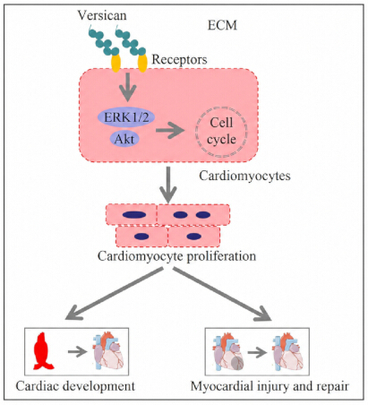
Figure 1: Proposed versican-associated signaling in cardiac development and myocardial repair. Versican in the ECM interacts with cardiomyocyte surface receptors and is associated with ERK1/2 and Akt signaling and cardiomyocyte cell-cycle activity. The schematic summarizes a candidate mechanistic framework; cell-cycle entry alone does not establish completed cytokinesis or functional myocardial regeneration. Abbreviations: ECM = extracellular matrix; ERK1/2 = extracellular signal-regulated kinase 1/2. Please click here to view a larger version of this figure.
Disclosures
The authors report no competing interests.
Acknowledgements
This work received support from the Hainan Province Natural Science Foundation of China (Grant 823RC572).
References
- Schlotter F, et al. Ventricular septal defect complicating acute myocardial infarction: diagnosis and management. A Clinical Consensus Statement of the Association for Acute CardioVascular Care (ACVC) of the ESC, the European Association of Percutaneous Cardiovascular Interventions (EAPCI) of the ESC and the ESC Working Group on Cardiovascular Surgery. Eur Heart J. 2024;45(28):2478-92.
- Beghini A, et al. 2024 update in heart failure. ESC Heart Fail. 2025;12(1):8-42.
- Nissen SE, et al. Bempedoic acid for primary prevention of cardiovascular events in statin-intolerant patients. JAMA. 2023;330(2):131-40.
- Johnson J, Mohsin S, Houser SR. Cardiomyocyte proliferation as a source of new myocyte development in the adult heart. Int J Mol Sci. 2021;22(15):7764.
- Zhu C, Yuan T, Krishnan J. Targeting cardiomyocyte cell cycle regulation in heart failure. Basic Res Cardiol. 2024;119(3):349-69.
- Garbern JC, Lee RT. Heart regeneration: 20 years of progress and renewed optimism. Dev Cell. 2022;57(4):424-39.
- Yaghoobi A, et al. Role of long noncoding RNAs in pathological cardiac remodeling after myocardial infarction: an emerging insight into molecular mechanisms and therapeutic potential. Biomed Pharmacother. 2024;172:116248.
- Dalil D, et al. The role of long noncoding RNAs in cardiomyocyte proliferation and heart regeneration after myocardial infarction: a systematic review. Cell Transplant. 2024;33:9636897241266725.
- Alharbi HO, Sugden PH, Clerk A. Mitogen-activated protein kinase signalling in rat hearts during postnatal development: MAPKs, MAP3Ks, MAP4Ks and DUSPs. Cell Signal. 2024;124:111397.
- Zheng K, et al. Effects and mechanisms of the myocardial microenvironment on cardiomyocyte proliferation and regeneration. Front Cell Dev Biol. 2024;12:1429020.
- Hilgendorf I, Frantz S, Frangogiannis NG. Repair of the infarcted heart: cellular effectors, molecular mechanisms and therapeutic opportunities. Circ Res. 2024;134(12):1718-51.
- Casarella S, et al. Focal adhesion's role in cardiomyocytes function: from cardiomyogenesis to mechanotransduction. Cells. 2024;13(8):664.
- Miao L, et al. β1 integrins regulate cellular behaviour and cardiomyocyte organization during ventricular wall formation. Cardiovasc Res. 2024;120(11):1279-94.
- Li X, et al. Inhibition of fatty acid oxidation enables heart regeneration in adult mice. Nature. 2023;622(7983):619-26.
- Shi Y, et al. α-Ketoglutarate promotes cardiomyocyte proliferation and heart regeneration after myocardial infarction. Nat Cardiovasc Res. 2024;3(9):1083-97.
- Katano W, et al. Sall1 and Sall4 cooperatively interact with Myocd and SRF to promote cardiomyocyte proliferation by regulating CDK and cyclin genes. Development. 2023;150(24):dev201913.
- Mahiny-Shahmohammady D, Hauck L, Billia F. Defining the molecular underpinnings controlling cardiomyocyte proliferation. Clin Sci (Lond). 2022;136(12):911-34.
- Su Q, et al. Single-cell insights: pioneering an integrated atlas of chromatin accessibility and transcriptomic landscapes in diabetic cardiomyopathy. Cardiovasc Diabetol. 2024;23(1):139.
- Sadek KM, Mahmoud SFE, Zeweil MF, Abouzed TK. Proanthocyanidin alleviates doxorubicin-induced cardiac injury by inhibiting NF-kB pathway and modulating oxidative stress, cell cycle, and fibrogenesis. J Biochem Mol Toxicol. 2021;35(4):e22716.
- Zhang Q, et al. Signaling pathways and targeted therapy for myocardial infarction. Signal Transduct Target Ther. 2022;7(1):78.
- Ireland J, Kilian KA. The importance of matrix in cardiomyogenesis: defined substrates for maturation and chamber specificity. Matrix Biol Plus. 2024;24:100160.
- Rienks M, Papageorgiou AP, Frangogiannis NG, Heymans S. Myocardial extracellular matrix: an ever-changing and diverse entity. Circ Res. 2014;114(5):872-88.
- Wight TN, et al. V3: an enigmatic isoform of the proteoglycan versican. Am J Physiol Cell Physiol. 2023;325(2):C519-C537.
- Wight TN, et al. Versican-a critical extracellular matrix regulator of immunity and inflammation. Front Immunol. 2020;11:512.
- Wight TN. Versican: a versatile extracellular matrix proteoglycan in cell biology. Curr Opin Cell Biol. 2002;14(5):617-23.
- Bassat E, Tzahor E. How can young extracellular matrix promote cardiac regeneration? Versi-Can! Circulation. 2024;149(13):1016-18.
- Feng J, et al. Versican promotes cardiomyocyte proliferation and cardiac repair. Circulation. 2024;149(13):1004-15.
- Mittal N, et al. Versican is crucial for the initiation of cardiovascular lumen development in medaka (Oryzias latipes). Sci Rep. 2019;9(1):9475.
- Song R, Zhang L. Cardiac ECM: its epigenetic regulation and role in heart development and repair. Int J Mol Sci. 2020;21(22):8610.
- Laflamme MA, Murry CE. Heart regeneration. Nature. 2011;473(7347):326-35.
- Weinberger M, Riley PR. Animal models to study cardiac regeneration. Nat Rev Cardiol. 2024;21(2):89-105.
- Zaragoza MV, et al. LMNA-related dilated cardiomyopathy: single-cell transcriptomics during patient-derived iPSC differentiation support cell type and lineage-specific dysregulation of gene expression and development for cardiomyocytes and epicardium-derived cells with lamin A/C haploinsufficiency. Cells. 2024;13(17):1479.
- Jang J, et al. Endocardial HDAC3 is required for myocardial trabeculation. Nat Commun. 2024;15(1):4166.
- Kern CB, et al. Proteolytic cleavage of versican during cardiac cushion morphogenesis. Dev Dyn. 2006;235(8):2238-47.
- Nandadasa S, Foulcer S, Apte SS. The multiple, complex roles of versican and its proteolytic turnover by ADAMTS proteases during embryogenesis. Matrix Biol. 2014;35:34-41.
- Foulcer SJ, et al. Determinants of versican-V1 proteoglycan processing by the metalloproteinase ADAMTS5. J Biol Chem. 2014;289(40):27859-73.
- Tang F, et al. Defining the versican interactome in lung health and disease. Am J Physiol Cell Physiol. 2022;323(2):C249-C276.
- Lenzi LGS, et al. Alterations in the structure, composition, and organization of galactosaminoglycan-containing proteoglycans and collagen correspond to the progressive stages of Dupuytren's disease. Int J Mol Sci. 2024;25(13):7192.
- Gupta N, et al. Targeting of stromal versican by miR-144/199 inhibits multiple myeloma by downregulating FAK/STAT3 signalling. RNA Biol. 2020;17(1):98-111.
- Ruiz-Rodriguez MJ, et al. Versican accumulation drives Nos2 induction and aortic disease in Marfan syndrome via Akt activation. EMBO Mol Med. 2024;16(1):132-57.
- Cooley MA, et al. Fibulin-1 is required during cardiac ventricular morphogenesis for versican cleavage, suppression of ErbB2 and Erk1/2 activation, and to attenuate trabecular cardiomyocyte proliferation. Dev Dyn. 2012;241(2):303-14.
- Wight TN, Kang I, Merrilees MJ. Versican and the control of inflammation. Matrix Biol. 2014;35:152-61.
- Elhashani S, et al. Expression of versican isoforms V0/V1 by pancreatic cancer associated fibroblasts increases fibroblast proliferation. Pancreatology. 2024;24(5):719-31.
- Barallobre-Barreiro J, et al. Extracellular matrix in heart failure: role of ADAMTS5 in proteoglycan remodeling. Circulation. 2021;144(25):2021-34.
- Lindsey SE, Butcher JT, Yalcin HC. Mechanical regulation of cardiac development. Front Physiol. 2014;5:318.
- Inai K. Atrioventricular valve abnormalities: from molecular mechanisms underlying morphogenesis to clinical perspective. In: Nakanishi T, Markwald RR, Baldwin HS, Keller BB, Srivastava D, Yamagishi H, editors. Etiology and Morphogenesis of Congenital Heart Disease: From Gene Function and Cellular Interaction to Morphology. Springer; Tokyo; 2016. p. 135-43.
- Hao DJ, et al. Hapln1 promotes dedifferentiation and proliferation of iPSC-derived cardiomyocytes by promoting versican-based GDF11 trapping. J Pharm Anal. 2024;14(3):335-47.
- Wu Y, et al. Oncostatin M promotes versican expression in cardiac fibroblasts to facilitate extracellular matrix remodeling. Biochim Biophys Acta Mol Basis Dis. 2026;1872(4):168153.
- Kuppe C, et al. Spatial multi-omic map of human myocardial infarction. Nature. 2022;608(7924):766-77.
- Huang X, et al. Resveratrol ameliorates high-phosphate-induced VSMCs to osteoblast-like cells transdifferentiation and arterial medial calcification in CKD through regulating Wnt/β-catenin signaling. Eur J Pharmacol. 2022;925:174953.
- Michaud ME, et al. Early injury landscape in vein harvest by single-cell and spatial transcriptomics. Circ Res. 2024;135(1):110-34.