This protocol provides a guide for live confocal imaging of microtubule dynamics using fluorescent microtubule markers in Drosophila larval sensory neurons.
Method Article
Imaging Microtubule Dynamics In Vivo in Drosophila Sensory Dendrites
* These authors contributed equally
In This Article
Summary
Abstract
Drosophila larval class IV sensory neurons are located just beneath the cuticle and are characterized by highly branched dendritic arbors that expand isotropically in two dimensions as the larvae develop. The growth of these arbors is driven by stochastic processes involving growth, shrinkage, and pausing of the dendritic terminal tips. To test the hypothesis that tip dynamics are driven by microtubule dynamics, protocols have been developed for imaging fluorescently labeled microtubules within dendrites. The method enables visualization of microtubule dynamics in fine terminal dendrites of class IV sensory neurons using confocal microscopy. It is designed to preserve dendritic arbor morphology during imaging while allowing microtubules to be visualized together with a membrane marker. This manuscript describes the procedures used to prepare Drosophila larvae for live imaging and to visualize fluorescently labeled microtubules in dendrites. The protocol, therefore, provides an approach to examining microtubule behavior in fine terminal dendrites while remaining compatible with membrane co-labeling.
Introduction
Neurons are highly polarized cells whose cytoskeletons define their architecture. Microtubules, one component of the neuronal cytoskeleton, provide structural support and serve as tracks for long-distance cargo transport1,2. The microtubule cytoskeleton is crucial for dendrite development3. Dendrites grow stochastically through a process of branching, elongation, and contraction4. Drosophila larval dendritic arborization (da) neurons are a powerful in vivo model system for studying dendritic morphogenesis. These sensory neurons are grouped into four classes (I–IV) based on increasing dendritic complexity3,5. Class IV da neurons have complex, highly branched dendrites and function as multimodal nociceptors6,7,8. The dendrites of these neurons form stereotyped receptive fields along the abdominal segments and lie just beneath the transparent cuticle9,10. This anatomy, together with the genetic tractability of Drosophila, makes class IV da neurons a suitable system for visualizing dendrite development in vivo with high spatial and temporal resolution. As the dendrites grow during development, their tips stochastically transition between states of growth, shrinkage, and pause11. Tip growth and shrinkage share many kinetic similarities with microtubule growth and shrinkage12. Therefore, visualizing microtubule dynamics in these cells is of considerable interest.
A common approach for visualizing microtubule dynamics in living cells, including those in Drosophila larvae, is to image fluorescently labeled end-binding proteins such as EB1 and EB313,14,15,16,17,18. However, interpretation of EB-GFP signals can be ambiguous. For example, a distally moving EB-GFP punctum, moving away from the cell body, could represent an outward-growing microtubule plus end or an inward-growing plus end of a microtubule sliding in the distal direction. Although EB-GFP predominantly labels the plus ends of growing microtubules, it can also label growing minus ends19, leading to additional ambiguity. Direct visualization of microtubules can reduce these ambiguities.
An alternative approach to EB-GFP is direct labeling of microtubules using GFP-tubulin20,21. However, fluorescent tubulin robustly labels proximal dendrites22, it fails to label distal dendrites23. Another difficulty with fluorescent tubulin is that the high concentration of soluble tubulin makes individual microtubules difficult to distinguish in narrow processes such as terminal dendrites. Therefore, imaging microtubule dynamics in intact neurons under normal physiological conditions remains challenging.
The Drosophila microtubule-associated protein (MAP) Jupiter, a structural homolog of mammalian Tau, MAP2, and MAP4, has been used to label microtubules in Drosophila tissues, including the ovary and embryos24. In class IV dendrites, mCherry-Jupiter and sfGFP-Jupiter have been used to quantify microtubules in proximal branches22,25,26. This protocol describes a live-imaging method that enables high-resolution, time-lapse observation of neuronal microtubule behavior in distal dendrites of Drosophila class IV neurons using fly lines expressing the fluorescently labeled tubulin-binding domain of Jupiter.
Protocol
This study used Drosophila melanogaster, an invertebrate model for which institutional animal ethics approval was not required. The tools used for the protocol are listed in the Table of Materials.
1. Generation of experimental fly lines
- Set up a cross to obtain the desired parental line. Use the GAL4/UAS binary system to drive mNeonGreen (mNG)-Jupiter in class IV dendritic arborization (da) neurons expressing GAL4 under the ppk promoter.
- Cross virgin females from a homozygous UAS-mNG-Jupiter line with males from a homozygous ppk-GAL4; CD4tdTomato line. Obtain F1 progeny carrying both ppk-GAL4; CD4tdTomato and UAS-mNG-Jupiter.
NOTE: F1 progeny carrying both ppk-GAL4; CD4tdTomato and UAS-mNG-Jupiter express mNG and CD4tdTomato strongly in class IV da neurons. Perform imaging using larvae derived from at least three independent genetic crosses, with a minimum of six larvae imaged per cross. - Keep the cross in a maintenance bottle for mating, with a dab of yeast paste applied to the wall. Incubate at 25 °C for 2–3 days.
NOTE: Feeding yeast boosts egg laying and oviposition. - Prepare the yeast paste by placing a spoonful of yeast in a small beaker and adding a few drops of water. Mix thoroughly with a spatula until the paste reaches a semisolid consistency, adding water dropwise as needed.
- Transfer the flies into a new maintenance bottle containing fresh yeast paste every day.
NOTE: Ensure the yeast paste is neither too soft nor too watery. If it becomes too soft, the flies may become stuck. For additional methodological details, see Hales et al.27 and Roote and Prokop28.
2. Egg laying on grape-agar plates, egg collection, and larval staging
- Prepare approximately 0.5 L of grape-agar medium by combining 376 mL of distilled water, 126 mL of grape juice, 15 g of agar, and 6 g of sucrose in a 1 L microwave-safe beaker.
- Microwave for 3–5 min, swirling every 1 min until the agar is completely dissolved. Allow the mixture to cool slightly, and then add 10 mL of ethanol and 5 mL of acetic acid.
- Mix thoroughly and pour the medium into 60 mm Petri dishes to prepare the grape-agar plates. Prepare the plates in advance, store them at 4 °C by stacking them upside down and wrapping them in Petri dish sleeves, and use them within 2 weeks. Prewarm the plates to room temperature before use.
- Transfer approximately 150 flies, comprising 50 males and 100 females, from the maintenance bottle to an egg-laying cage.
- Cover the egg-laying cage with a 60 mm grape-agar plate containing a dab of yeast paste.
- Allow the flies to lay eggs at 25 °C for 2 h on the grape-agar plate containing yeast paste. Replace the plate with a fresh grape-agar plate containing yeast paste and allow egg laying for another 2 h.
- Provide a third 60 mm grape-agar plate containing yeast paste and allow the flies to lay eggs for 2 h.
- Use the third plate as the experimental plate and discard the first two plates to minimize heterogeneity in embryo age.
NOTE: The initial egg collection often contains embryos at different developmental stages because female Drosophila can retain fertilized eggs in the reproductive tract for several hours before laying them. Discarding the initial plates helps obtain more synchronized eggs. - For staged imaging after egg laying (AEL), allow the embryos on the experimental plates to age at 25 °C until the desired stage is reached: Stage 1, 24 h; Stage 2, 48 h; Stage 3, 72 h; and Stage 4, 96–120 h.
3. Cleaning the larvae
- Wash the larvae with 5 mL of 5% sucrose solution, followed by 5 mL of dH₂O, to remove yeast debris from the surface and prevent desiccation.
- Transfer the larvae, typically three or four, to the lid of a 35 mm cell-culture dish using a 3/0 paintbrush. Dry the larvae with a laboratory tissue to remove excess water.
4. Anesthetization
- Clean and dry the larvae before anesthetization.
- Prepare the anesthetization setup in advance (Figure 1). Place the cell-culture dish within the cardboard-box enclosure to reduce air movement over the dish and minimize disruption of the isoflurane anesthesia.
CAUTION: Isoflurane is a volatile anesthetic. Handle and use isoflurane with appropriate laboratory ventilation and in accordance with institutional chemical-safety procedures. Immobilize the larvae by adding the measured volume of isoflurane to the anesthetic wand duct-taped inside a 35 mm cell-culture dish that serves as an evaporation reservoir. - Place the cell-culture dish lid containing the clean, dry larvae on top of the cell-culture dish.
- Anesthetize the larvae for 2–3 min.
NOTE: The isoflurane volume and duration of anesthesia depend on the larval stage and number of larvae. Determine the amount empirically. For example, use 10–20 µL of isoflurane for 2 min to anesthetize three 72 h larvae for approximately 30 min. Use 5 µL of isoflurane per larva per min as the starting dose for immobilization, regardless of larval stage.
5. Mounting
- Make a rectangular or circular enclosure with Vaseline on a clean glass microscope slide and leave a small gap in one corner (Figure 1C). Use a henna dispenser to draw the enclosure on the slide.
- Transfer the anesthetized larvae to the enclosure using a paintbrush. Mount the larvae dorsal side up in a drop of Halocarbon Oil 700.
- Place a No. 1.5 coverslip over the larva. Apply gentle pressure until the Vaseline seals the gap between the coverslip and slide and the larva is slightly flattened against the microscope slide.
- Image the sample immediately for up to 30–45 min from the transfer step described in step 5.2. Discontinue imaging if blebbing or neuronal damage, including dendritic breakage, is observed in the membrane channel; use a lower dose of anesthetic.
NOTE: Ensure that no bubbles are present in the Halocarbon Oil after placing the coverslip. Avoiding bubbles is required for high-quality dynamic videos and for minimizing drift. To minimize air-bubble formation, leave a small gap at one corner of the Vaseline enclosure to allow air to escape during coverslip placement. Seal the gap using a fine paintbrush if necessary.
6. Microscopy and image acquisition
- Perform live imaging of the larval samples using a spinning-disk confocal microscope equipped with a 50 µm pinhole and a perfect-focus system.
- Set the 488 nm and 561 nm laser lines to 30% of maximum power, assuming 100 mW lasers, to visualize the microtubule marker, such as mNG-Jupiter, and the membrane marker, CD4tdTomato.
- Set the exposure time to 200 ms. Capture the images using an sCMOS camera with 6.5 µm sensors and the associated image-acquisition software.
- Acquire all images using a 40× water-immersion objective with a numerical aperture of 1.25 and a pixel size of 0.1614 µm.
- Manually focus the sample to locate the second, third, and fourth abdominal hemisegments of the dorsal surface, where class IV neurons are readily accessible through the transparent cuticle.
- For each recording, acquire a set of z-stacks spanning up to 10 µm in depth at 1 µm per step. Acquire the stacks at 10 s intervals for 10 or 15 min.
NOTE: Unless otherwise specified, use larvae at 72 h AEL for imaging.
7. Image processing and analysis
- Import the acquired time-lapse movies in .ND2 format into FIJI (Fiji Is Just ImageJ) using the Bio-Formats importer. Convert the movies to TIFF format for downstream analysis.
- Stabilize the movies to correct for sample drift using the image stabilization plugin with affine transformation settings in FIJI.
- Exclude movies from the analysis if movement is excessive. Consider movement greater than 1 µm/min excessive and exclude those movies from the analysis. Also exclude movies if blebbing is observed in the membrane-marker channel (CD4tdTomato).
- Collapse the z-stacks into two-dimensional time series by generating maximum-intensity projections along the z-axis in FIJI.
- Draw linear regions of interest (ROIs) in FIJI along the feature of interest to quantify the dynamic properties of the tips and microtubules. Use the Multi Kymograph plugin in FIJI to generate kymographs. Generate the kymograph using a 3-pixel-wide line scan along the ROI and process it using the Multi Kymograph plugin in FIJI.
- Extract intensity profiles using the Plot Profile tool with a line width of 3 pixels. Generate a multicolumn CSV file in which alternate columns encode position and intensity along each line scan.
- Estimate the background intensity from a matched ROI in an empty region of the field. Subtract the background intensity from each frame.
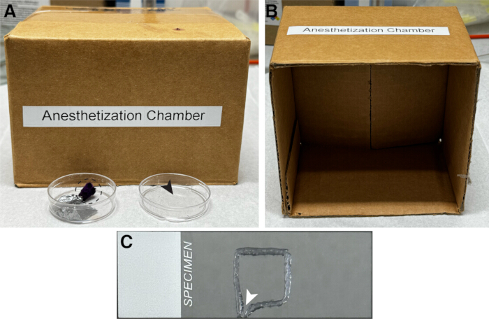
Figure 1: Components of the anesthetization setup. (A) A small cardboard box covering a 35 mm cell-culture dish containing an evaporation anesthetic wand secured with duct tape (black dashed circle). The lid of the cell-culture dish was used to hold clean, dry larvae before and during anesthesia (black arrowhead). (B) Inside view of the box. (C) A microscope slide with a square Vaseline enclosure prepared using a henna dispenser to contain the larval sample during imaging. A small gap (white arrowhead) facilitates air removal during coverslip placement. Please click here to view a larger version of this figure.
Results
Following the protocol described above, microtubules were successfully visualized in class IV dendritic arborization (da) neurons using mNG-Jupiter in combination with the membrane marker CD4tdTomato (Figure 2A–C). The membrane marker delineated the overall morphology of the dendritic arbor, whereas mNG-Jupiter labeled the microtubule-containing regions within the dendrites. At the distal ends of terminal dendrites, mNG-Jupiter fluorescence was weak or absent near the dendritic tips, whereas CD4tdTomato fluorescence remained strong in the corresponding terminals (Figure 2D–F).
The dendritic arbors showed no obvious morphological differences compared with larvae imaged with the membrane marker alone in earlier studies11. To quantitatively assess whether mNG-Jupiter expression caused morphological abnormalities, the number of dendritic tips was counted in mNG-Jupiter-expressing arbors and control arbors at 72 h AEL. In eight cells from eight mNG-Jupiter larvae, the number of tips in one quarter of the arbor was 153 ± 25 (mean ± SD), compared with 144 ± 27 (mean ± SD) in control larvae (Figure 2G). No significant difference was detected using Welch’s t-test (see Figure 2 legend). Therefore, mNG-Jupiter expression did not detectably alter the number of dendritic branches.
Time-lapse imaging demonstrated the dynamic behavior of the mNG-Jupiter signal within terminal dendrites. In the representative sequence shown in Figure 3A, the position of the bright mNG-Jupiter-labeled microtubule end changed over time. Successive line scans of the mNG-Jupiter signal were arranged over time to visualize changes in microtubule position and length. The resulting kymograph in Figure 3B shows alternating phases of extension and retraction of the fluorescent signal, consistent with the observed growth and shrinkage of the labeled microtubule in terminal dendrites (Videos 1 and 2). In thicker dendrites closer to the soma, the greater number of microtubules prevented visualization of the dynamics of individual microtubules.
Together, the representative images and time-lapse recordings show that the protocol enables visualization of both dendritic morphology and dynamic changes in microtubule-associated fluorescence in intact Drosophila larval sensory neurons. The membrane marker provides a reference for dendritic structure, whereas mNG-Jupiter allows microtubule behavior within the dendrite to be followed over time (Figures 2 and 3).
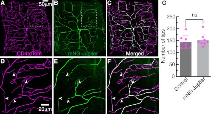
Figure 2: Dendrites in class IV neurons. The dendritic arbor of a class IV dendritic arborization (da) neuron imaged by spinning-disk confocal microscopy at 72 h after egg laying (AEL). Microtubules are visualized using the GAL4/UAS binary expression system. The class IV cell-specific driver ppk-GAL4 (pickpocket) facilitates expression of mNG-tagged Jupiter (UAS-mNG-Jupiter). Dendritic membranes are marked by direct ppk promoter-mediated expression of tdTomato fused to the CD4 membrane marker (ppk-CD4tdTomato). (A) Membrane marker, CD4tdTomato. (B) Microtubule marker, mNG-Jupiter. (C) Merged image. (D–F) Enlarged views of the regions outlined by white dashed boxes in A–C, respectively, illustrate the distribution of the microtubule and membrane markers. White arrowheads indicate the distribution of mNG-Jupiter in terminal dendrites. (G) Quantification of dendritic tip number in control and mNG-Jupiter-expressing neurons at 72 h AEL. Each dot represents the number of tips counted within one quarter of a single neuron from a distinct larva. A total of eight neurons from eight independent larvae were analyzed per group. Error bars represent mean ± SD. Groups were compared using a two-sided unpaired Welch’s t-test; ns, not significant (P > 0.05). Please click here to view a larger version of this figure.

Figure 3: Microtubule dynamics in a terminal dendrite. (A) Three images of a terminal dendrite expressing CD4tdTomato (upper) and mNG-Jupiter (lower) at the indicated time points. Arrowheads indicate the microtubule end. A dim mNG signal is visible beyond the bright microtubule signal. (B) Kymograph showing microtubule growth and shrinkage. Arrowheads indicate growth phases, and arrows indicate shrinkage phases. Successive 3-pixel-wide line scans of the mNG signal are arranged one below another, with time increasing in the downward direction. Please click here to view a larger version of this figure.
Video 1: Time-lapse imaging of mNG-Jupiter in class IV dendritic arborization neurons. Time-lapse sequence showing mNG-Jupiter fluorescence in Drosophila larval class IV dendrites. The sequence illustrates changes in microtubule-associated fluorescence over time within the dendritic arbor. Images were acquired using the live-imaging conditions described in the Protocol. Scale bar, 20 µm. Please click here to download this file.
Video 2: Time-lapse imaging of the CD4tdTomato membrane marker in class IV dendritic arborization neurons. Time-lapse sequence showing CD4tdTomato fluorescence in the corresponding Drosophila larval class IV dendritic arbor. The membrane marker provides a reference for dendritic morphology during the imaging period. Images were acquired using the live-imaging conditions described in the Protocol. Scale bar, 20 µm. Please click here to download this file.
Discussion
This protocol demonstrates the use of transgenic Jupiter fly lines for live imaging of microtubule dynamics in Drosophila sensory neurons using confocal microscopy. The method enables visualization of microtubule-associated fluorescence in intact larvae while preserving dendritic morphology, as assessed using a membrane marker11,29. A similar approach has also been used to image actin dynamics using GFP-actin and LifeAct-GFP in class IV neurons22,30,31,32. In principle, the same general approach may also be applied to imaging other microtubule-binding proteins in class IV cells and in other classes of dendritic arborization neurons (classes I, II, and III). In particular, combining fluorescent microtubule labeling with EB labeling may reduce ambiguity in interpreting microtubule growth. For example, the absence of an EB comet could indicate either the absence of microtubules or the presence of microtubules that are not actively growing; direct visualization of microtubules can help distinguish these possibilities. Thus, co-labeling mNG-Jupiter with fluorescent EB1 may enable interpretation of microtubule growth dynamics within the local context of microtubule organization, abundance, and inferred orientation.
Several steps are critical for obtaining suitable live-imaging data. The protocol uses the volatile anesthetic isoflurane for a defined, short immobilization regimen. The isoflurane dose must be carefully controlled during immobilization because insufficient anesthesia permits larval movement, whereas excessive anesthesia may lead to neuronal degradation. Air bubbles should also be avoided during mounting because they can interfere with image quality and contribute to sample drift. In addition, excessive pressure from the coverslip should be avoided because it may cause dendritic blebbing and subsequent neuronal damage. These steps are important for maintaining sample stability and dendritic morphology throughout imaging.
An important limitation of this protocol is that the immobilization and visualization method, involving the combination of anesthetic exposure and physical constraint by the coverslip, may influence microtubule dynamics. To determine whether microtubules are perturbed under these conditions, alternative immobilization methods can be used separately or in combination with this protocol. Such approaches include imaging embryos and using temperature-sensitive mutants, such as Shibirets 33, followed by comparison of kinetic parameters such as growth rates and catastrophe frequency.
With this limitation, the current protocol allows larvae to be imaged for approximately 30 minutes after mounting without dendritic blebbing, deterioration of morphology, or loss of detectable microtubule and tip dynamics. The method enables acquisition of high-resolution, time-lapse data from intact larvae and visualization of dynamic microtubule behavior in terminal dendrites.
Disclosures
The authors have no conflicts of interest to disclose.
Acknowledgements
The authors thank all members of the Howard laboratory, especially former laboratory member Dr. Maijia Liao, for initiating the microtubule-labeling project, and Dr. Sonal Shree for developing the protocol for visualizing dendritic dynamics in Drosophila within the Howard laboratory. Research reported in this publication was supported by the National Institutes of Health under Award Number R35 GM161634 (National Institute of General Medical Sciences) and Award Number R01 NS118884 (National Institute of Neurological Disorders and Stroke) to J.H. The content is solely the responsibility of the authors and does not necessarily represent the official views of the National Institutes of Health. This work was also supported by NSF Grant PHY-2210464 and PEW Charitable Trusts Award 38044 to J.H.
Materials
| Name | Company | Catalog Number | Comments |
|---|---|---|---|
| Acetic acid (glacial, ACS reagent, ≥99.7%) | Sigma-Aldrich | 695092 | Used to prepare grape-agar medium; 5 mL per approximately 0.5 L preparation |
| Agar (bactoagar) | Becton Dickinson | 214010 | Used to prepare grape-agar medium; 15 g per approximately 0.5 L preparation |
| Anesthetic wand | Carolina Biological Supply Co. | 173010 | Used to hold isoflurane within the evaporation reservoir during larval anesthetization |
| Beaker, 1 L, microwave-safe | Corning Inc. | 1000-1L | Used to prepare approximately 0.5 L of grape-agar medium |
| Beaker, 100 mL | Corning Inc. | 1000-100 | Used to prepare yeast paste |
| Bio-Formats importer | Open Microscopy Environment Project | Not applicable | FIJI plugin used to import .ND2 time-lapse files |
| Camera (sCMOS, Zyla 4.2 Plus) | Andor Technology | ZYLA-4.2P-USB3 | 6.5 µm sensor; used for image acquisition |
| Cardboard box (5 × 5 × 3 in) | Uline | S-4579 | Used to cover the anesthetization setup and reduce air movement over the cell-culture dish |
| Cell-culture dish | Corning Incorporated | 430165 | 35 × 10 mm; used for larval handling and as the isoflurane evaporation reservoir |
| Coverslip | Globe Scientific Inc. | 1404-15 | 22 × 22 mm, No. 1.5; placed over the mounted larva |
| Darwin chamber | Darwin Chambers | IN030-AA-LT-DA-MP | Used for maintaining fly crosses, egg laying, and larval staging at 25 °C |
| Distilled water, laboratory reagent grade | Research Products International (RPI) | Not applicable | Used for grape-agar preparation, yeast-paste preparation, and washing larvae |
| Duct tape | Uline | S-6519 | Used to secure the anesthetic wand inside the cell-culture dish |
| Egg-laying cage/embryo collection cage | Genesee Scientific | 59-100 | Small size; fits a 60 × 15 mm Petri dish; used for controlled egg collection |
| Ethanol (anhydrous, 200 proof) | Decon Labs Inc. | V1001G | Used to prepare grape-agar medium; 10 mL per approximately 0.5 L preparation |
| FIJI (Fiji Is Just ImageJ) | NIH | Version 2.16.0/1.54p | Used for image import, stabilization, maximum-intensity projection, ROI analysis, kymograph generation, and intensity-profile extraction |
| Fly lines: (1) w; ppk-Gal4; ppk-cd4-tdTom; (2) w; w+, UASt-mNeonGreen-Jupiter-RD/CyO | Bloomington Drosophila Stock Center(1); Howard Lab (2) | BDSC# 32078, BDSC# 35845 | Used to express CD4tdTomato and mNG-Jupiter in class IV dendritic arborization neurons |
| Grape juice, 100% Concord grape | Welch's | Not applicable | Used to prepare grape-agar medium; 126 mL per approximately 0.5 L preparation |
| Halocarbon oil 700 | Sigma-Aldrich | H8898 | Used to mount larvae for live imaging |
| Henna dispenser | ZEYER | ZEYER-1005 | Used to form the Vaseline enclosure on the microscope slide |
| Image Stabilizer | Developed by Kang Li | Not applicable | FIJI plugin used with affine transformation settings to correct sample drift |
| Isoflurane | Santa Cruz Biotechnology, Inc. | sc-470926 | Used to immobilize larvae before mounting and imaging |
| Laboratory tissue (Kimwipes) | Kimberly-Clark Professional | 34120 | Used to remove excess water from washed larvae before anesthetization |
| Maintenance bottle | Lab Express Inc. | 7001-PBT | Square-bottom polypropylene bottle, 6 oz; contains Fly Food B (Bloomington Recipe) and is used to maintain fly stocks and crosses |
| Microwave | Panasonic | NN-SN686S | Used to dissolve agar during preparation of grape-agar medium |
| Microscope, inverted | Nikon Instruments Inc. | 736228 | Ti Eclipse inverted microscope equipped with a perfect-focus system for live imaging |
| Microscope slide | Fisherbrand | 12550143 | Superfrost slide; used to mount larvae for imaging |
| Multi Kymograph | Developed by Jens Rietdorf, Arne Seitz, and Johannes Schindelin | Not applicable | FIJI plugin used to generate kymographs of microtubule dynamics |
| Nikon Elements software | Nikon Instruments Inc. | Version 5.21.03 | Used for acquisition of time-lapse microscopy images and generation of .ND2 files |
| Objective, water immersion | Nikon Instruments Inc. | MRD77400 | 40×, 1.25 NA; used for live imaging |
| Paintbrush | Dynasty | 2157r | Sizes 0 and 3/0; used to transfer larvae and seal the Vaseline enclosure |
| Petri dish | Falcon | 351007 | 60 × 15 mm; used for grape-agar plates |
| Spatula | VWR | 82027-532 | 22.9 cm (9 in); used to mix yeast and water during yeast-paste preparation |
| Spinning-disk confocal unit | Yokogawa CSU-W1 | SBW001 | 50 µm pinhole; used for live confocal imaging |
| Sucrose | Sigma-Aldrich | 57-50-1 | Used in grape-agar medium and as a 5% solution for washing larvae |
| Vaseline | Unilever | Not applicable | Used to form the enclosure between the microscope slide and coverslip |
| Yeast (Red Star Active Dry Yeast) | Lesaffre Corporation | Not applicable | Used to prepare yeast paste and support egg laying |
References
- Kapitein LC, Hoogenraad CC. Building the neuronal microtubule cytoskeleton. Neuron. 2015;87(3):492–506.
- Howard J, Hyman AA. Dynamics and mechanics of the microtubule plus end. Nature. 2003;422(6933):753–758.
- Jan YN, Jan LY. Branching out: mechanisms of dendritic arborization. Nature Reviews Neuroscience. 2010;11(5):316–328.
- Fujishima K, Horie R, Mochizuki A, Kengaku M. Principles of branch dynamics governing shape characteristics of cerebellar Purkinje cell dendrites. Development. 2012;139(18):3442–3455.
- Grueber WB, Jan LY, Jan YN. Tiling of the Drosophila. epidermis by multidendritic sensory neurons. Development. 2002;129(12):2867–2878.
- Hwang RY, et al. Nociceptive neurons protect Drosophila. larvae from parasitoid wasps. Current Biology. 2007;17(24):2105–2116.
- Grueber WB, et al. Dendrites of distinct classes of Drosophila. sensory neurons show different capacities for homotypic repulsion. Current Biology. 2003;13(8):618–626.
- Guo Y, et al. Identification of the peptide Vulnusin, a wound signal that mediates mechanical-injury-induced nociception in Drosophila. Neuron. 2025;113(20):3352–3362.e5.
- Han C, et al. Integrins regulate repulsion-mediated dendritic patterning of Drosophila. sensory neurons by restricting dendrites in a 2D space. Neuron. 2012;73(1):64–78.
- Grueber WB, et al. Projections of Drosophila. multidendritic neurons in the central nervous system: links with peripheral dendrite morphology. Development. 2007;134(1):55–64.
- Shree S, et al. Dynamic instability of dendrite tips generates the highly branched morphologies of sensory neurons. Science Advances. 2022;8(26):eabn0080. doi: 10.1126/sciadv.abn0080.
- Ouyang X, et al. Neurons exploit stochastic growth to rapidly and economically build dense dendritic arbors. Nature Communications. 2025;16(1):5903. doi: 10.1038/s41467-025-60800-7.
- Mimori-Kiyosue Y, Shiina N, Tsukita S. The dynamic behavior of the APC-binding protein EB1 on the distal ends of microtubules. Current Biology. 2000;10(14):865–868.
- Stepanova T, et al. Visualization of microtubule growth in cultured neurons via .the use of EB3-GFP (end-binding protein 3-green fluorescent protein). Journal of Neuroscience. 2003;23(7):2655–2664.
- Piehl M, Cassimeris L. Organization and dynamics of growing microtubule plus ends during early mitosis. Molecular Biology of the Cell. 2003;14(3):916–925.
- Ori-McKenney KM, Jan LY, Jan YN. Golgi outposts shape dendrite morphology by functioning as sites of acentrosomal microtubule nucleation in neurons. Neuron. 2012;76(5):921–930.
- Stone MC, Roegiers F, Rolls MM. Microtubules have opposite orientation in axons and dendrites of Drosophila. neurons. Molecular Biology of the Cell. 2008;19(10):4122–4129.
- Zheng Y, et al. Dynein is required for polarized dendritic transport and uniform microtubule orientation in axons. Nature Cell Biology. 2008;10(10):1172–1181.
- Zanic M, Stear JH, Hyman AA, Howard J. EB1 recognizes the nucleotide state of tubulin in the microtubule lattice. PLoS One. 2009;4(10):e7585. doi: 10.1371/journal.pone.0007585.
- Carminati JL, Stearns T. Microtubules orient the mitotic spindle in yeast through dynein-dependent interactions with the cell cortex. Journal of Cell Biology. 1997;138(3):629–641.
- Straight AF, Marshall WF, Sedat JW, Murray AW. Mitosis in living budding yeast: anaphase A but no metaphase plate. Science. 1997;277(5325):574–578.
- Nanda S, Bhattacharjee S, Cox DN, Ascoli GA. Distinct relations of microtubules and actin filaments with dendritic architecture. iScience. 2020;23(12):101865. doi: 10.1016/j.isci.2020.101865.
- Lee HH, Jan LY, Jan YN. Drosophila. IKK-related kinase Ik2 and Katanin p60-like 1 regulate dendrite pruning of sensory neuron during metamorphosis. Proceedings of the National Academy of Sciences of the United States of America. 2009;106(15):6363–6368.
- Karpova N, et al. Jupiter, a new Drosophila. protein associated with microtubules. Cell Motility and the Cytoskeleton. 2006;63(5):301–312.
- Poe AR, et al. Dendritic space-filling requires a neuronal type-specific extracellular permissive signal in Drosophila. Proceedings of the National Academy of Sciences of the United States of America. 2017;114(38):E8062–E8071.
- Nanda S, Bhattacharjee S, Cox DN, Ascoli GA. Local microtubule and F-actin distributions fully constrain the spatial geometry of Drosophila. sensory dendritic arbors. International Journal of Molecular Sciences. 2023;24(7):6741. doi: 10.3390/ijms24076741.
- Hales KG, Korey CA, Larracuente AM, Roberts DM. Genetics on the fly: a primer on the Drosophila. model system. Genetics. 2015;201(3):815–842.
- Roote J, Prokop A. How to design a genetic mating scheme: a basic training package for Drosophila. genetics. G3: Genes|Genomes|Genetics. 2013;3(2):353–358.
- Shree S, Howard J. Protocol for imaging the same class IV neurons at different stages of development. Bio-protocol. 2024;14(16):e5052. doi: 10.21769/BioProtoc.5052.
- Stürner T, et al. The branching code: a model of actin-driven dendrite arborization. Cell Reports. 2022;39(4):110746. doi: 10.1016/j.celrep.2022.110746.
- Nithianandam V, Chien CT. Actin blobs prefigure dendrite branching sites. Journal of Cell Biology. 2018;217(10):3731–3746.
- Kilo L, Stürner T, Tavosanis G, Ziegler AB. Drosophila. dendritic arborisation neurons: fantastic actin dynamics and where to find them. Cells. 2021;10(10):2777. doi: 10.3390/cells10102777.
- Liao M, Liang X, Howard J. The narrowing of dendrite branches across nodes follows a well-defined scaling law. Proceedings of the National Academy of Sciences. 2021;118(27):e2022395118. doi: 10.1073/pnas.2022395118.
Reprints and Permissions
This article has been published
Video Coming Soon