Heligmosomoides polygyrus 是一种具有强大免疫调节能力的小鼠线虫,其特性与人类高发的蠕虫感染非常相似。本文介绍了一种用于长期维持该线虫的实验方案 H. polygyrus 生命周期.
本文内容
摘要
摘要
Heligmosomoides polygyrus(以前称为Nematospiroides dubius,也被部分研究者称为H. bakeri)是一种肠道线虫,可通过多种免疫调节机制在小鼠体内建立慢性感染,其感染特征与人类常见的蠕虫感染非常相似。H. polygyrus在蠕虫来源的免疫调节研究领域已被广泛研究,发现其无论是通过主动感染还是分离的分泌产物,均能强烈抑制过敏和自身免疫疾病的实验模型。本文所述方案详细介绍了H. polygyrus生命周期的管理方法,包括稳定生产L3幼虫、成虫寄生虫的回收,以及其排泄-分泌产物(HES)的收集。
引言
Heligmosomoides polygyrus 是一种自然感染小鼠的胃肠道线虫,与人类中高发的线虫寄生虫密切相关1。与其他线虫模型(如Nippostrongylus brasiliensis)相比,H. polygyrus 能在小鼠体内持续建立慢性感染,这直接归因于其采用的多种强大的免疫调节机制,这些机制可抑制宿主的免疫应答2。
H. polygyrus 具有直接生活史:感染性 L3 期幼虫通过粪-口途径被宿主摄入(在实验室条件下可通过口服灌胃给予),随后迁移至十二指肠的浆膜下层并形成包囊,约在初次感染后 8 天返回肠腔发育为成虫。交配和产卵发生在感染后第 10 天,自感染后第 14 天起即可收集成虫用于培养,并获取其排泄-分泌产物3。H. polygyrus 还与共生微生物群发生相互作用,在易感小鼠感染后,其体内乳杆菌属(Lactobacilli)水平升高4–6;而小鼠暴露于乳杆菌后,H. polygyrus 的感染水平亦会增加6。
已有研究表明,H. polygyrus 的活动性感染可在多种自身免疫性疾病7-10、结肠炎11,12和过敏13-16的动物模型中保护机体免受免疫病理损伤。因此,人们对来源于该寄生虫的排泄-分泌分子(“HES”)在体内下调病理反应的潜力产生了极大关注17,18。事实上,已有研究发现,在给予小鼠HES产物处理后可产生保护效应19,相关作用通路也正在被逐步阐明18,20。本文中,我们描述了一种可靠制备Heligmosomoides polygyrus并获取其分泌产物的实验方案,这些产物可进一步用于多种功能性生化及免疫学研究。
方案
注意:本方案中的所有操作均须遵循英国政府内政部和爱丁堡大学兽医服务中心制定的指导方针。
1. 灌胃法感染小鼠
- 将Heligmosomoides polygyrus L3幼虫保存在蒸馏水中,置于4 °C条件下,最长可保存六个月。
- 使用前,用蒸馏水清洗L3幼虫三次:每次以300 × g离心10分钟(开启刹车),除去上清液,仅保留500 μl水(避免扰动沉淀的L3幼虫),然后重新悬浮沉淀物。
- 第三次清洗时,将水补至精确体积(通常为40 ml),用剪去尖端以扩大开口的200 μl吸头吸取20 μl。将两份20 μl样品置于60 mm培养皿表面,计数L3幼虫(通常具有活动性,最好在解剖显微镜下以50倍放大倍率观察)。将最终的沉淀物重悬于蒸馏水中,使浓度达到每毫升2,000条L3幼虫。
- 用于生活周期繁殖时,通过口服灌胃法对8周龄F1代(C57BL/6xCBA)小鼠感染400条H. polygyrus L3幼虫(将小鼠颈部皮肤固定,使其保持直立姿势,轻轻将钝头灌胃针经口腔和食道插入胃中),灌胃体积为200 μl蒸馏水。每次感染前需充分混匀(幼虫在水中迅速沉降),用1 ml注射器吸取200 μl,使用专用的圆头灌胃针。对于较年轻的实验小鼠(6–8周龄)或其他近交系小鼠(例如 C57BL/6或BALBc),每只小鼠感染200条L3幼虫。
2. 多形螺旋线虫(H. polygyrus)的传代与维持
- 将木炭置于一个大塑料盆中央,用冷水冲洗至少30分钟(未清洗的活性炭对L3幼虫有毒性)。排干盆中水分,将木炭放在两层吸水纸上,暴露于室温空气中直至完全干燥。
- 如需获取虫卵,先用镊子(必要时配合剪刀)从结肠中刮取粪便。如需大量L3幼虫,将小鼠置于金属网架上,连续数天收集其粪粒。
- 按至少1:1的比例将粪便与颗粒状木炭混合,调整至湿润度恰好能使混合物黏附于滤纸。在培养皿中取一张湿润的滤纸,在其中心涂抹一层薄薄的混合物,然后将其放入潮湿箱内(加入湿润的纸巾和一皿水),避光培养12–14天。
- 从第7天开始取出L3幼虫,并在丢弃滤纸前至少收集两次。幼虫会在滤纸边缘形成一圈,将滤纸从培养皿中取出,用移液器吸取每皿5 ml无菌水冲洗残留于培养皿上的幼虫,收集至50 ml离心管中。
- 取下滤纸,用蒸馏水将残留在培养皿上的剩余幼虫冲洗至50 ml离心管中。将悬液在300 × g下离心10分钟。用蒸馏水洗涤幼虫三次,随后将其保存于最多50 ml蒸馏水中,置于4 °C冰箱中备用。
注意:幼虫在4 °C下至少6个月内保持活性和感染能力。
3. 成虫H. polygyrus虫体的收集
- 提前准备好改良的贝尔曼装置,如图所示 图1.
- 感染后14天处死小鼠。
- 用70%乙醇清洗腹部。剪开腹部皮肤并向两侧拉开,暴露前腹壁。沿中线做切口进入腹腔。
- 将整个小肠和大肠(从近端十二指肠至远端直肠)完整切除,放入干燥的培养皿中。
- 将肠道全长拉直,切除含有粪便的结肠,并将其放入另一个培养皿中,以备后续制备虫卵使用。
- 切除含有成虫的近端20 cm小肠——通过十二指肠相对较厚的肠壁以及因管腔内存在虫体而常呈现的红色外观进行识别。将标本放入盛有5 ml、预热至37 °C的Hanks’溶液的培养皿(直径100 mm)中(每皿放置两个标本)。
- 用剪刀纵向剪开含有虫体的近端肠道部分(最好使用圆头剪刀),然后用两张载玻片刮擦肠壁内侧以清除虫体,随后弃去清洁的肠壁组织。
- 将蠕虫放入小块棉布袋中,用订书机封口,并用回形针固定在玻璃漏斗边缘图1).
- 将Hanks’溶液倒入漏斗中,并向每个漏斗中加入约4个培养皿的线虫。
- 将装置置于37 °C培养箱中孵育1–2小时,在孵育中途轻轻振荡以使可能堵塞棉布滤膜的肠道组织碎片脱落。注意避免碎片溢出棉布袋外,否则将导致最终HES制备物受到污染。成虫应已缓慢穿过棉布并沉积于玻璃试管底部。在水槽上方小心将试管从连接的橡胶软管上取下(注意避免在此过程中丢失虫体)。
- 使用塑料移液管将蠕虫转移至50 ml离心管中,用Hanks’ Solution洗涤六次(让蠕虫依靠重力沉降,用移液器吸除上清液,加入40 ml Hanks’ Solution,重复此步骤五次)。
注意:从此步骤开始,线虫培养必须保持无菌。- 转移到超净工作台,用添加了100 U/ml青霉素和100 µg/ml链霉素的无菌Hanks’溶液洗涤六次,备用 体外 培养。
- 用剪开尖端以扩大开口的黄色枪头吸取两份20 µl样品,计数回收的成虫;预期成虫数量约为接种幼虫数的50%。
4. HES 培养体系的建立:培养基制备与成虫洗涤
- 将3.11步骤中的虫体浸入约10 ml添加了10%庆大霉素的RPMI培养基中,持续20分钟,将离心管倾斜放置以确保虫体完全浸没。
- 在超净工作台中操作:用Hanks溶液(添加5 U/ml青霉素和5 μg/ml链霉素)再清洗六次。
- 制备H. polygrus培养基。
- 在超净工作台中保持无菌条件,向500 ml RPMI1640培养基中加入11.1 ml 45%葡萄糖(终浓度为1.2%,因RPMI1640本身含0.2%葡萄糖)、5 ml 100倍青霉素-链霉素溶液(终浓度为5 U/ml青霉素,5 μg/ml链霉素)、5 ml L-谷氨酰胺(终浓度为2 mM)以及5 ml庆大霉素(终浓度为1%)。不要添加胎牛血清(FCS)。
- 将虫体分装至带透气盖的T25培养瓶中,每瓶约1,000条虫体,加入15 ml H. polygyrus培养基,直立放置于37 °C培养箱(含5% CO2)中培养3周。
5. HES 的制备
- 每隔不超过每周两次的时间,从培养物中收集含 HES 的培养基,首次收集应在培养 24 小时后进行(由于脂多糖(LPS)污染和宿主蛋白含量较高,可单独处理或弃去)。每次收集的样品应分开保存,并清晰标注日期和批次编号。每次更换等体积的 H. polygyrus 培养基。
- 将含 HES 的培养基在 400 × g 条件下离心 5 分钟,然后通过 0.2 µm 低蛋白结合滤膜过滤除菌,收集至 50 ml 离心管中。在 -20 °C 冰箱中保存,并清晰标注采虫日期和 HES 收集日期。
- 在连续收集 HES 21 天后,弃去虫体。
- 合并 500–1,000 ml 的 HES 上清液(通常来自冷冻储存的样品,不包括最初 24 小时的收集物),在超滤装置中使用 3,000 分子量截留值(MWCO)的滤膜,在氮气压力下进行浓缩。
注意:务必小心,避免滤膜干涸。- 准备滤膜装置时,先将 3 kDa 滤膜有光泽的一面朝下,置于 1 升烧杯中,用蒸馏水搅拌清洗 3 次,每次 20 分钟。按照制造商说明组装超滤装置,滤膜有光泽的一面朝上。将装置置于 4 °C 环境中,在开始浓缩合并的 HES 样品前,先让 50 ml 蒸馏水通过滤膜。
- 根据需要将每管 HES 样品加入超滤装置中(每天 100–140 ml),直至体积浓缩至 2–5 ml。
- 为去除含 HES 培养基中的污染物,向滤膜装置中加入 50 ml 无热原的 PBS,然后浓缩至约 2 ml。重复此步骤两次(共使用 150 ml PBS)。将 HES 转移至 15 ml 离心管中,在超净台中用 0.2 μm 滤膜过滤除菌,并使用分光光度计(E280 = 10)或 Bradford 法测定蛋白浓度。
- 分装,标注批次编号和日期,于 -80 °C 冻存。
- 在使用每批 HES 前,按照制造商说明书进行显色法 LAL 检测。若每 1 µg 蛋白质中 LPS 含量超过 1 U,则不建议将该批次用于体内(in vivo)实验或体外(in vitro)培养。
- 将 24 小时收集的 HES 样品以相同方法单独处理;该样品含有 LPS 和部分宿主蛋白,虽不适合功能性实验,但可作为通过单克隆抗体亲和纯化分离特定分子的有用来源。
结果
感染易感性 H. polygyrus 在很大程度上受小鼠品系的遗传背景控制(表1);C57BL/6 和 CBA 小鼠具有高度易感性 21,22为了维持寄生虫的生活周期,选择这两种品系的F1杂交后代,因其能够耐受远高于任一亲本品系的蠕虫负荷,且不出现明显发病(如过度的肠上皮损伤)。通过口服灌胃给予400条L3期幼虫,以在F1小鼠中维持生活周期(所得成虫负荷如图所示) 图2),而通常对纯合近交系进行实验时使用200条L3幼虫作为感染剂量(例如,C57BL/6 或 BALBc)。然而,该剂量可能需要根据动物设施之间不同的环境共因子(如肠道菌群的差异)进行调整。
HES 批次在功能测定和蛋白质组成方面已证明具有可重复的效力;此外,当分析连续培养每周的上清液(最长至4周)时,发现其蛋白质谱非常相似(图3). 当通过Bradford法测定HES浓度时(参见 5.5),总蛋白浓度通常约为 1 mg/ml(图4).
浓缩HES的另一种方法是使用离心浓缩器(例如Vivaspin 3-kDa截留分子量膜),将样品在摆动转子离心机中以最高4,000 g离心,从而去除缓冲盐和低分子量组分。离心浓缩最适合小体积样品处理(1-10 ml),其最大浓缩倍数约为30倍。
在收集HES时,避免污染至关重要。为防止宿主分子污染,我们弃去从鼠肠中分离成虫后最初24小时内的含HES培养基。此外,我们使用显色法LAL试剂盒(见5.7)对每批HES中的LPS污染水平进行定量。1 U LPS约等于100 pg LPS,低于此水平的污染被视为可忽略。 23在我们实验室的检测中,大多数批次的HES中内毒素含量显著低于此限值,HES中内毒素的平均浓度为0.23 U/µg (图5)。LPS 的作用在 体内 病理模型(例如抑制哮喘反应)至少需要10 ng的LPS 23因此 体内 给药 5 µg HES(每批含100 pg LPS)/µg HES 将包含 500 pg LPS,远低于 LPS 产生问题的有效浓度。
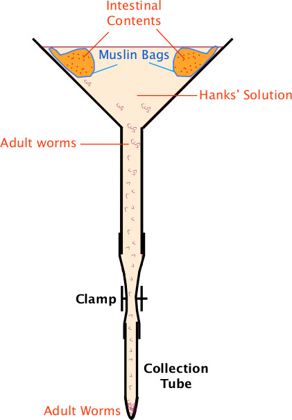
图 1:Baermann 装置 用于收集成虫 H. polygyrus 的 Baermann 装置设置(见第 2 节描述)。
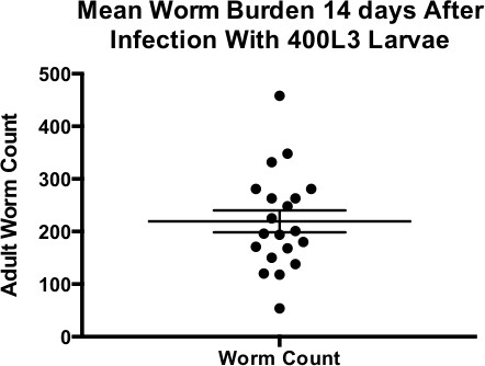
图 2:感染 400 条 L3 幼虫后 14 天的平均蠕虫负荷 感染 400 条 L3 幼虫后 14 天的平均蠕虫负荷。图中数据点来自 19 次独立的 C57BL/6xCBA 小鼠感染实验。误差线表示均值 ± 标准误(SEM)。
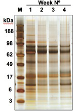
图3:连续培养周数中HES的蛋白质谱分析 对连续培养各周收集的HES蛋白质进行SDS-PAGE分析的结果。
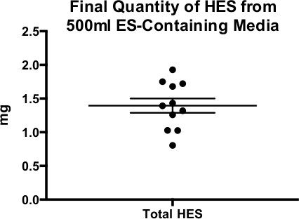
图4:来自500 ml含ES培养基的HES最终产量 从约500 ml上清液中提取的11个不同批次的HES蛋白产量。数据以均值±标准误表示。
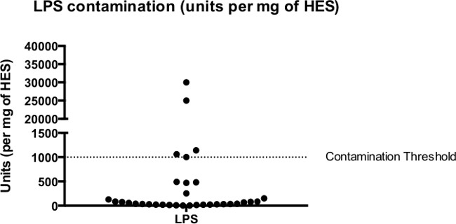
图 5:HES 的内毒素污染 通过鲎试剂细胞裂解物试验测定的 41 批 HES 中的内毒素污染水平。HES 中内毒素的中位浓度为 86 U/mg。
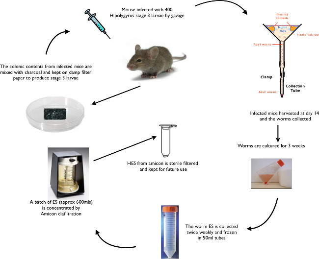
图6: 秀丽小杆线虫(H. polygyrus)生活周期动态示意图 从L3期幼虫经口灌胃感染开始,至幼虫及成虫回收,再到HES的分离,概述其关键生活周期阶段。
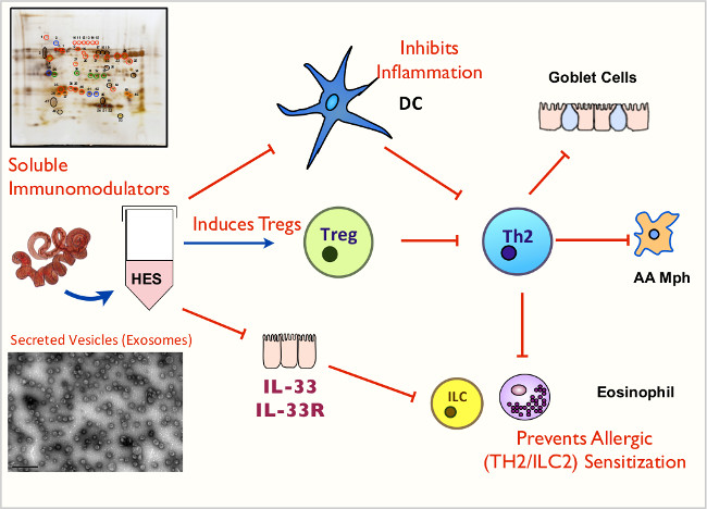
图7: HES及其功能的动画示意图 HES中所含可溶性介质和外泌体的关键免疫调节作用。
| 基因型(及背景品系) | 主要感染表型 | 参考文献 |
| 近交系 | ||
| A/J, CBA, C3H | 高度易感 | 22,29 |
| C57BL/6 和 C57BL/10 | 易感 | 22,30 |
| BALB/c, DBA/2, 129/J | 中等易感性 | 22,31 |
| NIH, SJL, SWR | 低易感性 | 22,32 |
| 细胞因子或细胞因子受体转基因 | ||
| IL-1β-/- | 更易感 | 33 |
| IL-1R-/- | 易感性降低(a);易感性无变化但肉芽肿增多(b) | (a) 33 |
| (b) 34 | ||
| IL-2Rβ 转基因(C57BL/6) | 抗性 | 35 |
| IL-4-/- | 繁殖力更高 | 36 |
| IL-4R-/-(C57BL/6 或 BALB/c) | 高度易感 | 22 |
| IL-6–/–(BALB/c 或 C57BL/6) | 高度抗性 | 37 |
| IL-9 转基因(FVB) | 抗性 | 38 |
| IL-21R –/–(C57BL/6) | Th2 缺陷,肉芽肿形成减少 | 39,40 |
| IL-25-/-(BALB/c) | 更易感 | 33 |
| IL-33R(T1/ST2)-/-(BALB/c) | 易感性无变化 | 33 |
| TGFβRIIdn(C57BL/6) | Th1 水平升高,易感性增加 | 41,42 |
| T 细胞标志物转基因 | ||
| CD28–/–(BALB/c) | 繁殖力轻微升高 | 43 |
| CD86(B7-2)–/–(BALB/c) | 繁殖力更高 | 44 |
| OX40L–/–(BALB/c) | 繁殖力更高 | 45 |
| 先天免疫位点转基因 | ||
| Ⅰ型干扰素受体(IFNAR)-/-(C57BL/6) | 繁殖力更高,肉芽肿更多 | 34 |
| MyD88–/–(C57BL/6) | 更抗性,肉芽肿更多 | 34 |
| C-KitW/Wv(WBB6) | 更易感 | 46,47 |
表1:不同遗传背景及基因靶向小鼠品系中 H. polygyrus的初次感染情况。
讨论
H. polygyrus 的生命周期以稳定且一致的方式进行。在第0天经自然摄入或口服灌胃感染L3期幼虫后,到第5天十二指肠浆膜下开始形成囊肿,随后幼虫蜕皮发育,至第10天起成虫进入肠腔。从第14天起可在粪便中观察到虫卵,从第21天起可在十二指肠浆膜表面观察到肉芽肿。上述方案(详见图6)不仅可稳定回收具有活性的L3期幼虫,用于后续实验及生命周期感染,还可高效制备H. polygyrus排泄-分泌产物(HES)。
H. polygyrus 感染在依赖卵清蛋白或屋尘螨过敏原(Der p 1)的哮喘模型中已被证明具有保护作用14此外,抑制气道炎症可通过CD4+CD25+ 调节性T细胞 14 或 CD19+CD23你好 调节性B细胞 24 来自未致敏的, H. polygyrus感染的小鼠。在自身免疫模型中, H. polygyrus 感染已被证明具有抑制作用 多发性硬化实验性自身免疫性脑脊髓炎(EAE)模型 9,且这种抑制作用可通过来自感染小鼠的CD4+ T细胞或CD19+ B细胞进行转移 24.
Heligmosomoides polygyrus 的排泄-分泌产物(HES)可通过多种机制调节宿主免疫反应,并抑制Th2介导的炎症(如图7所示),包括:a)抑制树突状细胞对刺激的应答能力25;b)诱导CD4+Foxp3+调节性T细胞的产生18;以及c)阻断IL-33的生成20。研究表明,在OVA-氢氧化铝哮喘模型中,于致敏或激发阶段给予HES可发挥保护作用19;同样,在与链格孢(Alternaria)提取物过敏原联合经鼻腔给予时,HES也可通过抑制早期IL-33的释放而产生保护效应20。避免HES被脂多糖(LPS)污染对于后续免疫学实验的成功往往至关重要。在本方案中,实现这一目标的关键步骤包括:确保巴ermann装置中粗布袋内的残渣不进入最终的HES制备物(见2.10),以及弃去培养最初24小时收集的ES溶液(见5.1)。
近年来,HES 已在蛋白质组水平上得到全面表征,已鉴定出超过 370 种不同的蛋白质26,27。此外,第四阶段幼虫的分泌物(ES)也已接受蛋白质组学分析28。目前正在进行的研究包括表征 HES 中主要的糖类成分(已知具有强免疫原性)3,以及被包裹在 50–100 nm 囊泡或外泌体中的分泌型微小 RNA(micro-RNA)(Buck et al.,已投稿待发表)。随着可重复的 HES 收集方案的建立,以及大量蛋白质组数据明确了 HES 的分子组成,目前的研究已为鉴定并验证来自H. polygyrus的单个分子在宿主免疫系统中介导关键效应的治疗潜力奠定了基础。
致谢
作者的研究由惠康基金会通过爱丁堡临床学术培训项目(C.J.C.J.)以及项目资助(Y.H. 和 R.M.M.)和研究员资助(J.R.G.)、英国生物技术和生物科学研究生理事会(G.C.)、美国哮喘基金会(E.R.、H.J.McS. 和 R.M.M.)、英国哮喘协会(H.J.McS.)以及拉宁基金会(D.J.S. 和 R.M.M.)提供经费支持。
材料
| 姓名 | 公司 | 目录编号 | 评论 |
|---|---|---|---|
| Amicon 浓缩器 | Millipore | 5112 | 型号 8050 50 ml http://www.emdmillipore.com/INTL/en/product/Stirred-Cell-Model-8050,-50%C2%A0mL,MM _NF-5122 |
| Hanks’ 平衡盐溶液 | Sigma | H9394 | 500 ml http://www.sigmaaldrich.com/catalog/product/sigma/h9394?lang=en®ion=GB |
| 青霉素-链霉素 | Invitrogen | 15070-063 | 100 ml http://www.lifetechnologies.com/order/catalog/product/15070063 |
| L-谷氨酰胺 | Invitrogen | 25030-081 | 200 MM 100 ml https://www.lifetechnologies.com/order/catalog/product/25030081?ICID=search-25030081 |
| 活性炭 | Merck | 1.09624.0500 | 18-35 目 http://www.merckmillipore.com/GB/en/product/Charcoal-activated,MDA_CHEM-109624 |
| 葡萄糖溶液 | Sigma | G8769 | 100 ml 45% 溶液 http://www.sigmaaldrich.com/catalog/product/sigma/g8769?lang=en®ion=GB |
| Falcon 管 | Sarstedt | 62.547.254 | 50 ml http://www.scribd.com/doc/124320105/Sarstedt-Falcon |
| T25 培养瓶 | Sigma | CLS430639 | 25 cm 透气培养瓶 http://www.sigmaaldrich.com/catalog/product/sigma/cls430639?lang=en®ion=GB |
| 显色法 LAL 检测 | Lonza | 50-647U | QCL-1000 检测试剂盒 http://www.lonza.com/products-services/pharma-biotech/endotoxin-detection/endotoxin-detection-assays/endpoint-chromogenic-lal-assay.aspx |
| 庆大霉素 | Gibco | 15710-049 | 100 ml http://www.lifetechnologies.com/order/catalog/product/15710072 |
| Roswell Park Memorial Institute (RPMI) 1640 | Gibco | 11875093 | 500 ml;不含 L-谷氨酰胺 http://www.lifetechnologies.com/order/catalog/product/11875093 |
参考文献
- Monroy, F. G., Enriquez, F. J. Heligmosomoides polygyrus : a model for chronic gastrointestinal helminthiasis. Parasitol. Today. 8, 49-54 (1992).
- Maizels, R. M., et al. Immune modulation and modulators in Heligmosomoides polygyrus infection. Exp. Parasitol. 132, 76-89 (2012).
- Hewitson, J. P., et al. Heligmosomoides polygyrus elicits a dominant nonprotective antibody response directed at restricted glycan and peptide epitopes. J Immunol. 187, 4764-4777 (2011).
- Walk, S. T., Blum, A. M., Ewing, S. A., Weinstock, J. V., Young, V. B. Alteration of the murine gut microbiota during infection with the parasitic helminth Heligmosomoides polygyrus. Inflamm Bowel Dis. 16, 1841-1849 (2010).
- Rausch, S., et al. Small intestinal nematode infection of mice Is associated with increased enterobacterial loads alongside the intestinal tract. PLoS ONE. 8, e74026(2013).
- Reynolds, L. A., et al. Commensal-pathogen interactions in the intestinal tract: Lactobacilli promote infection with, and are promoted by, helminth parasites. Gut Microbes. 5, 10-19 (2014).
- Saunders, K. A., Raine, T., Cooke, A., Lawrence, C. E. Inhibition of autoimmune type 1 diabetes by gastrointestinal helminth infection. Infect Immun. 75, 397-407 (2007).
- Liu, Q., et al. Helminth infection can reduce insulitis and type 1 diabetes through CD25- and IL-10-independent mechanisms. Infect Immun. 77, 5347-5358 (2009).
- Donskow-Łysoniewska, K., Krawczak, K., Doligalska, M. Heligmosomoides polygyrus: EAE remission is correlated with different systemic cytokine profiles provoked by L4 and adult nematodes. Exp Parasitol. 132, 243-248 (2012).
- Mishra, P. K., Patel, N., Wu, W., Bleich, D., Gause, W. C. Prevention of type 1 diabetes through infection with an intestinal nematode parasite requires IL-10 in the absence of a Th2-type response. Mucosal Immunol. 6, 297-308 (2013).
- Sutton, T. L., et al. Anti-Inflammatory mechanisms of enteric Heligmosomoides polygyrus infection against trinitrobenzene sulfonic acid-induced colitis in a murine model. Infect Immun. 76, 4772-4782 (2008).
- Hang, L., et al. Heligmosomoides polygyrus bakeri infection activates colonic Foxp3+ T cells enhancing their capacity to prevent colitis. J Immunol. 191, 1927-1934 (2013).
- Bashir, M. E., Andersen, P., Fuss, I. J., Shi, H. N., Nagler-Anderson, C. An enteric helminth infection protects against an allergic response to dietary antigen. J Immunol. 169, 3284-3292 (2002).
- Wilson, M. S., et al. Suppression of allergic airway inflammation by helminth-induced regulatory T cells. J. Exp. Med. 202, 1199-1212 (2005).
- Kitagaki, K., et al. Intestinal helminths protect in a murine model of asthma. J Immunol. 177, 1628-1635 (2006).
- Hartmann, S., et al. Gastrointestinal nematode infection interferes with experimental allergic airway inflammation but not atopic dermatitis. Clin Exp Allergy. 39, 1585-1596 (2009).
- Pritchard, D. I., Lawrence, C. E., Appleby, P., Gibb, I. A., Glover, K. Immunosuppressive proteins secreted by the gastrointestinal nematode parasite Heligmosomoides polygyrus. Int. J. Parasitol. 24, 495-500 (1994).
- Grainger, J. R., et al. Helminth secretions induce de novo T cell Foxp3 expression and regulatory function through the TGF-β pathway. J Exp Med. 207, 2331-2341 (2010).
- McSorley, H. J., et al. Suppression of type 2 immunity and allergic airway inflammation by secreted products of the helminth Heligmosomoides polygyrus. Eur. J. Immunol. 42, 2667-2682 (2012).
- McSorley, H. J., Blair, N. F., Smith, K. A., McKenzie, A. N. J., Maizels, R. M. Blockade of IL-33 release and suppression of type 2 innate lymphoid cell responses by helminth secreted products in airway allergy. Mucosal Immunol. 7, 1068-1078 (2014).
- Behnke, J. M., Menge, D. M., Noyes, H. Heligmosomoides bakeri: a model for exploring the biology and genetics of restance to chronic gastrointestinal nematode infections. Parasitology. 136, 1565-1580 (2009).
- Filbey, K. J., et al. Innate and adaptive type 2 immune cell responses in genetically controlled resistance to intestinal helminth infection. Immunology and Cell Biology. 92, 436-448 (2014).
- Bortolatto, J., et al. Toll-like receptor 4 agonists adsorbed to aluminium hydroxide adjuvant attenuate ovalbumin-specific allergic airway disease: role of MyD88 adaptor molecule and interleukin-12/interferon-gamma axis. Clin Exp Allergy. 38, 1668-1679 (2008).
- Wilson, M. S., et al. Helminth-induced CD19+CD23hi B cells modulate experimental allergic and autoimmune inflammation. Eur J Immunol. 40, 1682-1696 (2010).
- Segura, M., Su, Z., Piccirillo, C., Stevenson, M. M. Impairment of dendritic cell function by excretory-secretory products: A potential mechanism for nematode-induced immunosuppression. Eur J Immunol. 37, 1887-1904 (2007).
- Hewitson, J. P., et al. Proteomic analysis of secretory products from the model gastrointestinal nematode Heligmosomoides polygyrus reveals dominance of Venom Allergen-Like (VAL) proteins. J Proteomics. 74, 1573-1594 (2011).
- Moreno, Y., et al. Proteomic analysis of excretory-secretory products of Heligmosomoides polygyrus assessed with next-generation sequencing transcriptomic information. PLoS Negl Trop Dis. 5, e1370(2011).
- Hewitson, J. P., et al. Secretion of protective antigens by tissue-stage nematode larvae revealed by proteomic analysis and vaccination-induced sterile immunity. PLOS Pathogens. 9, e1003492(2013).
- Prowse, S. J., Mitchell, G. F. On the choice of mice for dissection of strain variations in the development of resistance to infection with Nematospiroides dubius. Austral J Exp Biol Med. 58, 603-605 (1980).
- Behnke, J. M., Robinson, M. Genetic control of immunity to Nematospiroides dubius: a 9-day anthelmintic abbreviated immunizing regime which separates weak and strong responder strains of mice. Parasite Immunol. 7, 235-253 (1985).
- Zhong, S., Dobson, C. Heligmosomoides polygyrus: resistance in inbred, outbred, and selected mice. Exp. Parasitol. 82, 122-131 (1996).
- Behnke, J. M., et al. Genetic variation in resistance to repeated infections with Heligmosomoides polygyrus bakeri, in inbred mouse strains selected for the mouse genome project. Parasite Immunol. 28, 85-94 (2006).
- Zaiss, M. M., et al. IL-1β suppresses innate IL-25 and IL-33 production and maintains helminth chronicity. PLoS Pathog. 9, e1003531(2013).
- Reynolds, L. A., et al. MyD88 signaling inhibits protective immunity to the gastrointestinal helminth parasite Heligmosomoides polygyrus. J Immunol. , (2014).
- Morimoto, M., Utsumiya, K. Enhanced protection against Heligmosomoides polygyrus in IL-2 receptor β-chain overexpressed transgenic mice with intestinal mastocytosis. J Vet Med Sci. 73, 849-851 (2011).
- Urban, J. F., Katona, I. M., Paul, W. E., Finkelman, F. D. Interleukin 4 is important in protective immunity to a gastrointestinal nematode infection in mice. Proc. Natl. Acad. Sci. USA. 88, 5513-5517 (1991).
- Smith, K. A., Maizels, R. M. IL-6 controls susceptibility to helminth infection by impeding Th2 responsiveness and altering the Treg phenotype in vivo. Eur J Immunol. 44, 150-161 (2014).
- Hayes, K. S., Bancroft, A. J., Grencis, R. K. Immune-mediated regulation of chronic intestinal nematode infection. Immunol Rev. 201, 75-88 (2004).
- Fröhlich, A., et al. IL-21 receptor signaling is integral to the development of Th2 effector responses in vivo. Blood. 109, 2023-2031 (2007).
- King, I. L., Mohrs, K., Mohrs, M. A nonredundant role for IL-21 receptor signaling in plasma cell differentiation and protective type 2 immunity against gastrointestinal helminth infection. J Immunol. 185, 6138-6145 (2010).
- Ince, M. N., et al. Role of T cell TGF-β signaling in intestinal cytokine responses and helminthic immune modulation. Eur J Immunol. 39, 1870-1878 (2009).
- Reynolds, L. A., Maizels, R. M. Cutting Edge: In the absence of TGF-β signaling in T cells, fewer CD103+ regulatory T cells develop, but exuberant IFN-γ production renders mice more susceptible to helminth infection. J Immunol. 189, 1113-1117 (2012).
- Ekkens, M. J., et al. Memory Th2 effector cells can develop in the absence of B7-1/B7-2, CD28 interactions, and effector Th cells after priming with an intestinal nematode parasite. J Immunol. 168, 6344-6351 (2002).
- Greenwald, R. J., et al. B7-2 is required for the progression but not the initiation of the type 2 immune response to a gastrointestinal nematode parasite. J. Immunol. 162, 4133-4139 (1999).
- Ekkens, M. J., et al. The role of OX40 ligand interactions in the development of the Th2 response to the gastrointestinal nematode parasite Heligmosomoides polygyrus. J. Immunol. 170, 384-393 (2003).
- Hashimoto, K., et al. Immunity-mediated regulation of fecundity in the nematode Heligmosomoides polygyrus--the potential role of mast cells. Parasitology. 137, 881-887 (2010).
- Hepworth, M. R., et al. Mast cells orchestrate type 2 immunity to helminths through regulation of tissue-derived cytokines. Proc Natl Acad Sci U S A. 109, 6644-6649 (2012).
重印与许可
