开发了一种集成的细胞操作平台,可与单探针质谱装置联用,用于在常温条件下对单个悬浮细胞进行在线分析。
本文内容
摘要
摘要
单细胞质谱技术(SCMS)能够在单个细胞水平上实现对多种细胞组分的高灵敏度检测和准确定量分析。单探针是一种微尺度取样与电离装置,可与质谱仪联用,用于在常温常压条件下对细胞成分进行在线、快速的SCMS分析。此前,单探针SCMS技术主要用于固定在基底上的细胞检测,限制了可研究的细胞类型。在本研究中,单探针SCMS技术已与一种常用于体外受精的细胞操作系统相结合。该集成化的细胞操控与分析平台利用细胞选择探针捕获已识别的悬浮单细胞,并将其转移至单探针尖端进行微尺度裂解,随后立即开展质谱分析。该捕获与转移过程在分析前将细胞从周围溶液中分离,最大限度减少了基质分子对质谱分析的干扰。该集成系统能够对来源于体液样本(如尿液、血液、唾液等)中的患者来源靶细胞进行SCMS分析,为SCMS技术在人类医学与疾病生物学中的应用提供了可能。
引言
人类生物学,尤其是疾病生物学,正日益被理解为单个细胞水平上活动的结果。然而,传统的分析方法(如液相色谱-质谱联用技术,LCMS)通常用于分析由细胞群体制备的样品,所获得的分子信息无法准确反映单个细胞水平上的化学过程。这些常规的传统方法无法识别细胞异质性对分析测量结果的影响,而为了制备裂解液所进行的细胞破坏和混合过程,可能导致细胞组分的改变或丢失1,2。这些传统方法的局限性在患者细胞分析中尤为突出,因为所获得的样品可能包含多种不同类型细胞的复杂混合物。为了克服这些不足,单细胞分子分析方法(包括单细胞质谱技术,SCMS)正不断被开发并应用于生物分析,特别是细胞代谢物和小分子生物分子的分析3,4。
最早开发的单细胞质谱分析(SCMS)技术采用基于真空的方法,在非环境条件下进行分析2,5,6,7,8,9,10,11。非环境条件下的SCMS技术能够分析细胞脂质和代谢物,但需要在人工条件下进行样品预处理,因此不适用于实时分析。非环境分析的样品制备过程包括添加基质成分,而该制备过程可能改变细胞组分在其天然环境中的状态12。因此,环境质谱(ambient mass spectrometry, MS)技术被用于在接近天然的环境中分析细胞,该技术在取样时无需真空环境。无需真空环境使得实验设计更加灵活:可加入摄像设备以监测细胞过程,并可将更温和的电离技术与分离技术联用,从而从每次单细胞实验中获得更丰富的信息4,12,13,14,15,16,17,18,19,20,21,22,23,24,25,26,27,28,29,30,31,32,33,34,35,36,37,38,39,40,41,42。
单探针SCMS方法是一种常温技术,可在接近天然的环境中分析活体哺乳动物癌细胞系21,43,44,45,46。此外,单探针装置已被用于其他质谱应用,包括多细胞球状体中外源分子的分析以及组织的质谱成像47,48,49,50,51,52。然而,由于该方法需要将细胞固定在基底上,因此无法直接分析悬浮细胞3,53。因此,单探针SCMS系统无法直接用于采样非贴壁单细胞,例如非贴壁细胞系或从患者血液及其他体液中分离的悬浮细胞54。在本研究中,将集成细胞操作平台(ICMP)与单探针SCMS技术相结合,以实现对活体悬浮细胞的在线分析,并最大限度减少样品前处理步骤(图1)46。该ICMP包括一台倒置显微镜用于监控细胞选择、一根玻璃细胞选择探针、一个用于捕获单个漂浮细胞的显微注射器、一块用于维持细胞温度的加热板、两套细胞操作系统用于控制玻璃细胞选择探针和单探针的空间移动,以及一台数字显微镜用于观察细胞从细胞选择探针尖端转移到单探针尖端的过程。单探针的制备方法已在先前的文献中详细描述,此处不再赘述21,48。ICMP与单探针系统连接至一台高分辨率质谱仪。该集成装置可从复杂的生物样品中采样特定的单个细胞,同时最大程度减少基质分子的干扰。
方案
1. 玻璃细胞挑选探针的制备
- 将单孔玻璃管加工成尖端锋利的锥形探针。
- 将一根单孔玻璃管(内径:0.3 mm,外径:1.1 mm)放入垂直移液管夹持器的夹具中,使玻璃管中心对准加热线圈,并拧紧以固定玻璃管。加热线圈由一段18号镍铬电阻丝(长约60 mm)围绕一根金属棒(直径 = 3.90 mm)缠绕2.5圈构成。
- 将玻璃管设置为温度程序19.5(厂家单位)。此参数可根据具体仪器进行调整。
- 将电磁铁推杆设置为4(厂家单位)。此参数可根据具体仪器进行调整。
- 触发电磁铁以拉伸玻璃管。此步骤会在尖端形成两个融合在一起的探针。
- 使用镊子在每个探针尖端约1 mm处剪断,使探针尖端形成直径约10 µm的开口。
- 弯曲玻璃探针,以便于与ICMP/单探针SCMS装置耦合。
- 将拉制好的玻璃探针放入微加工仪中,使探针尖端位于铂加热丝上方约3 mm处。
- 将铂丝加热功率调至最高温度的30%。
- 将探针从原始位置弯曲约45°(图2)。
2. 细胞整合操作平台的组装
- 将倒置显微镜、显微注射仪和两个细胞操作系统放置在电动平台上,以便于与质谱仪轻松耦合。
- 通过将其中一个细胞操作系统末端更换为臂夹,以适配单探针。
- 使用带针头的塑料注射器向显微注射仪中注入矿物油。避免管路中产生气泡,否则会影响吸液效果。
- 将倒置显微镜的载物台插入件更换为加热板。分析前将加热板温度设定为 37 °C。
- 安装玻璃细胞选择装置。
- 将玻璃细胞选择探针插入显微注射仪的金属支架中,使探针长端(非弯曲端)插入毛细管支架,并拧紧螺丝以固定探针。调整探针尖端角度,使其与加热板平行。
警告:玻璃探针非常尖锐且易碎,容易断裂。在将探针插入显微注射仪时,请保护眼睛并格外小心。 - 将显微注射仪的金属支架固定在细胞操作系统上。调整探针尖端位置,使其靠近倒置显微镜光源的中央区域。
- 将玻璃细胞选择探针插入显微注射仪的金属支架中,使探针长端(非弯曲端)插入毛细管支架,并拧紧螺丝以固定探针。调整探针尖端角度,使其与加热板平行。
3. 为质谱仪进样口创建一个延长的离子传输管
- 使用金属切割器截取一段长约 250 mm 的不锈钢管(外径:0.0625(1/16)英寸,内径:0.021 英寸)。
- 从管端测量 135 mm 处并安装金属卡箍,使约 135 mm 长度暴露在空气中,约 115 mm 长度位于质谱仪内部。使用两个扳手拧紧卡箍以固定其位置。
4. 将ICMP与单探针装置耦合
- 将含有单探针的玻片固定在细胞操作系统的臂夹中。
注意:单探针的制备依据先前发表的方案48,但在本研究中进行了两项微小修改:纳米电喷雾电离(nano-ESI)发射端被加长,以便于与质谱仪耦合;单探针被粘接到玻片右侧,以避免干扰玻璃细胞选择装置的空间移动(图2)。 - 通过将供液毛细管插入塑料卡套的套管(1/16 英寸 × 0.005 英寸)中并用手拧紧接头,将其连接至导电接头。
- 将导电接头的另一端连接一根毛细管(内径:40 µm,外径:150 µm),该毛细管连接装有采样溶剂的注射器;将毛细管插入套管(1/32 英寸 × 0.007 英寸)并拧紧接头。本实验中使用含 0.1% 甲酸的乙腈作为采样溶剂。
注意:采样溶剂可调整,但应主要含有乙腈(或含甲酸的乙腈以获得更好的离子化效果),以实现快速的微尺度细胞裂解。 - 将注射器固定在质谱仪的注射泵上。
- 将电离电压线连接到附着于导电接头的铜线上。
- 将导电接头的另一端连接一根毛细管(内径:40 µm,外径:150 µm),该毛细管连接装有采样溶剂的注射器;将毛细管插入套管(1/32 英寸 × 0.007 英寸)并拧紧接头。本实验中使用含 0.1% 甲酸的乙腈作为采样溶剂。
- 将纳米电喷雾电离(nano-ESI)发射端定位至延伸离子传输管入口约 1 mm 处。
- 使用细胞操作系统控制单探针的空间移动,使纳米电喷雾电离发射端位于延伸离子传输管前端中央位置。
5. 悬浮细胞样品制备
- 分析前一天(约 18–24 小时),在细胞培养瓶(T25)中接种待测细胞。本研究中使用 K562 人髓系白血病细胞作为模型。
- 将 1× 磷酸盐缓冲液(PBS)和添加了 10% 合成胎牛血清(FBS)及 1% 青霉素-链霉素的 Roswell Park Memorial Institute(RPMI)培养基在 37 °C 下预热 30 分钟。
- 通过将细胞与预热的培养基混合,在总体积为 10 mL 的条件下接种约 1 × 10⁶ 个细胞。通常,使用 10 mL 移液器将 8 mL RPMI 培养基加入细胞培养瓶中,然后使用 2 mL 移液器将 2 mL 融合状态的 K562 细胞加入培养基中,以获得约 1 × 10⁶ 个细胞。
- 将细胞在 37 °C 和 5% CO2 条件下孵育至进行分析。
- 准备用于分析的细胞。
- 将细胞从细胞培养瓶中用移液器转移至 15 mL 离心管中。
- 在 400 × g 和 37 °C 条件下离心 5 分钟,弃去上清液。
- 将细胞重悬于 4 mL 含有药物化合物的 RPMI 培养基中,药物浓度为所需的处理浓度。
注:对于对照组细胞的分析,将细胞重悬于 4 mL RPMI 培养基中,并直接跳至 步骤 6。 - 在 37 °C 和 5% CO2 条件下孵育细胞,孵育时间根据处理需求而定。
- 在 400 × g 和 37 °C 条件下离心 5 分钟,吸除上清液。
- 将细胞重悬于 10 mL PBS 中,在 400 × g 和 37 °C 条件下离心 5 分钟。离心后弃去上清液。重复此步骤 3 次,以最大程度减少细胞外成分中残留药物的检测干扰。
- 将细胞重悬于 4 mL PBS 中用于后续分析。
6. 使用ICMP/单探针装置进行SCMS测量
- 为质谱仪的实验定制参数。
- 在仪器软件的扫描模式标题下,选择定义扫描。在m/z 400处使用60,000 m/∆m的分辨率,1次微扫描,最大注入时间100 ms,开启自动增益控制(AGC)。实验中采用的质量范围(m/z)为100–1000。可根据仪器型号调整参数。
- 在注射泵下,选择150 nL/min的流速。每次实验均需优化流速。
- 选择NSI离子源并施加约4.5 kV的电压。该参数也需针对每次实验进行优化。
- 打开倒置显微镜(顶部平板和底部物镜均选择40倍放大),并将其连接至笔记本电脑的USB端口以获取实时视频。打开加热板并将其设定为37 °C。
- 在计算机上,进入采集数据选项卡,并在采集时间下选择连续采集。
- 准备用于分析的样品。
- 用移液器将2–3 mL样品加入小型培养皿(35 mm × 12 mm)的盖子中。
- 将样品置于加热板上倒置显微镜光源的中央位置。
- 准备玻璃细胞选择探针用于分析。使用细胞操作系统移动探针,使其尖端在倒置显微镜下与细胞处于同一焦平面。
- 选择单个细胞进行分析。
- 使用细胞操作系统将细胞选择探针的尖端移至目标细胞。此过程通过倒置显微镜进行监控。
注:如果细胞选择探针的尖端无法与细胞处于同一焦平面,可能是探针的弯曲部分角度不正确。调整细胞选择探针的位置,直至显微镜下可同时清晰聚焦探针尖端和细胞。 - 轻轻旋转微注射器的手柄,调节管路内石蜡油的位置。微注射器提供轻微负压,将目标细胞牢固吸附在细胞选择探针的尖端。
注:若通过负压无法捕获细胞,请检查细胞选择探针是否已完全插入毛细管夹持器中。此外,检查微注射器和管路中的石蜡油液位,如有气泡应予以排出。 - 使用细胞操作系统将附着在细胞选择探针尖端的细胞移至单探针尖端,并利用聚焦于单探针尖端的数码显微镜监控该过程。当接触时,单探针尖端上的微小乙腈液滴会迅速裂解细胞,随后细胞裂解物立即被离子化,用于在线质谱分析。
注:由于所选细胞通过轻微负压固定在细胞选择探针尖端,在转移至单探针尖端的过程中可能脱落。因此,若在5秒内未观察到典型细胞脂质的离子信号(见下文代表性结果),则可能细胞已脱落,需重新选择另一个细胞。
- 使用细胞操作系统将细胞选择探针的尖端移至目标细胞。此过程通过倒置显微镜进行监控。
结果
首先,使用未经处理的 K562 细胞建立实验方法。在典型的单细胞质谱分析(SCMS)实验中,可在细胞转移过程中、细胞内容物检测期间以及测量结束后观察到质谱的明显变化(图 S1)。监测三个常见的细胞脂质峰(磷脂酰胆碱,PC),包括 PC(34:4)(m/z 754.536)、PC(36:4)(m/z 782.567)和 PC(38:5)(m/z 808.583),以确认细胞已成功转移并检测到细胞内容物(图 S2)21,43,46,55,56。如果在 5 秒内未观察到脂质峰,则需调整显微注射器中的矿物油液位,以降低细胞选择探针尖端吸附细胞的负压;需注意避免将矿物油从细胞选择探针中推出。通过在未经处理的细胞裂解液样本上进行串联质谱(MS/MS)分析,确认了 m/z 750–850 质量范围内多种 PC 的身份(图 3、图 S2、表 1)46。
K562 细胞还经过多种药物化合物处理,以拓展该方法的适用性。K562 细胞分别与吉西他滨(1 µM)和紫杉醇(1 µM)孵育 1 小时,与 OSW-1 孵育 4 小时(100 nM)和 2 小时(1 µM)。随后用 PBS 洗涤细胞,以尽量减少细胞外成分中药物化合物的检测信号。基质(例如细胞培养基、PBS 和溶剂中的离子)对细胞内容物质谱的干扰可通过数据相减法消除,因其离子信号存在显著差异(图 S3)。使用 ICMP/单探针质谱装置可检测到全部三种药物化合物(图 S4)46。这些结果表明,该方法可用于在接近天然的环境中,对溶液中的单个细胞进行细胞内脂质、药物及代谢物的研究。
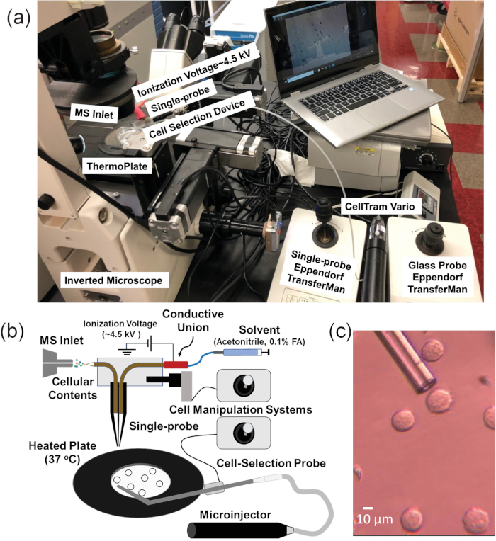
图1. 单个悬浮细胞质谱实验的实验装置。 (A)集成细胞操作平台(ICMP)与质谱仪的耦合系统。(B)悬浮细胞分析的示意图。(C)使用细胞选择探针筛选K562细胞的实验图像。经Standke等46许可转载。版权所有 2019 美国化学会。 请点击此处查看该图的放大版本。
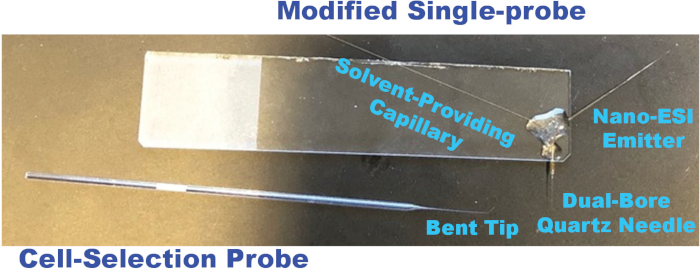
图2. 用于单个悬浮细胞质谱实验的改良单探针和细胞选择探针照片。 请点击此处查看该图的放大版本。
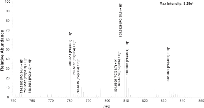
图 3. 单个细胞的局部放大质谱图,显示代表性物种(m/z 750–850)。化学结构通过 MS/MS 分析确认(图 S1)。经 Standke 等人46 许可转载。版权所有 2019 年美国化学会。 请点击此处查看此图的放大版本。
| 药物分子* | m/z | 质量误差 (ppm) |
| [Gemcitabine + H]+ | 264.076 | 11.32 |
| [Taxol + Na]+ | 876.318 | 2.74 |
| [OSW-1 + Na]+ | 895.445 | 0.89 |
| 细胞脂质 | ||
| [PC(34:4) + H]+ | 754.535 | 3.71 |
| [PC(34:3) + H]+ | 756.551 | 3.44 |
| [PC(34:2) + H]+ | 758.569 | 0.66 |
| [PC(36:5) + H]+ | 780.551 | 3.07 |
| [PC(36:4) + H]+ | 782.568 | 2.17 |
| [PC(36:3) + H]+ | 784.585 | 0.64 |
| [PC(38:7) + H]+ | 804.551 | 4.1 |
| [PC(38:6) + H]+ | 806.567 | 2.48 |
| [PC(38:5) + H]+ | 808.583 | 2.72 |
| [PC(38:4) + H]+ | 810.601 | 0 |
| [PC(40:7) + H]+ | 832.583 | 3.12 |
表1. 使用ICMP/单探针装置鉴定的细胞组分。 通过将MS/MS结果与标准化合物进行比对,确认了所有药物化合物的检出。
讨论
集成的细胞操作与分析平台旨在拓展单探针质谱(MS)方法的多功能性,实现在接近天然的环境中对非贴壁细胞进行在线、快速分析。该技术的主要优势在于所需样品前处理极少,因此细胞可在模拟其标准状态的条件下被分析。特别是,可对感兴趣的单个细胞进行可视化识别与选择,从而在维持细胞天然环境的同时,最大限度地减少基质效应对质谱电离效率的影响,使分析结果更能代表细胞的天然状态(图S3)。该技术未来有望用于研究悬浮于生物流体中的患者细胞。该技术的另一优势在于采样溶剂的选择具有灵活性。采样溶剂中应包含乙腈作为主要成分,以实现快速的微尺度裂解。此外,可将内标物(例如同位素标记的药物化合物)加入采样溶剂中,用于定量单个细胞中目标分子(例如药物分子),这些应用未来可能在推动个体化药物治疗方面发挥关键作用54。
尽管该集成系统可方便地用于分析多种类型的细胞,但该方法的一个局限性在于,单探针和细胞选择探针目前均无商业供应,因此每次实验前都需要对多个参数(例如流速、电压、纳米电喷雾发射器与离子传输管之间的距离等)进行优化。此外,由于单探针和细胞选择探针尺寸微小,环境扰动(如气流)可能导致两个探针之间难以形成稳定连接。一种短期解决方案是将细胞选择探针末端附近部分轻微弯曲,以缩短锥形部分的长度。未来的工作将包括开发一个外壳,以封装装置中的关键部件,从而最大限度地减少环境因素的影响。由于单个细胞内含物的量有限,且采集时间较短(约2–3秒),质谱/质谱(MS/MS)分析仅适用于相对丰度较高的分子种类。其他影响检测灵敏度的因素还包括:伴随细胞引入的基质导致离子化效率受到抑制,以及离子在较长的离子传输管中可能发生损失。
披露
作者无任何利益冲突需要披露。
致谢
作者感谢 Naga Rama Kothapalli 在建立悬浮细胞和细胞裂解物实验的样品制备方法方面所做的工作。此外,作者感谢美国国立卫生研究院(NIH)提供经费支持(项目编号:R01GM116116 和 R21CA204706)。
材料
| 姓名 | 公司 | 目录编号 | 评论 |
|---|---|---|---|
| 乙腈 | Millipore Co. | AX0145-1 | 采样溶剂 |
| CellTram Vario | Eppendorf | 6221 | ICMP |
| 铜线 | stores.ebay.com/jewelerheaven | Dead soft, round, 20 guage, 25 ft | 导电连接装置 |
| 数字体视显微镜 | Shenzhen D&F Co. | Supereyes T004 | 分析 |
| 一次性移液器,1-5 µL | Rochester Scientific | 5065 | 细胞选择探针制备 |
| 双孔石英毛细管,1.120"x0.005"x12" | Friedrich & Dimmock, Inc. | MBT-005-020-2Q | 单探针制备 |
| 环氧树脂 | Devcon | Part No. 20945 | 单探针制备 |
| Eppendorf 细胞操作系统 | Eppendorf | Transferman NK517800397-U.R. | ICMP |
| 外螺母 | VALCO*CHEMINERT | EN1 | 离子传输管制备 |
| 甲酸 | Sigma-Aldrich | 399388-500ML | 采样溶剂 |
| 熔融石英毛细管,内径:40 µm,外径:100 µm | Polymicro Technologies | TSP040105 | 单探针制备,导电连接装置 |
| 熔融石英毛细管,内径:50 µm,外径:150 µm | Polymicro Technologies | 1068150015 | 导电连接装置 |
| HyClone 合成胎牛血清(FBS) | Fischer Sci | SH3006603 | 细胞培养 |
| 在线微滤器 | IDEX Health & Science LLC | M-520 | 导电连接装置 |
| 激光拉制仪 | Sutter Instrument Co. | Model P-2000 | 单探针制备 |
| LED 紫外灯 | Foshan Liang Ya Dental Equipment | LY-C240 | 单探针制备 |
| LTQ Orbitrap 质谱仪 | Thermo Scientific | LTQ Orbitrap XL | 分析 |
| 微电极拉制仪 | Narishige, Co. | MF-9 | 细胞选择探针制备 |
| 微型连接器 | IDEX Health & Science LLC | M-539 | 导电连接 |
| PEEK 毛细管,1/32x0.005x 5ft | IDEX Health & Science LLC | 1576 | 导电连接装置 |
| PEEK 毛细管,1/32x0.007x 5ft | IDEX Health & Science LLC | 1577 | 导电连接装置 |
| 青霉素/链霉素 | Gibco/Life Technologies | 15140-122 | 细胞培养 |
| 培养皿,35x10 mm | VWR | 25382-334 | 样品制备 |
| 磷酸盐缓冲液(PBS) | VWR | 0780-50L | 细胞培养 |
| 铂丝 | Narishige, Co. | Model PT-A | 微电极拉制仪 |
| 电源 | Nikon | PSM-2120 | ICMP |
| RPMI,含 Corning 谷氨酰胺的 1X 溶液 | Corning | 10-104-CV | 细胞培养 |
| 单孔毛细管 | Boralex | 5065 | 细胞选择探针制备 |
| 不锈钢卡套,适用于外径 1/16" | IDEX Health & Science LLC | VHP-200-01x | 离子传输管制备 |
| 不锈钢毛细管,1/32x 205 µm x30 cm | IDEX Health & Science LLC | U-1128 | 离子传输管制备 |
| 注射器,250 µL | Hamilton | 1725LTN250UL | 采样注射器 |
| T25 培养瓶 | CellStar | 690160 | 细胞培养 |
| Thermo LTQ XL 离子源接口法兰 | New Objective | PB5500 | 分析 |
| ThermoPlate | TokaiHit | 55R30N | ICMP |
| TrypLE Express | Gibco | 12605-010 | 细胞培养 |
| 管子切割器,适用于 1/16" 不锈钢管 | SUPELCO | 58692-U | 离子传输管制备 |
| USB 数码显微镜 | dx.com | SO2 25~500X | 分析 |
| 紫外固化树脂 | Prime Dental | Item No. 006.030 | 单探针制备 |
| 垂直移液器拉制仪 | David Kopf Instruments | Model 720 | 细胞选择探针制备 |
| 电压外壳 | PicoChip | PCH-A00120 | ICMP/MS 接口 |
| 线剪 | Craftsman | 4 1/2 in end nipper | 导电连接装置 |
参考文献
- Mao, S., et al. In Scatheless Cell Detachment Reveals Correlation between Adhesion Strength and Viability at Single-Cell Resolution. Angewandte Chemie International Edition. 57 (1), 236-240 (2017).
- Rubakhin, S. S., Romanova, E. V., Nemes, P., Sweedler, J. V. Profiling metabolites and peptides in single cells. Nature Methods. 8 (4), S20-S29 (2011).
- Linwen, Z., Akos, V. Single-Cell Mass Spectrometry Approaches to Explore Cellular Heterogeneity. Angewandte Chemie International Edition. 57 (17), 4466-4477 (2017).
- Chen, X., et al. Single-cell analysis at the threshold. Nature Biotechnology. 34, 1111(2016).
- Fu, Q., Tang, J., Cui, M., Xing, J., Liu, Z., Liu, S. Application of porous metal enrichment probe sampling to single cell analysis using matrix-assisted laser desorption ionization time of flight mass spectrometry (MALDI-TOF-MS). Journal of Mass Spectrometry. 51, (2016).
- Passarelli, M. K., Ewing, A. G., Winograd, N. Single-Cell Lipidomics: Characterizing and Imaging Lipids on the Surface of Individual Aplysia californica Neurons with Cluster Secondary Ion Mass Spectrometry. Analytical Chemistry. 85 (4), 2231-2238 (2013).
- Passarelli, M. K., et al. Single-Cell Analysis: Visualizing Pharmaceutical and Metabolite Uptake in Cells with Label-Free 3D Mass Spectrometry Imaging. Analytical Chemistry. 87 (13), 6696-6702 (2015).
- Ostrowski, S. G., Kurczy, M. E., Roddy, T. P., Winograd, N., Ewing, A. G. Secondary Ion MS Imaging To Relatively Quantify Cholesterol in the Membranes of Individual Cells from Differentially Treated Populations. Analytical Chemistry. 79 (10), 3554-3560 (2007).
- Ibáñez, A. J., et al. Mass spectrometry-based metabolomics of single yeast cells. Proceedings of the National Academy of Sciences of the United States of America. 110 (22), 8790-8794 (2013).
- Lanni, E. J., Rubakhin, S. S., Sweedler, J. V. Mass spectrometry imaging and profiling of single cells. Journal of Proteomics. 75 (16), 5036-5051 (2012).
- Ong, T. -H., Tillmaand, E. G., Makurath, M., Rubakhin, S. S., Sweedler, J. V. Mass spectrometry-based characterization of endogenous peptides and metabolites in small volume samples. Biochimica et biophysica acta. 1854 (7), 732-740 (2015).
- Yang, Y., Huang, Y., Wu, J., Liu, N., Deng, J., Luan, T. Single-cell analysis by ambient mass spectrometry. TrAC Trends in Analytical Chemistry. 90, 14-26 (2017).
- Sims, C. E., Allbritton, N. L. Analysis of single mammalian cells on-chip. Lab on a Chip. 7 (4), 423-440 (2007).
- Mizuno, H., Tsuyama, N., Harada, T., Masujima, T. Live single-cell video-mass spectrometry for cellular and subcellular molecular detection and cell classification. Journal of Mass Spectrometry. 43 (12), 1692-1700 (2008).
- Fujii, T., et al. Direct metabolomics for plant cells by live single-cell mass spectrometry. Nature Protocols. 10, 1445(2015).
- Esaki, T., Masujima, T. Fluorescence Probing Live Single-cell Mass Spectrometry for Direct Analysis of Organelle Metabolism. Analytical Sciences. 31 (12), 1211-1213 (2015).
- Tsuyama, N., Mizuno, H., Tokunaga, E., Masujima, T. Live Single-Cell Molecular Analysis by Video-Mass Spectrometry. Analytical Sciences. 24 (5), 559-561 (2008).
- Mizuno, N., Harada, T., Masujima, T., T, H. Live single-cell video-mass spectrometry for cellular and subcellular molecular detection and cell classification. Journal of Mass Spectrometry. , (2008).
- Phelps, M., Hamilton, J., Verbeck, G. F. Nanomanipulation-coupled nanospray mass spectrometry as an approach for single cell analysis. Review of Scientific Instruments. 85 (12), 124101(2014).
- Phelps, M. S., Verbeck, G. F. A lipidomics demonstration of the importance of single cell analysis. Analytical Methods. 7 (9), 3668-3670 (2015).
- Pan, N., Rao, W., Kothapalli, N. R., Liu, R., Burgett, A. W. G., Yang, Z. The Single-Probe: A Miniaturized Multifunctional Device for Single Cell Mass Spectrometry Analysis. Analytical Chemistry. 86 (19), 9376-9380 (2014).
- Gong, X., et al. Single Cell Analysis with Probe ESI-Mass Spectrometry: Detection of Metabolites at Cellular and Subcellular Levels. Analytical Chemistry. 86 (8), 3809-3816 (2014).
- Lorenzo Tejedor, M., Mizuno, H., Tsuyama, N., Harada, T., Masujima, T. In Situ Molecular Analysis of Plant Tissues by Live Single-Cell Mass Spectrometry. Analytical Chemistry. 84 (12), 5221-5228 (2012).
- Shimizu, T., et al. Live Single-Cell Plant Hormone Analysis by Video-Mass Spectrometry. Plant and Cell Physiology. 56 (7), 1287-1296 (2015).
- Yamamoto, K., et al. Cell-specific localization of alkaloids in Catharanthus roseus stem tissue measured with Imaging MS and Single-cell MS. Proceedings of the National Academy of Sciences. 113 (14), 3891(2016).
- Date, S., Mizuno, H., Tsuyama, N., Harada, T., Masujima, T. Direct Drug Metabolism Monitoring in a Live Single Hepatic Cell by Video Mass Spectrometry. Analytical Sciences. 28 (3), 201(2012).
- Hiyama, E., et al. Direct Lipido-Metabolomics of Single Floating Cells for Analysis of Circulating Tumor Cells by Live Single-cell Mass Spectrometry. Analytical Sciences. 31 (12), 1215-1217 (2015).
- Masuda, K., Abouleila, Y., Ali, A., Yanagida, T., Masujima, T. Live Single-Cell Mass Spectrometry (LSC-MS) for Plant Metabolomics. BT - Plant Metabolomics: Methods and Protocols. , 269-282 (2018).
- Ferreira, C. R., Eberlin, L. S., Hallett, J. E., Cooks, R. G. Single oocyte and single embryo lipid analysis by desorption electrospray ionization mass spectrometry. Journal of Mass Spectrometry. 47 (1), 29-33 (2012).
- Ferreira, C. R., Pirro, V., Eberlin, L. S., Hallett, J. E., Cooks, R. G. Developmental phases of individual mouse preimplantation embryos characterized by lipid signatures using desorption electrospray ionization mass spectrometry. Analytical and Bioanalytical Chemistry. 404 (10), 2915-2926 (2012).
- Lee, J. K., Jansson, E. T., Nam, H. G., Zare, R. N. High-Resolution Live-Cell Imaging and Analysis by Laser Desorption/Ionization Droplet Delivery Mass Spectrometry. Analytical Chemistry. 88 (10), 5453-5461 (2016).
- Bergman, H. -M., Lanekoff, I. Profiling and quantifying endogenous molecules in single cells using nano-DESI MS. Analyst. 142 (19), 3639-3647 (2017).
- Yin, L., Zhang, Z., Liu, Y., Gao, Y., Gu, J. Recent advances in single-cell analysis by mass spectrometry. Analyst. 144 (3), 824-845 (2019).
- González-Serrano, A. F., et al. Desorption Electrospray Ionization Mass Spectrometry Reveals Lipid Metabolism of Individual Oocytes and Embryos. PLoS ONE. 8 (9), e74981(2013).
- Liu, Y., et al. Study on Variation of Lipids during Different Growth Phases of Living Cyanobacteria Using Easy Ambient Sonic-Spray Ionization Mass Spectrometry. Analytical Chemistry. 86 (14), 7096-7102 (2014).
- Shrestha, B., et al. Subcellular Metabolite and Lipid Analysis of Xenopus laevis Eggs by LAESI Mass Spectrometry. PLoS ONE. 9 (12), e115173(2014).
- Stolee, J. A., Shrestha, B., Mengistu, G., Vertes, A. Observation of Subcellular Metabolite Gradients in Single Cells by Laser Ablation Electrospray Ionization Mass Spectrometry. Angewandte Chemie International Edition. 51 (41), 10386-10389 (2012).
- Zhang, L., et al. In Situ metabolic analysis of single plant cells by capillary microsampling and electrospray ionization mass spectrometry with ion mobility separation. Analyst. 139 (20), 5079-5085 (2014).
- Shrestha, B., Nemes, P., Vertes, A. Ablation and analysis of small cell populations and single cells by consecutive laser pulses. Applied Physics A. 101 (1), 121-126 (2010).
- Stolee, J. A., Vertes, A. Toward Single-Cell Analysis by Plume Collimation in Laser Ablation Electrospray Ionization Mass Spectrometry. Analytical Chemistry. 85 (7), 3592-3598 (2013).
- Zhang, L., Vertes, A. Single-Cell Mass Spectrometry Approaches to Explore Cellular Heterogeneity. Angewandte Chemie International Edition. 57 (17), 4466-4477 (2018).
- Onjiko, R. M., Moody, S. A., Nemes, P. Single-cell mass spectrometry reveals small molecules that affect cell fates in the 16-cell embryo. Proceedings of the National Academy of Sciences. 112 (21), 6545 LP-6550 (2015).
- Liu, R., Pan, N., Zhu, Y., Yang, Z. T-Probe: An Integrated Microscale Device for Online In Situ Single Cell Analysis and Metabolic Profiling Using Mass Spectrometry. Analytical Chemistry. 90 (18), 11078-11085 (2018).
- Pan, N., Rao, W., Standke, S. J., Yang, Z. Using Dicationic Ion-Pairing Compounds To Enhance the Single Cell Mass Spectrometry Analysis Using the Single-Probe: A Microscale Sampling and Ionization Device. Analytical Chemistry. 88 (13), 6812-6819 (2016).
- Sun, M., Tian, X., Yang, Z. Microscale Mass Spectrometry Analysis of Extracellular Metabolites in Live Multicellular Tumor Spheroids. Analytical Chemistry. 89 (17), 9069-9076 (2017).
- Standke, S. J., Colby, D. H., Bensen, R. C., Burgett, A. W. G., Yang, Z. Mass Spectrometry Measurement of Single Suspended Cells Using a Combined Cell Manipulation System and a Single-Probe Device. Analytical Chemistry. 91 (3), 1738-1742 (2019).
- Rao, W., Pan, N., Yang, Z. High Resolution Tissue Imaging Using the Single-probe Mass Spectrometry under Ambient Conditions. Journal of The American Society for Mass Spectrometry. 26 (6), 986-993 (2015).
- Rao, W., Pan, N., Yang, Z. Applications of the Single-probe: Mass Spectrometry Imaging and Single Cell Analysis under Ambient Conditions. Journal of Visualized Experiments. (112), 53911(2016).
- Rao, W., Pan, N., Tian, X., Yang, Z. High-Resolution Ambient MS Imaging of Negative Ions in Positive Ion Mode: Using Dicationic Reagents with the Single-Probe. Journal of the American Society for Mass Spectrometry. 27 (1), 124-134 (2016).
- Liu, R., Zhang, G., Yang, Z. Towards rapid prediction of drug-resistant cancer cell phenotypes: single cell mass spectrometry combined with machine learning. Chemical Communications. 55 (5), 616-619 (2019).
- Sun, M., Yang, Z. Metabolomic Studies of Live Single Cancer Stem Cells Using Mass Spectrometry. Analytical Chemistry. 91 (3), 2384-2391 (2019).
- Tian, X., Zhang, G., Shao, Y., Yang, Z. Towards enhanced metabolomic data analysis of mass spectrometry image: Multivariate Curve Resolution and Machine Learning. Analytica Chimica Acta. 1037, 211-219 (2018).
- Hu, P., Zhang, W., Xin, H., Deng, G. Single Cell Isolation and Analysis. Frontiers in Cell and Developmental Biology. 4, 116(2016).
- Wu, C., Wu, P., Zhao, H., Liu, W., Li, W. Clinical Applications of and Challenges in Single-Cell Analysis of Circulating Tumor Cells. DNA and Cell Biology. 37 (2), 78-89 (2017).
- Schober, Y., Guenther, S., Spengler, B., Römpp, A. Single Cell Matrix-Assisted Laser Desorption/Ionization Mass Spectrometry Imaging. Analytical Chemistry. 84 (15), 6293-6297 (2012).
- Pulfer, M., Murphy, R. C. Electrospray mass spectrometry of phospholipids. Mass Spectrometry Reviews. 22 (5), 332-364 (2003).
重印与许可
