由于缺乏足够的工具和方法,神经退行性疾病中神经元与胶质细胞之间的相互作用尚未被充分理解。在此,我们描述了从人多能干细胞获得诱导神经元、少突胶质前体细胞以及少突胶质细胞的优化方案,并举例说明这些方法在理解阿尔茨海默病中细胞类型特异性贡献方面的应用价值。
需要JoVE订阅才能观看此内容。 请登录或开始免费试用
方法文章
由多能干细胞生成人神经元和少突胶质细胞以模拟神经元-少突胶质细胞相互作用 多能干细胞, 神经元, 少突胶质细胞, 细胞分化, 神经发育, 共培养, 神经退行性疾病, 人源细胞模型, 髓鞘形成, 神经胶质生物学
* These authors contributed equally
本文内容
勘误通知
Important: There has been an erratum issued for this article. View Erratum Notice
摘要
摘要
在阿尔茨海默病(AD)及其他神经退行性疾病中,少突胶质细胞功能障碍是一种常见的早期病理特征,但其如何参与疾病的发生与发展,特别是在大脑灰质中的作用,目前仍知之甚少。少突胶质细胞谱系细胞的功能障碍主要表现为髓鞘形成缺陷以及少突胶质前体细胞(OPCs)自我更新能力受损。这两种缺陷至少部分是由于随着病理过程的积累,神经元与少突胶质细胞之间的相互作用遭到破坏所致。在中枢神经系统发育过程中,OPCs可分化为具有髓鞘形成能力的少突胶质细胞。在成熟的脑皮层中,OPCs是主要的增殖性细胞(约占脑细胞总数的5%),并以神经活动依赖的方式调控新髓鞘的形成。此类神经元-少突胶质细胞间的通讯机制迄今研究仍十分不足,尤其是在AD等神经退行性疾病的背景下,主要受限于缺乏合适的工具。近年来,我们课题组及其他研究团队已在优化现有技术方面取得显著进展,能够分别从人多能干细胞高效诱导生成功能性神经元和少突胶质细胞。在本论文中,我们详细描述了经过优化的实验流程,包括建立共培养体系以模拟神经元与少突胶质细胞之间的连接。我们的示例结果提示,OPCs/少突胶质细胞可能对脑内淀粉样蛋白沉积及突触完整性具有意料之外的作用,并凸显了该方法在AD研究中的应用价值。这种还原论方法有助于从大脑固有的复杂性中解析出特定的异细胞间相互作用。本文所述的实验方案有望推动未来关于少突胶质细胞缺陷在神经退行性疾病发病机制中作用的深入研究。
引言
少突胶质细胞谱系细胞——包括少突胶质前体细胞(OPCs)、形成髓鞘的少突胶质细胞以及介于两者之间的过渡类型——是人类脑细胞中的一个重要类别1,在神经发育和衰老过程中积极参与中枢神经系统的正常运作与维持的多种关键功能2,3,4。尽管少突胶质细胞因生成髓鞘以促进神经元活动传导并支持白质中轴突健康而广为人知,但OPCs在髓鞘化稀少的灰质中却十分丰富(约占5%),并执行依赖神经活动的信号传导功能,调控学习行为与记忆形成5,6,7,8。然而,少突胶质细胞在阿尔茨海默病(AD)及其他与年龄相关的神经退行性疾病发病机制中如何发挥功能及其功能障碍,目前研究仍不充分9。造成这一研究空白的主要原因在于缺乏合适的模型系统,以及缺乏指导实验路径的普遍性知识。
鉴于近年来在从多能干细胞(包括胚胎干细胞(ES)和诱导多能干细胞(iPS))中获取人类脑细胞方面的最新突破,这....
访问受限。请登录或开始试用以查看此内容。
方案
1. 由人多能干细胞诱导人神经元
- 慢病毒制备(约5天,详细方案如先前所述16)
- 将约100万个HEK293T细胞接种至每个T75培养瓶中,使转染时细胞融合度约为40%。使用表达四环素诱导型Ngn2和嘌呤霉素抗性基因(PuroR;在同一TetO启动子控制下)、rtTA以及三种辅助质粒pRSV-REV、pMDLg/pRRE和VSV-G的质粒进行转染(每瓶加入12 µg慢病毒载体DNA和每种辅助质粒DNA各6 µg)。每次慢病毒制备至少准备三个培养瓶。使用PEI进行转染,操作遵循制造商说明书。转染16小时后更换培养基并弃去旧培养基。
- 连续3天每天收集含病毒颗粒的培养上清,并更换新鲜培养基。将收集的上清液合并用于病毒纯化。通过0.22 µm滤膜过滤病毒液,然后在49,000 × g条件下离心90分钟。将沉淀重悬于适当体积的PBS-葡萄糖溶液中(约150 µL)。
- 神经元诱导(约5天)
注意:该诱导方案(图1A;流程图)对经验证具有多能性的iPS细胞和ES细胞均非常有效(可通过免疫组织化学染色检测明确的多能性标志物进行验证;图1B)。- 使用商业化H1人源ES细胞(传代52代,见材料表
访问受限。请登录或开始试用以查看此内容。
结果
直接从人多能干细胞生成人诱导神经元
起始使用的人多能干细胞必须具有高度的多能性,这对于成功生成诱导神经元(iNs)或诱导少突胶质前体细胞/少突胶质细胞(iOPCs/iOLs)至关重要。因此,在启动本论文所述的任一诱导方案之前,应使用特定标志物(如 Oct4 和 SOX2)对细胞进行染色鉴定(图1A)。本研究采用人 H1 细胞,依据 Zhang 等人先前发表的方案并加以部分修改,获得诱导性兴奋性前脑神经元(图1C)12,16,17,18。本文介绍了一种在第2天将iNs重新接种于基质溶液(见材料表)中进行纯培养的方案,无需任何饲养层(如胶质细胞或成纤维细胞)。除此前已发表的方案外,我们还观察到,.......
访问受限。请登录或开始试用以查看此内容。
讨论
除了通过髓鞘化为突触结构提供物理和代谢支持并促进跳跃式信号传导外,少突胶质细胞系细胞还能通过与神经元之间快速而动态的相互作用,调控神经元活动模式5,6,7。在阿尔茨海默病(AD)病理过程中,少突胶质细胞的反应最初被认为仅仅是继发于炎症和氧化应激;但现在已有令人信服的证据表明,髓鞘完整性受损是在Aβ聚集和tau蛋白过度磷酸化出现之前发生的早期致病事件9。此外,在AD中,通过少突胶质前体细胞(OPC)自我更新实现的髓鞘修复过程尤为脆弱38,而这一过程高度依赖于神经元活动。因此,阐明支持健康神经元-少突胶质细胞信号传导的机制,为发现新的治疗靶点提供了重要机遇。
单转录因子 Ngn2 方案是目前最常用的干细胞来源人神经元的生成技术之一,本文所述步骤进一步优化了该方法,以获得高纯度的神经元培养物。我们的 iOPC.......
访问受限。请登录或开始试用以查看此内容。
披露
作者无任何利益冲突需要披露。
致谢
本工作获得了美国国立卫生研究院(R00 AG054616 资助 Y.A.H.,T32 GM136566 资助 K.C.)、斯坦福大学医学院以及Siebel奖学金(授予 S.C.)的资助。Y.A.H. 是布朗转化科学研究所转化神经科学中心的GFL转化教授。
....访问受限。请登录或开始试用以查看此内容。
材料
| 姓名 | 公司 | 目录编号 | 评论 |
|---|---|---|---|
| Accutase | STEMCELL Technologies | 7920 | |
| B27添加剂 | ThermoFisher | 17504044 | |
| bFGF | ThermoFisher | PHG 0266 | |
| cAMP | MilliporeSigma | A9501 | |
| 氯马斯汀 | MilliporeSigma | SML0445 | |
| DMEM/F12培养基 | STEMCELL Technologies | 36254 | |
| DMSO | ThermoFisher | D12345 | |
| 强力霉素 | MilliporeSigma | D3072 | |
| 胎牛血清 | ScienCell | 10 | |
| H1人胚胎干细胞 | WiCell | WA01 | |
| Matrigel | Corning | 354234 | |
| mTeSR plus | STEMCELL Technologies | 5825 | |
| N2添加剂 | ThermoFisher | 17502001 | |
| Neurobasal A培养基 | ThermoFisher | 10888-022 | |
| 非必需氨基酸 | ThermoFisher | 11140-050 | |
| PDGF-AA | R&D Systems | 221-AA-010 | |
| PEI | VWR | 71002-812 | |
| pMDLg/pRRE | Addgene | 12251 | |
| 聚凝胺 | MilliporeSigma | TR-1003-G | |
| pRSV-REV | Addgene | 12253 | |
| 嘌呤霉素 | ThermoFisher | A1113803 | |
| ROCK抑制剂Y-27632 | STEMCELL Technologies | 72302 | |
| SAG | Tocris | 4366 | |
| STEMdiff神经前体细胞冻存培养基 | STEMCELL Technologies | 5838 | |
| STEMdiff SMADi神经诱导试剂盒 | STEMCELL Technologies | 8581 | |
| T3三碘甲腺原氨酸 | MilliporeSigma | T6397 | |
| Tempo-iOlogo:人iPSC来源的少突胶质前体细胞 | Tempo BioScience | SKU102 | |
| TetO-Ng2-Puro | Addgene | 52047 | |
| VSV-G | Addgene | 12259 |
参考文献
- Pelvig, D. P., Pakkenberg, H., Stark, A. K., Pakkenberg, B. Neocortical glial cell numbers in human brains. Neurobiology of Aging. 29 (11), 1754-1762 (2008).
- Barres, B. A. The mystery and magic of glia: a perspective on their roles in health and disease. Neuron. 6....
访问受限。请登录或开始试用以查看此内容。
重印与许可
勘误
Formal Correction: Erratum: Generation of Human Neurons and Oligodendrocytes from Pluripotent Stem Cells for Modeling Neuron-Oligodendrocyte Interactions
Posted by JoVE Editors on 12/29/2020. Citeable Link.
An erratum was issued for: Generation of Human Neurons and Oligodendrocytes from Pluripotent Stem Cells for Modeling Neuron-Oligodendrocyte Interactions. The Representative Results section has been updated.
Figure 3 was updated from:
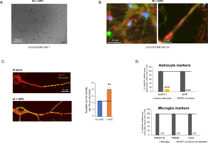
Figure 3: Co-culture of iNs and iOPCs. (A) Representative bright field image of co-cultured iNs and iOPCs at Day 7, showing a proper density for further maturation. (B) Representative immunofluorescence image of iNs and iOPCs co-cultured for 28 days. Axonal marker neurofilament NF is shown in green and oligodendrocytic marker MBP in red. Right, a segment of iN axon ensheathed by iOL process (MBP+). (C) Synapse formation assayed in 4-week-old co-cultures. Cells were stained for Synapsin 1 (Syn1, green) and MAP2 (red), and synaptic puncta were quantified by confocal analysis of density along the dendritic segments as described17,18. (D) In our co-cultures of iNs and iOPCs (7 days of co-culturing), the expression of astrocyte markers, ALDHL1 and GFAP, is minimal (top), and the expression of microglia markers, TMEM119, TREM2, and CD33, is not detected (N.D.) by qPCR. The contamination from these two glial cell types is thus excluded. Please click here to view a larger version of this figure.
to:
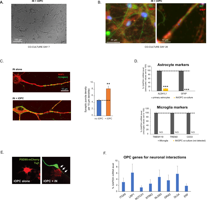
Figure 3: Co-culture of iNs and iOPCs. (A) Representative bright field image of co-cultured iNs and iOPCs at Day 7, showing a proper density for further maturation. (B) Representative immunofluorescence image of iNs and iOPCs co-cultured for 28 days. Axonal marker neurofilament NF is shown in green and oligodendrocytic marker MBP in red. Right, a segment of iN axon ensheathed by iOL process (MBP+). (C) Synapse formation assayed in 4-week-old co-cultures. Cells were stained for Synapsin 1 (Syn1, green) and MAP2 (red), and synaptic puncta were quantified by confocal analysis of density along the dendritic segments as described17,18. (D) In our co-cultures of iNs and iOPCs (7 days of co-culturing), the expression of astrocyte markers, ALDHL1 and GFAP, is minimal (top), and the expression of microglia markers, TMEM119, TREM2, and CD33, is not detected (N.D.) by qPCR. The contamination from these two glial cell types is thus excluded. (E) Coculturing iOPC with iN leads to the formation of neuron-OPC synapses. The fluorescence-tagged post-synaptic marker PSD95-mCherry is expressed only in OPCs, and display a diffuse pattern in single cultures (left) but aggregate to form puncta in cocultures (right, indicated by arrows; Tuj1, neuronal marker). (F) The expression of well-characterized oligodendroglial genes that can sense and respond to neuronal activities in the pure cultures of iOPCs at Day 14. Please click here to view a larger version of this figure.
The fourth paragraph was updated from:
Co-culturing of iNs and iOPCs
This protocol is optimized specifically for co-culturing iNs and iOPCs and allow our real-time monitoring of the inter-cellular communications between these two cell types along the course of neural development. The ideal plating densities for both cell types need to be decided with a series of cell number titration to achieve proper differentiation (Figure 3A). After 4 weeks in co-cultures, the iOPCs are expected to be adequately differentiated into OLs that are positive for specific markers such as MBP and extend processes to ensheath axons (Figure 3B). The co-culture system can robustly boost up the number of synapses, indicating that the iOPCs provide a neuronal support through physical contacts or release of trophic factors (Figure 3C). We can maintain the co-cultures in acceptable health condition for up to 6 weeks and observe that the synapse number and other neuronal attributes plateau around the fifth week. Of note, astrocytes and microglia are not present in our preparations and their absence can be documented by checking the expression of specific markers (Figure 3D).
to:
Co-culturing of iNs and iOPCs
This protocol is optimized specifically for co-culturing iNs and iOPCs and allow our real-time monitoring of the inter-cellular communications between these two cell types along the course of neural development. The ideal plating densities for both cell types need to be decided with a series of cell number titration to achieve proper differentiation (Figure 3A). After 4 weeks in co-cultures, the iOPCs are expected to be adequately differentiated into OLs that are positive for specific markers such as MBP and extend processes to ensheath axons (Figure 3B). The co-culture system can robustly boost up the number of synapses, indicating that the iOPCs provide a neuronal support through physical contacts or release of trophic factors (Figure 3C). We can maintain the co-cultures in acceptable health condition for up to 6 weeks and observe that the synapse number and other neuronal attributes plateau around the fifth week. Of note, astrocytes and microglia are not present in our preparations and their absence can be documented by checking the expression of specific markers (Figure 3D). The iOPCs express a good number of well-characterized genes that can potentially respond to and mediate the activity-dependent signals from neighboring neurons, in a paracrine (e.g. neurotrophins and metabolites) and/or a synaptic manner (Figure 3E and 3F).
