本方案描述了人诱导性多能干细胞衍生的类似2型肺泡上皮细胞(iAT2s)。这些细胞可在三维培养条件下以自我更新的球体形式进行培养,或适应于气液界面(ALI)培养条件。
方法文章
生成人诱导性多能干细胞来源的2型肺泡上皮细胞的三维球体及二维气液界面培养模型
* These authors contributed equally
本文内容
摘要
摘要
在肺部,肺泡上皮构成一道抵御环境刺激的物理屏障,在维持稳态和疾病发生中发挥关键作用。Ⅱ型肺泡上皮细胞(AT2s)是远端肺上皮的兼性前体细胞。AT2s的功能障碍和损伤可导致并加剧多种肺部疾病。因此,深入理解AT2s的生物学特性对于阐明肺部生物学及疾病机制至关重要。然而,原代人源AT2s通常难以分离且来源有限。为克服这些局限,可通过模拟肺发育过程的定向分化方案,由人诱导性多能干细胞(iPSC)分化获得Ⅱ型肺泡上皮细胞(iAT2s),从而提供一种替代来源 体内 肺发育。iAT2s 在无饲养层条件下生长,其转录组程序与人类成体原代AT2细胞相似,并能执行AT2细胞的关键功能,例如表面活性物质的合成、包装和分泌。本方案详细描述了通过连续传代在三维(3D)培养体系中维持具有自我更新能力的iAT2s,或将iAT2s适应于气液界面(ALI)培养体系的方法。在接种前,需制备iAT2s的单细胞悬液,接种于3D溶解态基底膜基质(下文简称 "基质"),它们会自组装成单层上皮细胞球体。在三维培养中,iAT2细胞可被连续解离为单细胞悬液,用于传代或接种至二维气液界面(ALI)培养系统。在ALI培养中,iAT2细胞形成具有极性的单层结构,其顶面暴露于空气中,使该体系易于进行环境暴露实验。因此,本方案可提供无限供应的iAT2细胞,产量可达1 × 10以上30 每输入细胞在15次传代过程中产生的细胞数,同时维持由SFTPC指示的AT2细胞程序tdTomato 表达。所获得的细胞代表了一种可重复且具有相关性的研究平台,可用于研究基因突变、模拟环境暴露或进行药物筛选。
引言
在肺部,气道和肺泡上皮细胞是首先接触吸入性环境暴露物的细胞,这些暴露物包括通过吸入气溶胶传播的病原体以及香烟烟雾等有害刺激物。2型肺泡上皮细胞(AT2s)在维持肺部稳态中起关键作用,因其是远端肺上皮的兼性前体细胞,能够产生表面活性物质以降低肺泡表面张力,并对吸入性暴露物启动固有免疫反应。1,2然而,肺部损伤或在AT2细胞中选择性表达的基因发生突变(例如)均可导致AT2功能障碍 SFTPC, SFTPB,以及 ABCA33,4,5. 以往研究这些基因突变的方法主要依赖于小鼠模型6 或导入永生化细胞系的含突变工程载体7因此,需要能够模拟遗传和环境干扰对生理相关细胞类型中AT2细胞影响的平台,并以高效的方式 体外 需要进一步的系统研究来深入理解AT2细胞在健康与疾病中的作用。
在模拟空气传播暴露方面,气液界面(ALI)培养的原代气道上皮细胞已被成功用于揭示香烟烟雾引起的关键分子反应8 以及模拟气道感染9,10与气道上皮模型系统相比,作为疾病感染或损伤关键部位的肺泡上皮,其相应的气液界面(ALI)培养系统发展程度要低得多。已采用永生化细胞系在气液界面进行培养,以模拟肺泡上皮11,12,但这些细胞系在转录组水平上与原代肺泡上皮细胞存在差异13 缺乏关键的细胞机制,例如在气液界面处分泌表面活性剂或形成紧密连接的能力12原代人AT2细胞可在成纤维细胞存在下以三维球体形式培养1,14 但存在局限性,包括难以从肺组织外植体中获取,以及在迄今为止大多数培养条件下易发生衰老或失去细胞表型。这些特性限制了原代AT2细胞的广泛应用 体外 研究,尽管近期在优化无饲养层的三维培养体系以扩增原代AT2细胞方面已取得进展15,16,17,18.
定向分化方案已被开发用于模拟 体内 生成人诱导性多能干细胞(iPSC)来源的2型肺泡上皮细胞(iAT2s)的发育里程碑19iAT2细胞在含有CHIR99021、KGF、地塞米松、cAMP和3-异丁基-1-甲基黄嘌呤(IBMX)的无血清、三维限定培养基中,以自我更新的球状体形式生长,该培养基称为 "CK + DCI"19,无需成纤维细胞饲养层,且可进行长期培养 >20个片段20,21此外,iAT2细胞与人类成体原代AT2细胞具有相似的转录组程序,能够形成板层小体,并产生和包装表面活性物质19,21,22该方案详细描述了通过将细胞解离为单细胞悬液对iAT2s进行连续传代的方法。在此阶段,iAT2s可重新接种并进一步在三维培养体系中扩增,或接种于二维气液界面(ALI)培养体系中。23这些方法可用于研究AT2细胞在稳态及疾病状态下的内在生物学特性20,22 并利用可扩展的、符合生理相关性的平台来探究化合物或刺激物的作用效果23,24,这一点先前已有报道。
方案
所有涉及人iPSC系分化的实验均遵循波士顿大学机构审查委员会的规定(方案编号H33122)。用于重编程为iPSC的真皮成纤维细胞来自已签署书面知情同意书的供体,获取过程经密苏里州圣路易斯市华盛顿大学医学院人类研究保护办公室批准。重编程的iPSC由马萨诸塞州波士顿市波士顿大学和波士顿医疗中心再生医学中心制备。
1. 肺泡球体解离
- 根据表1中所述的成分配制完全无血清分化培养基(cSFDM)。
- 根据表2,在已配制的cSFDM基础培养基中配制CK + DCI培养基。
- 根据实验需要,将2D(适用于人胚胎干细胞的)和/或3D(生长因子减少型)基质置于冰上解冻。
- 使用移液器或带真空的抽吸移液器,从12孔板中含由定向分化衍生的肺泡球体(alveolospheres)的3D基质液滴中吸除全部CK + DCI培养基19。
- 每滴加入1 mL分散酶(dispase,2 mg/mL)。使用P1000移液器轻轻将液滴吹打混入分散酶中。37 °C孵育1小时,于30分钟后轻轻吹打上下混合一次。
- 将步骤1.5中解离的类器官(来自一个基质液滴)转移至15 mL锥形管中。加入10 mL Iscove改良Dulbecco培养基(IMDM,参见材料表)进行洗涤。
- 室温下以300 x g离心5分钟。使用移液器或带真空的抽吸移液器吸除上清液,尽可能残留最少的上清。
注意:必须彻底去除所有分散酶,因为残留的分散酶可能会溶解后续接种细胞所用的基质。若在沉淀物上方观察到明显的浑浊层,说明分散酶尚未完全溶解基质,可向沉淀物中再添加分散酶,并在37 °C继续孵育20-30分钟。 - 每滴用1 mL 0.05%胰蛋白酶重悬细胞,并转移回12孔板。37 °C孵育12-15分钟。在显微镜下观察解离情况。此阶段应避免过度吹打细胞。
注意:孵育结束时,使用P1000移液器吹打3-5次后,细胞应达到单细胞悬液状态。若需将iAT2细胞传代至气液界面培养(ALI,步骤3),则胰酶消化时间应尽量缩短(最长12分钟),使细胞呈2-3个细胞的小团簇状态,而非完全单细胞悬液,以便接种至细胞培养小室时更适宜。 - 加入等体积含胎牛血清(FBS)的培养基(DMEM中含10%适用于胚胎干细胞的FBS)以终止胰蛋白酶作用。室温下以300 x g离心5分钟。
- 用10 mL IMDM洗涤细胞一次。室温下以300 x g离心5分钟。
- 用适量体积的缓冲液重悬细胞用于计数,并使用血细胞计数板对细胞进行计数(参见材料表)。
注意:从一个初始接种密度为400个细胞/µL、已完全融合的50 µL基质液滴中,预期每滴可获得500,000至1.5 x 10⁶个细胞。 - 使用iAT2细胞的单细胞悬液,通过接种于3D基质中生成肺泡球体(步骤2)和/或接种于细胞培养小室上进行气液界面(ALI)培养(步骤3)。
2. iAT2细胞的3D包埋培养
- 计数后(步骤 1.11),确定需重新接种到 3D 基质中的细胞数量(每 12 孔板孔中接种 50–100 µL 的 3D 基质液滴,细胞密度为 400 个细胞/µL 基质)。在室温下以 300 × g 离心 5 分钟,然后用移液器尽可能吸除上清液。
- 将细胞重悬于 3D 基质中。如需防止基质聚合(温度升高时会发生聚合),应快速操作并置于冰上。
- 使用 P200 移液器将一个 3D 基质液滴加入预热的 12 孔板的每个孔中。移液时需小心,避免在基质液滴中产生气泡。在连续加样多个液滴时,不要让细胞悬液发生沉降。
- 将培养板放入 37 °C 培养箱中孵育 20–30 分钟,使基质液滴完成聚合。
- 每孔加入 1 mL CK + DCI + 10 µM Y-27632 培养基(参见材料表),以完全覆盖基质液滴。
- 72 小时后,更换为不含 10 µM Y-27632 的 CK + DCI 培养基。
- 每 48–72 小时更换一次新鲜的 CK + DCI 培养基。
注意:iAT2 细胞通常需要每 10–14 天传代一次,具体时间取决于细胞系和接种密度。
3. 将iAT2细胞传代至气液界面(ALI)
- 使用前1小时,将2D基质按照生产商说明书要求用杜氏改良伊格尔培养基/营养混合物F-12(DMEM/F12)稀释至工作浓度,新鲜包被6.5 mm细胞培养小室 材料表然后,向每个6.5 mm细胞培养小室中加入100 µL稀释后的基质。将基质置于37 °C培养箱中孵育30分钟,或在室温下放置1小时,使其聚合。
- 用移液器或连接真空泵的抽吸移液器吸除细胞培养小室中多余的基质,并用 DMEM/F12 洗涤一次。在加入细胞前立即吸除该洗涤液。
- 计数后,确定在培养小室中接种所需数量细胞的细胞数目(520,000 个活细胞/cm²)。2,相当于每6.5 mm细胞培养插件中172,000个活细胞)。以300 x g 室温下孵育5分钟。尽可能吸除上清液。
- 将细胞重悬于适当体积的溶液中,使每孔细胞培养插入物接种100 µL CK + DCI + 10 µM Y-27632(细胞悬液浓度应为1,720 个细胞/µL)。取100 µL加入细胞培养插入物的顶室。以十字形轻轻振荡培养板,确保细胞在细胞培养插入物表面均匀分布,并在4倍物镜下显微镜观察确认。
注意:接种密度至关重要,可能需要优化,范围为每6.5 mm细胞培养小室160,000–300,000个细胞。钙黄绿素绿(见 材料表可将细胞膜通透性染料加入细胞中,随后在倒置荧光显微镜下观察,以在接种后对细胞进行成像。 - 向每个细胞培养小室的基底外侧加入500 µL CK + DCI + 10 µM Y-27632。
- 吸弃接种后48 h的顶端CK + DCI + 10 µM Y-27632培养基,使用移液器开始气液界面(ALI)培养(称为 "气升式").
注意:此时细胞应达到100%汇合。如果细胞未达到100%汇合,仍应在指定时间点执行步骤3.7–3.8;汇合的细胞单层可能需要更长时间才能形成。 - 72小时后,将基底侧培养基从含10 µM Y-27632的CK + DCI更换为不含10 µM Y-27632的CK + DCI。
注意:应尽可能减少,若存在,应极少, "泄漏" 向顶端加入培养基。然而,如果在最初几天内发生泄漏,则需继续每天吸出顶端液体,直至不再有进一步的泄漏。 - 每48-72小时用新鲜的CK + DCI更换基底外侧培养基。
注意:用于气液界面(ALI)培养的iAT2细胞在接种后5-14天达到最大程度的成熟。 - 如有更长期实验需求,可在接种后对细胞进行长达28天的培养,并密切监测。
注意:在培养时间过长时,可能会观察到气液界面(ALI)失效的可见迹象,例如细胞从插入物边缘脱落或单层细胞出现孔洞,此时该ALI模型已无法用于实验。
结果
将iAT2细胞传代为单细胞悬液,随后重铺于3D基质中或铺在细胞培养小室上进行气液界面(ALI)培养(图1)。单细胞解离后,通过流式细胞术分析iAT2细胞。简言之,将细胞重悬于含1%胎牛血清(FBS)的磷酸盐缓冲液(PBS)中,并加入钙黄素蓝活性染料(1:1000),在流式细胞仪上进行分析,设门筛选非碎片、单细胞及tdTomato阳性细胞。此处以SPC2细胞系(SPC2-ST-B2克隆20)为例,该细胞系携带靶向内源性SFTPC位点的tdTomato报告基因,便于在培养过程中对AT2细胞程序进行可视化观察与追踪(图2A)。当iAT2细胞重铺于3D基质形成类球体时(图2B),或铺于细胞培养小室上时(图2C),SFTPC-tdTomato的表达均得以维持。可通过跨上皮电阻(TEER)测定评估ALI培养模型的完整性(图2D)。测定TEER时使用伏特-欧姆计(见材料表),并在顶端室中加入100 µL CK + DCI培养基。未接种细胞但涂有Matrigel的细胞培养小室作为空白对照。每个孔在三个不同位置进行测量读数。
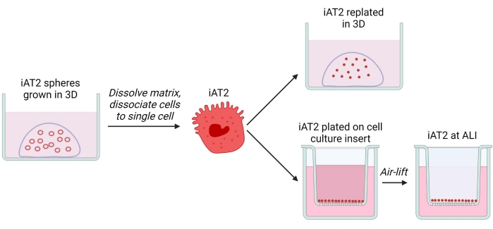
图 1:实验方案流程的示意图。 请点击此处查看该图的放大版本。
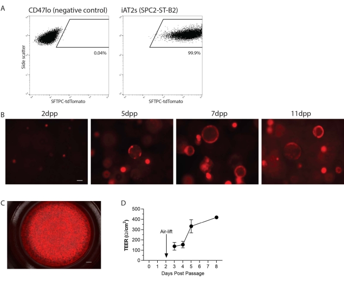
图 2:实验方案的代表性结果。(A)在三维培养条件下,来源于人诱导性多能干细胞的2型肺泡上皮样细胞(iAT2s)中SFTPC-tdTomato的代表性流式细胞术结果。所示阴性对照为非肺内胚层细胞(CD47lo)。(B)iAT2s在三维培养中不同传代后时间点(dpp)的代表性活细胞成像(SFTPC-tdTomato,比例尺 = 50 µm)。(C)iAT2s在气液界面接种后的代表性活细胞成像(SFTPC-tdTomato,比例尺 = 500 nm)。(D)iAT2s在气液界面接种后的代表性跨上皮电阻值。n = 3,误差条表示标准差。请点击此处查看该图的放大版本。
| 试剂 | 500 mL 所需体积 | 终浓度 |
| Iscove's Modified Dulbecco's Medium (IMDM) | 375 mL | 75% |
| Ham's F-12 Nutrient Mixture (F12) | 125 mL | 25% |
| B-27 (含 RA) 添加剂 | 5 mL | 1% |
| N-2 添加剂 | 2.5 mL | 0.50% |
| BSA (7.5% 储存液) | 3.3 mL | 0.05% |
| Primocin (50 mg/mL 储存液) | 1 mL | 100 µg/mL |
| Glutamax (100x 储存液) | 5 mL | 1x |
| 抗坏血酸 (50 mg/mL 储存液) | 500 µL | 50 µg/mL |
| 1-巯基甘油 (MTG) (由 26 µL 溶于 2 mL IMDM 配制) | 1.5 mL | 4.5 x 10⁻⁴ M |
表1:完全无血清分化培养基(cSFDM)的成分。
| 试剂 | 终浓度 |
| CHIR99021 | 3 µM |
| rhKGF | 10 ng/mL |
| Dexamethasone | 50 nM |
| 8-溴腺苷-3',5'-单磷酸钠盐 (cAMP) | 0.1 mM |
| 3-异丁基-1-甲基黄嘌呤 (IBMX) | 0.1 mM |
表2:CK + DCI 培养基的成分组成。
讨论
AT2细胞维持肺部稳态,这些关键肺泡细胞的功能障碍既可导致也可源于多种肺部疾病。由于获取和分离原代人AT2细胞存在困难,因此可生成iAT2细胞。通过应用 elsewhere25 所述的定向分化方法以及本文所述的扩增与细胞接种技术,可将来自任何个体的iPSC分化为具有强大自我更新能力的iAT2细胞,从而为生物医学研究提供患者特异性的细胞,包括基础生物医学研究、发育研究、疾病建模、细胞治疗或药物筛选。本方案详细描述了一种将iAT2细胞解离为单细胞悬液的方法,该悬液可重新接种于3D Matrigel中以生成肺泡球用于细胞扩增,或接种于2D空气-液体界面(ALI)培养系统中用于进一步实验。
该方案包含多个关键步骤,以确保在三维(3D)和气液界面(ALI)培养中成功传代和重新接种。必须尽量减少机械吹打,过度移液会降低细胞活力。此外,iAT2s在3D和2D ALI培养中的接种密度至关重要;对于3D培养,通常每微升基质中接种400个细胞/µ3D Matrigel 的 L 值对大多数 iPSC 系系均较为适宜。然而,细胞密度在 100–500 个细胞/µL有时是成功的,但不同细胞系可能需要优化密度。对于气液界面(ALI)培养,优化iAT2s在细胞培养小室上的接种密度也至关重要,以确保接种后48小时(即空气抬升当天)形成融合的单层细胞。某些iAT2细胞系每小室需要更多细胞;因此,若需 troubleshooting,建议在24孔细胞培养小室中每小室接种160,000–300,000个细胞。3D培养也可通过保持接种密度为400个细胞/µ3D基质的体积及将基质液滴大小减小至25 L µL 和 20 µL,分别。可通过在每个插件上包被30 μL基质,将ALI培养扩增至96孔细胞培养插件中 µ30 µL 基质中接种 80,000 个细胞 µL 每插孔,每次换液加入 150 µ每孔加入 L 的基底外侧培养基。对于其他孔板规格,需相应调整并优化接种密度以及基质和培养基的体积。
本方案的一个局限性在于,用于气液界面(ALI)接种的细胞必须充分纯化,达到NKX2-1+和SFTPC+19,20,21,23水平,才能形成iAT2的ALI结构,且不同细胞系之间的ALI培养形成能力可能存在差异。此外,本方案仅支持AT2细胞的扩增与培养,提供了一种基于单一关键肺泡细胞类型的简化模型系统。重要的是,这些平台中缺少其他相关肺泡细胞类型,例如肺泡I型细胞。其他研究团队已成功将原代人AT2细胞以类球体或类器官形式进行培养,无论是否添加成纤维细胞1,14,15,16,17;然而,原代人AT2细胞在无ALI条件的常规二维(2D)培养中 tends to 失去关键表面活性物质的表达26。此外,最近的研究表明,与新鲜未培养的原代人AT2细胞相比,iAT2细胞表现出更高的增殖标志物表达和较低的AT2成熟标志物水平27。尽管iAT2细胞与新鲜原代人AT2细胞之间存在这些差异,但我们发现即使是培养后的原代AT2细胞,其转录组也与新鲜未培养的原代AT2细胞存在差异27,因此我们认为这些体外模型在模拟体内肺部生物学方面各有优势与局限。
iAT2 平台可用于研究源自患者来源的诱导性多能干细胞(iPSC)的基因突变19,20。该系统还可大幅扩增,以满足高通量药物筛选的需求。此外,iAT2 气液界面(ALI)培养模型适用于多种环境暴露研究,例如病毒感染、细菌感染,或香烟烟雾、电子烟雾及其他气溶胶的暴露23。总之,本项生成 iAT2 的三维(3D)和二维气液界面(2D ALI)培养方案,可实现与疾病相关细胞类型的长期培养,提供一种生理相关性强、可扩展的实验平台,支持这些细胞在多种研究中的应用。
披露
作者无任何利益冲突需要披露。
致谢
我们衷心感谢Wilson、Kotton和Hawkins实验室成员提供的有益讨论。我们还非常感谢Greg Miller(CReM实验室主管)和Marianne James(iPSC核心实验室主管)提供的宝贵支持。我们感谢Brian Tilton及波士顿大学医学中心流式细胞术核心实验室在细胞分选方面的技术协助(该工作由美国国立卫生研究院资助项目#1S10OD021587-01A1支持)。本研究得到了澳大利亚国家健康与医学研究委员会授予RBW的CJ Martin早期职业研究员奖学金、NIH资助项目F30HL147426(授予KMA)、肺纤维化基金会颁发的I.M. Rosenzweig青年研究者奖,以及通过波士顿大学临床与转化科学研究所获得的整合试点资助项目的支持。 & 转化科学研究所(1UL1TR001430)资助 KDA;瑞士国家科学基金会(P2ELP3_191217)资助 ABA;美国国立卫生研究院(NIH)基金 U01HL148692、U01HL134745、U01HL134766 和 R01HL095993 资助 DNK;美国国立卫生研究院(NIH)基金 U01TR001810、R01DK101510 和 R01DK117940 资助 AAW;iPSC 的维持、保藏与共享由 NIH 基金 NO1 75N92020C00005 支持。图1使用 Biorender.com 制作。
材料
| 姓名 | 公司 | 目录编号 | 评论 |
|---|---|---|---|
| 3-异丁基-1-甲基黄嘌呤(IBMX) | Sigma | I5879 | 用于 CKDCI |
| 12 孔细胞培养板 | Corning | 3513 | 用于细胞培养 |
| 1-硫代甘油(MTG) | M6145 | Sigma | 用于 CKDCI |
| 2D Matrigel(基质)—适用于人胚胎干细胞的 Matrigel 基质 | Corning | 8774552 | 用于包被 ALI Transwell |
| 3D Matrigel(基质)—生长因子减少型 Matrigel | Corning | 356230 | 用于培养 iAT2 类球体 |
| 8-溴腺苷-3',5'-单磷酸钠盐(cAMP) | Sigma | B7880 | 用于 CKDCI |
| 抗坏血酸 | A4544 | Sigma | 用于 CKDCI |
| 不含视黄酸的 B27 | Life Technologies | 12587-010 | 用于 CKDCI |
| 牛血清白蛋白(BSA)(7.5%) | Thermo Fisher | 15260037 | 用于 CKDCI |
| 钙黄绿素蓝 | Life Technologies | C1429 | 用于流式细胞术中的活/死细胞区分 |
| 钙黄绿素绿 | Life Technologies | C1430 | 可选,用于细胞培养插件上细胞的可视化 |
| 细胞培养插件 — Costar 6.5 mm 透明 Transwell,孔径 0.4 µm | Millipore-Sigma | CLS3470-48EA | ALI Transwell |
| CHIR99021 | Tocris | 4423 | 用于 CKDCI |
| 地塞米松 | Sigma | D4902 | 用于 CKDCI |
| 分散酶(2 mg/mL) | Thermo Fisher | 354235 | 用于溶解 Matrigel |
| 杜氏改良伊格尔培养基(DMEM) | Thermo Fisher | 11995 | 用于配制胰酶失活培养基(含 10% FBS 的 DMEM) |
| 杜氏改良伊格尔培养基/营养混合物 F-12(DMEM/F12) | Thermo Fisher | A4192002 | 用于洗涤 |
| 杜氏磷酸盐缓冲液(PBS) | Thermo Fisher | 14190 | 用于流式细胞术分析 |
| 胎牛血清(FBS)(Hyclone 鉴定级) | GE Healthcare Life Sciences | SH30071.03 | 用于配制胰酶失活培养基(含 10% FBS 的 DMEM) |
| Glutamax | Life Technologies | 35050-061 | 用于 CKDCI |
| Ham's F-12 营养混合物(F12) | Cellgro | 10-080-CV | 用于 CKDCI |
| 血细胞计数板 | Fisher | 02-671-6 | 用于细胞计数 |
| Invivogen Primocin 1 G(50 mg/mL) | Fisher Scientific | NC9392943 | 用于 CKDCI |
| 伊氏改良杜氏培养基(IMDM) | Thermo Fisher | 12440053 | 用于 CKDCI |
| Millicell ERS-2 伏特计 | Millipore | MERS00002 | 用于测量跨上皮电阻 |
| N2 补充剂 | Life Technologies | 17502-048 | 用于 CKDCI |
| 重组人 KGF | R&D Systems | 251-KG-010 | 用于 CKDCI |
| 视黄酸 | Sigma | R2625 | 用于 CKDCI |
| 台盼蓝 | Thermo Fisher | 15250061 | 用于传代过程中的细胞计数 |
| 胰酶-EDTA(0.05%) | Gibco | 25-300-062 | 用于解离 iAT2 类球体 |
| Y-27632 二盐酸盐 | Tocris | 1254 | 传代后加入细胞中 |
参考文献
- Barkauskas, C. E., et al. Type 2 alveolar cells are stem cells in adult lung. Journal of Clinical Investigation. 123 (7), 3025-3036 (2013).
- Mason, R. J., Williams, M. C. Type II alveolar cell. Defender of the alveolus. American Review of Respiratory Disease. 115 (6), Pt 2 81-91 (1977).
- Nogee, L. M., et al. A mutation in the surfactant protein C gene associated with familial interstitial lung disease. New England Journal of Medicine. 344 (8), 573-579 (2001).
- Nogee, L. M., et al. A mutation in the surfactant protein B gene responsible for fatal neonatal respiratory disease in multiple kindreds. Journal of Clinical Investigation. 93 (4), 1860-1863 (1994).
- Shulenin, S., et al. ABCA3 gene mutations in newborns with fatal surfactant deficiency. New England Journal of Medicine. 350 (13), 1296-1303 (2004).
- Nureki, S. I., et al. Expression of mutant Sftpc in murine alveolar epithelia drives spontaneous lung fibrosis. Journal of Clinical Investigation. 128 (9), 4008-4024 (2018).
- Katzen, J., et al. An SFTPC BRICHOS mutant links epithelial ER stress and spontaneous lung fibrosis. Journal of Clinical Investigation Insight. 4 (6), 126125(2019).
- Mathis, C., et al. Human bronchial epithelial cells exposed in vitro to cigarette smoke at the air-liquid interface resemble bronchial epithelium from human smokers. American Journal of Physiology-Lung Cellular and Molecular Physiology. 304 (7), 489-503 (2013).
- Purkayastha, A., et al. Direct exposure to SARS-CoV-2 and cigarette Smoke increases infection severity and alters the stem cell-derived airway repair response. Cell Stem Cell. 27 (6), 869-875 (2020).
- Matrosovich, M. N., Matrosovich, T. Y., Gray, T., Roberts, N. A., Klenk, H. D. Human and avian influenza viruses target different cell types in cultures of human airway epithelium. Proceedings of the National Academy of Sciences. 101 (13), 4620-4624 (2004).
- Tollstadius, B. F., Silva, A. C. G. d, Pedralli, B. C. O., Valadares, M. C. Carbendazim induces death in alveolar epithelial cells: A comparison between submerged and at the air-liquid interface cell culture. Toxicology in Vitro. 58, 78-85 (2019).
- Winton, H. L., et al. Cell lines of pulmonary and non-pulmonary origin as tools to study the effects of house dust mite proteinases on the regulation of epithelial permeability. Clinical and Experimental Allergy. 28 (10), 1273-1285 (1998).
- Kanagaki, S., et al. Hydroxypropyl cyclodextrin improves amiodarone-induced aberrant lipid homeostasis of alveolar cells. American Journal of Respiratory Cell and Molecular Biology. 64 (4), 504-514 (2021).
- Sucre, J. M. S., et al. Successful establishment of primary type 2 alveolar epithelium with 3D organotypic co-culture. American Journal of Respiratory Cell and Molecular Biology. 59 (2), 158-166 (2018).
- Kobayashi, Y., et al. Persistence of a regeneration-associated, transitional alveolar epithelial cell state in pulmonary fibrosis. Nature Cell Biology. 22, 934-946 (2020).
- Choi, J., et al. Inflammatory signals induce AT2 cell-derived damage-associated transient progenitors that mediate alveolar regeneration. Cell Stem Cell. 27 (3), 366-382 (2020).
- Salahudeen, A. A., et al. Progenitor identification and SARS-CoV-2 infection in human distal lung organoids. Nature. 588 (7839), 670-675 (2020).
- Katsura, H., et al. Human lung stem cell-based alveolospheres provide insights into SARS-CoV-2-mediated interferon responses and pneumocyte dysfunction. Cell Stem Cell. 27 (6), 890-904 (2020).
- Jacob, A., et al. Differentiation of human pluripotent stem cells into functional lung alveolar epithelial cells. Cell Stem Cell. 21 (4), 472-488 (2017).
- Alysandratos, K. -D., et al. Patient-specific iPSCs carrying an SFTPC mutation reveal the intrinsic alveolar epithelial dysfunction at the inception of interstitial lung disease. Cell Reports. 36 (9), 109636(2021).
- Hurley, K., et al. Reconstructed single-cell fate trajectories define lineage plasticity windows during differentiation of human PSC-derived distal lung progenitors. Cell Stem Cell. 26 (4), 593-608 (2020).
- Sun, Y. L., et al. Heterogeneity in human iPSC-derived alveolar epithelial type II cells revealed with ABCA3/SFTPC reporters. American Journal of Respiratory Cell and Molecular Biology. 65 (4), 442-460 (2021).
- Abo, K. M., et al. Human pluripotent stem cell-derived alveolar type 2 cells mature and respond to environmental exposures in air-liquid interface culture. Journal of Clinical Investigation Insight. 7 (6), (2022).
- Huang, J., et al. SARS-CoV-2 infection of pluripotent stem cell-derived human lung alveolar type 2 cells elicits a rapid epithelial-intrinsic inflammatory response. Cell Stem Cell. 27 (6), 962-973 (2020).
- Jacob, A., et al. Derivation of self-renewing lung alveolar epithelial type II cells from human pluripotent stem cells. Nature Protocols. 14 (12), 3303-3332 (2019).
- Beers, M. F., Moodley, Y. When is an alveolar type 2 cell an alveolar type 2 cell? A conundrum for lung stem cell biology and regenerative medicine. American Journal of Respiratory Cell and Molecular Biology. 57 (1), 18-27 (2017).
- Alysandratos, K. D., et al. Impact of cell culture on the transcriptomic programs of primary and iPSC-derived human alveolar type 2 cells. bioRxiv. , (2022).
重印与许可
