迄今为止,尚无能够从同一只小鼠中同时分离出所有主要中枢神经系统常驻细胞类型的实验方案。本方案适用于正常小鼠及实验性自身免疫性脑脊髓炎小鼠,可用于研究神经炎症期间复杂的细胞网络,同时减少所需实验小鼠的数量。
方法文章
从成年自身免疫性脑脊髓炎小鼠中同时分离主要的中枢神经系统常驻细胞类型
* These authors contributed equally
本文内容
摘要
摘要
实验性自身免疫性脑脊髓炎(EAE)是研究多发性硬化症(MS)最常用的小鼠模型,常用于进一步阐明MS尚未明确的病因,从而开发新的治疗策略。髓鞘少突胶质细胞糖蛋白肽35-55(MOG35-55)诱导的EAE模型可在免疫后10天内重现一种自限性单相病程,表现为进行性上升性瘫痪。小鼠每日通过临床评分系统进行评估。MS由多种具有特定时间模式的致病机制驱动,因此研究中枢神经系统(CNS)驻留细胞类型在疾病进展中的作用具有重要意义。本方案的独特之处在于可同时分离成年EAE小鼠和健康小鼠中所有主要的CNS驻留细胞类型(小胶质细胞、少突胶质细胞、星形胶质细胞和神经元)。首先分离成年小鼠的脑和脊髓组织,随后通过磁珠激活细胞分选(MACS)技术分离小胶质细胞、少突胶质细胞、星形胶质细胞和神经元。采用流式细胞术对纯化后的单细胞悬液进行质量分析,确认细胞分离后的存活率,并显示每种细胞类型的纯度均约为90%。综上所述,该方案为分析健康和EAE小鼠中复杂的细胞网络提供了一种精确且全面的方法。此外,由于可从同一只小鼠中同时分离出四种细胞类型,所需小鼠数量可显著减少。
引言
多发性硬化症(MS)是一种中枢神经系统(CNS)的慢性炎症性自身免疫性疾病,其特征为脱髓鞘、轴突损伤、胶质增生和神经退行性变。尽管该领域已有大量研究,MS 的病理生理机制仍未完全阐明1,2,3,4。目前研究 MS 最常用的动物模型是髓鞘少突胶质细胞糖蛋白肽 35-55(MOG35-55)诱导的实验性自身免疫性脑脊髓炎(EAE),该模型在临床表现和病理生理特征上与 MS 具有诸多相似之处5,6,7,8,9。该模型基于免疫系统对中枢神经系统特异性抗原的反应,从而引发炎症、脱髓鞘以及神经轴突的退行性变。实验性自身免疫性脑脊髓炎(EAE)是研究 MS 中神经炎症通路和信号级联反应的合适模型。
目前针对多发性硬化症(MS)的治疗方案仅部分有效,且主要针对疾病早期的炎症阶段。然而,MS的神经退行性病变成分似乎是长期治疗策略面临的主要挑战。因此,需要可重复且精确的细胞分离方案,以全面研究自身免疫性疾病中的分子和细胞机制。尽管目前已存在一些针对单一细胞类型分离的方案10,11,12,13,14,15,但仍缺乏能够同时分离多种中枢神经系统(CNS)常驻细胞群体的完善方法。以往的CNS常驻细胞分离方案在维持细胞功能性和纯度方面存在不足,导致与邻近细胞共培养16,17,18,或难以适用于对细胞内网络进行复杂的离体(ex vivo)分析19,20,21,22。
本实验方案旨在建立一种可重复且全面的方法,用于同时分离成年健康小鼠和EAE小鼠中枢神经系统中所有主要常驻细胞类型的纯度高、具有活性的单细胞悬液。不同细胞类型通过磁珠激活细胞分选技术(MACS)23进行分离。细胞分离可通过阳性选择实现,即对细胞类型特异性表面标志物进行磁性标记;也可通过阴性选择实现,即对所有非目标细胞进行生物素标记并清除。采用流式细胞术检测,确保所分离的单细胞悬液纯度高于90%,活性至少达到80%。
总之,主要目标是建立一种同时分离中枢神经系统所有主要常驻细胞类型的方案,作为一种多功能工具,用于研究神经炎症通路,从而在健康小鼠和EAE小鼠中对复杂的细胞网络及生化信号级联进行系统而精确的分析。
方案
所有EAE实验均在10至12周龄的雌性C57BL/6J小鼠中进行,并已获得当地主管部门(北莱茵-威斯特法伦州自然、环境与消费者保护局)的批准。实验全过程均严格遵守德国及欧盟动物保护相关法律法规。所有小鼠均饲养于独立通风笼具的动物饲养环境中。
注意:以下试剂体积适用于一个成年小鼠的脑和脊髓组织,下文将其统称为中枢神经系统(CNS)细胞悬液,其重量约为 20 mg 至 500 mg。若计划解离多个 CNS 细胞悬液,所有试剂体积和材料均需相应放大。建议在整个实验过程中将含钙和镁的杜氏磷酸盐缓冲液(D-PBS;1x),并添加 1 g/L 葡萄糖和 36 mg/L 丙酮酸钠,持续置于冰上保存。若后续计划进行细胞培养,应使用生物安全柜并在无菌条件下操作所有步骤;否则,以下各实验步骤均无需在生物安全柜内进行。所有缓冲液应置于冰上保存,仅使用预冷的溶液,并在整个实验过程中避免涡旋振荡。整体操作流程见图1。
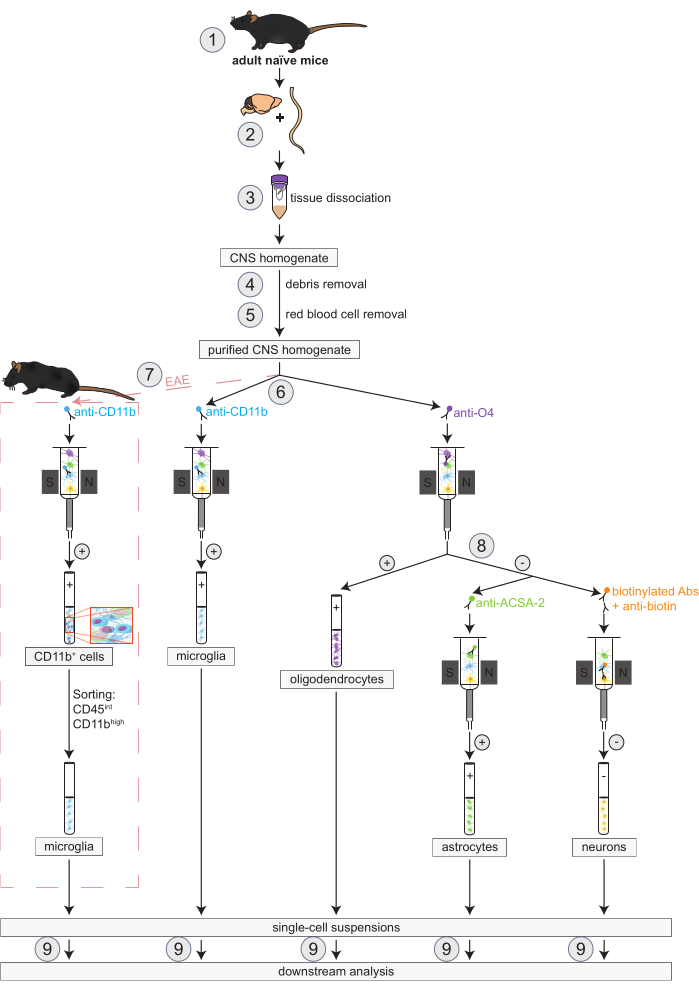
图1:在正常小鼠和EAE小鼠中同时分离少突胶质细胞、小胶质细胞、星形胶质细胞和神经元的工作流程。 该工作流程的前几个步骤在正常小鼠和EAE小鼠中是相同的。如果需要使用EAE模型小鼠,则需预先进行EAE诱导(1)。简而言之,本方案首先对小鼠脑和脊髓组织进行解剖(2)和解离(3),随后去除碎片(4)和红细胞(5)。接着,将获得的纯化中枢神经系统(CNS)细胞悬液分为两部分,用于通过磁珠激活细胞分选技术(MACS)同时分离少突胶质细胞和小胶质细胞(6)。小胶质细胞通过抗CD11b微珠进行捕获,而少突胶质细胞则使用抗O4微珠进行分离(均为阳性选择)。从少突胶质细胞分选后的阴性流出液中(8),使用抗ACSA-2微珠阳性选择分离星形胶质细胞,并通过生物素标记并耗尽所有非神经元细胞的方式实现神经元的阴性选择分离。在EAE小鼠中,CD11b+细胞的分离后,需进一步通过荧光激活细胞分选技术(FACS)对CD45intCD11bhigh细胞进行分选,以排除其他参与EAE病程中神经炎症过程的CD11b+免疫细胞,如巨噬细胞、树突状细胞、单核细胞、粒细胞和自然杀伤细胞(7)27, 28, 48。完成不同CNS常驻细胞类型的分离后,可进行纯度分析(9)。缩写:Abs = 抗体;ACSA-2 = 星形胶质细胞表面抗原-2;CD11b = 周期素依赖性激酶11B;CD45 = 受体型酪氨酸蛋白磷酸酶C;CNS = 中枢神经系统;EAE = 实验性自身免疫性脑脊髓炎;MACS = 磁珠激活细胞分选;O4 = 少突胶质细胞标记物O4。本图改编自49。 请点击此处查看此图的放大版本。
1. 主动诱导实验性自身免疫性脑脊髓炎(EAE)
- 试剂准备
- 用于细胞分离:配制 PB 缓冲液,并在 2-8 °C 下保存,最长不超过 1 周。配制储存液时,加入 475 mL 不含添加剂的 1× PBS(pH 7.2)和 25 mL 0.5% 牛血清白蛋白(BSA)。使用时以 BSA 溶液进行 1:20 稀释。
- 用于流式细胞术和荧光激活细胞分选(FACS):配制 FACS 缓冲液,即含 2% 胎牛血清(FCS)和 2 mM EDTA 的 PBS,并在 2-8 °C 下保存。配制方法为加入 500 mL 不含添加剂的 1× PBS、10 mL FCS 和 2 mL EDTA(来自 0.5 M EDTA 储存液)。
- 根据 Bittner 等人5的方案进行免疫。简言之,通过皮下注射含有 200 µg MOG35-55 肽和 200 µL 含 200 µg Mycobacterium tuberculosis 的完全弗氏佐剂乳剂诱导实验性自身免疫性脑脊髓炎(EAE)。
- 使用配备异氟烷蒸发器的麻醉舱,以 2% 异氟烷麻醉小鼠。麻醉期间在动物眼部涂抹兽用眼膏以防止角膜干燥。
- 免疫后 2 小时,按照 Huntemann 等人24的方案,腹腔注射 100 ng 百日咳毒素(PTx),溶于 100 µL 1× PBS 中。在免疫后第 2 天重复注射一次 PTx。
注意:需持续观察每只动物,直至其恢复足够的意识以维持胸卧位。接受注射操作的小鼠在完全恢复前不得与其他小鼠合笼。对于 Mycobacterium tuberculosis 和 PTx:避免吸入、摄入以及皮肤和眼睛接触。Mycobacterium tuberculosis 是先天免疫系统的激活剂,PTx 具有多种生物学效应。 - 每日监测 EAE 病程进展,由两名不知分组情况的研究人员分别监测体重并进行临床检查。
- 采用以下评分系统:0 分—无 EAE 临床症状;1 分—尾部部分轻瘫;2 分—尾部完全轻瘫;3 分—后肢中度无力;4 分—后肢完全无力并伴有共济失调步态;5 分—轻度截瘫;6 分—截瘫;7 分—截瘫;8 分—四肢轻瘫;9 分—四肢瘫痪;10 分—死亡。
- 实验中进一步参与的排除标准为临床评分 > 7 或体重下降超过初始体重的 20%。
- 为解剖脑和脊髓,在 EAE 诱导后第 16 天处死小鼠,此时疾病达到高峰。
2. 中枢神经系统组织制备(耗时:每只小鼠约10分钟)
- 用二氧化碳处死小鼠后,用 20 mL 的 1× PBS 对每只小鼠进行经心脏灌注。再重复一次,使用 20 mL 的 1× PBS 进行灌注。
- 将小鼠置于仰卧位,并用套管固定四肢。用 75% 乙醇擦拭动物的前部身体。此时无需采取其他无菌措施。
- 用剪刀沿皮肤和筋膜纵向切开,打开腹腔和胸腔。
- 从两侧剪断肋骨,并向上翻开胸腔,以充分暴露心脏。用套管将向上翻开的胸腔固定。
- 用剪刀剪开右心房。通过套管向左心室注入 20 mL 的 1× PBS,使血液从切开的右心房流出,以冲洗血液。
- 沿小鼠头部正中纵向切开皮肤,暴露颅骨,并用镊子将头部周围的皮肤向后剥离。用剪刀沿矢状缝切开颅骨。
- 将镊子尖端沿切口插入,撬开颅盖。用镊子移除残留的颅骨碎片,使脑组织完全暴露。
- 小心取出脑组织,并将其放入小鼠脑组织切片模具中。使用剃刀刀片将脑组织切成 1 mm 厚的矢状切片。
- 用剪刀在髂嵴上方剪断脊柱,以便将注射器插入椎管。
注意:取出脊髓最简便的方法是用 PBS 将其从椎管中冲出。否则,必须用剪刀逐个打开椎弓,然后取出脊髓。 - 使用装有 1× PBS 的 20 mL 注射器和 20G 针头,从尾端向头端方向将脊髓从椎管中冲洗出来。用手术刀将脊髓切成每段 0.5 cm 长的节段。
- 将每只小鼠的脑组织和相应脊髓组成的中枢神经系统细胞悬液分别置于一个独立的培养皿中,每个培养皿内加入约 3 mL 冷 D-PBS。将培养皿置于冰上,待后续处理。
3. 中枢神经系统组织解离(耗时:约1-1.5小时,具体取决于中枢神经系统细胞悬液的数量)
注意:通过将机械解离与酶解降解细胞外基质相结合,可实现成年小鼠神经组织的解离。该方法能够保持组织的结构完整性,所得细胞悬液可用于后续的细胞分离操作。
- 为每份中枢神经系统(CNS)细胞悬液配制适量体积的酶混合液1,其中包含50 µL酶P和1,900 µL缓冲液Z。两种试剂均来自成年动物脑组织解离试剂盒。
- 为每份中枢神经系统(CNS)细胞悬液配制适量体积的酶混合液2,其中包含10 µL酶A和20 µL缓冲液Y。两种试剂均来自成年动物脑组织解离试剂盒。
- 将1,950 µL酶混合液1转移至C管中,随后加入一份CNS细胞悬液的组织块。每只小鼠使用一根C管。
- 向每根C管中加入30 µL酶混合液2。紧密封闭C管,并将其倒置固定在带加热装置的组织解离仪套筒上。
- 运行名为37C_ABDK_01的程序(持续30分钟)。至少在程序开始的前5分钟进行观察,以确保所有管子以相同速度旋转。运行过程中可能出现错误。之后,继续进行步骤6。
- 在程序最后2分钟时,为每份解离后的CNS细胞悬液在50 mL离心管上放置一个70 µm细胞筛网。用2 mL D-PBS预先润湿这些筛网。
- 程序结束后,将C管从解离仪上取下并放入离心机。在300 x g 和4 °C条件下离心1分钟,使样品沉降至管底。
- 重悬样品并将其转移至预先润湿的细胞筛网上。向空的C管中加入10 mL冷D-PBS并封闭管口,轻轻振荡后将悬液转移至相应的筛网上。
- 弃去细胞筛网并封闭50 mL离心管。再次在300 x g 和4 °C条件下离心10分钟。随后,小心吸除全部上清液。
4. 去除碎片(耗时:约1.5–2小时,具体取决于中枢神经系统细胞悬液的数量)
注意:组织解离常产生髓鞘和细胞碎片,可能影响后续分析。通过添加碎片去除溶液,可有效去除中枢神经系统细胞悬液中的此类碎片。
- 用 3,100 µL 的 D-PBS 小心重悬每份 CNS 细胞悬液中的细胞沉淀。切勿涡旋震荡。
- 若同时处理多于一份 CNS 细胞悬液,可将来自同一条件或实验组的最多两份 CNS 细胞悬液合并至一个 15 mL 离心管中。
- 向一份 CNS 细胞悬液中加入 900 µL 成人脑组织解离试剂盒中的去碎片溶液;若为两份合并的 CNS 细胞悬液,则加入 1,800 µL 去碎片溶液。
- 颠倒混匀离心管,轻轻混悬细胞。随后,非常轻柔地在上方覆盖 4 mL 冷的 D-PBS。应可见清晰的梯度分层(图 2A)。
- 在 4 °C 条件下,以 3000 × g 离心 10 分钟,设置为全速加速且不使用刹车。
- 若分离效果理想,将形成三个相层(图 2C)。完全吸除最上层的两个相层(图 2C-1,2)并弃去。需确保无髓鞘残留(图 2E)。
注意:若梯度分离未成功但急需细胞,则不要吸除上层两个相层。应将冷的 D-PBS 加至 15 mL 离心管中至满刻度,反复颠倒混匀数次。随后在 4 °C 条件下以 1000 × g 离心 10 分钟,设置为全速加速且不使用刹车。吸除上清液后,重复步骤 4.1–4.4。 - 将离心管加冷的 D-PBS 至 14 mL 体积并盖紧。在实验台面上用力颠倒离心管,直至细胞沉淀从管底完全脱落。切勿涡旋震荡。
- 再次在 4 °C 条件下以 1000 × g 离心 10 分钟,设置为全速加速和全速刹车。小心且彻底地吸除上清液。
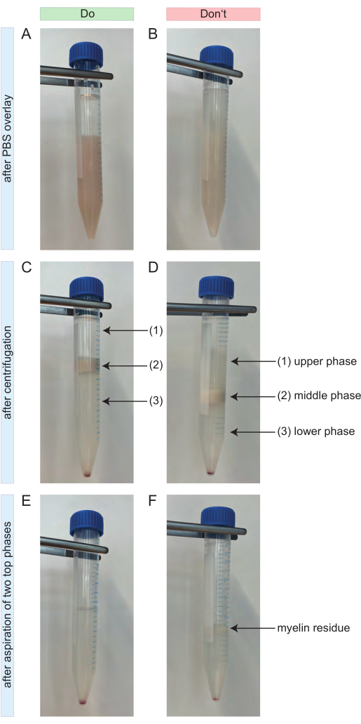
图 2:去碎片过程中的注意事项。(A)叠加4 mL PBS后形成梯度的正面示例。上层相由4 mL PBS组成,与下层含碎片去除液的中枢神经系统细胞悬液相之间界限清晰。(B)叠加4 mL PBS后形成梯度的负面示例。PBS与下方细胞悬液之间缺乏清晰的分界,部分PBS已扩散进入细胞悬液。(C)离心后梯度的正面示例。可清晰区分三个独立的相。在梯度的上层相(1)和下层相(3)中均未见髓鞘残留物。中间相包含全部髓鞘(2)。细胞沉淀可见于15 mL离心管底部。(D)离心后梯度的负面示例。三个相之间无法实现准确分离,部分髓鞘残留物可见于上层相(1)和下层相(3)中。(E)吸除顶部两相后梯度的正面示例。所得样品仅包含细胞沉淀及上方澄清的上清液,无髓鞘残留。(F)吸除顶部两相后梯度的负面示例。样品中仍残留部分髓鞘(黑色箭头)。缩写:CNS = central nervous system;PBS = phosphate-buffered saline 请点击此处查看该图的放大版本。
5. 红细胞去除(耗时:约1小时,具体时间取决于中枢神经系统细胞悬液的数量)
注意:此步骤可防止后续红细胞污染,并确保红细胞得到充分裂解,同时对从中枢神经系统组织分离的其他细胞类型影响最小。以下体积适用于来自100 mg至1 g神经组织的细胞悬液,相当于两只成年小鼠的脑和脊髓组织。若处理超过两份中枢神经系统细胞悬液,应相应地同比增加所有试剂和总体积。
- 开始制备红细胞去除液(RBCRS):每两份合并的中枢神经系统(CNS)细胞悬液配制一份。将成人脑组织解离试剂盒中的红细胞去除储存液(10x)100 µL 加入 900 µL 的 ddH2O 中稀释,得到最终 1:10 的稀释液。
- 将 RBCRS 储存于 2–8 °C,待用。当日未用完的溶液应丢弃。
- 将最多两份 CNS 细胞悬液的细胞沉淀重悬于 1 mL RBCRS 中。避免涡旋震荡。在 4 °C 下孵育 10 分钟。
- 向两份合并的细胞悬液中加入 10 mL 冷的 PB 缓冲液。在 4 °C、300 × g 条件下离心 10 分钟,随后完全吸除上清液。
- 用移液器缓慢吹打,将每份 CNS 细胞悬液的细胞沉淀重悬于 80 µL PB 缓冲液中。相应地,若为两份 CNS 细胞悬液的细胞沉淀,则使用 160 µL PB 缓冲液进行重悬。
- 当处理来自相同实验条件的多份 CNS 细胞悬液时,应将所有细胞悬液合并。
- 测定细胞数量,例如使用改良型血细胞计数板。通常先将细胞悬液用 PB 缓冲液稀释 1:50,再用 0.4% 台盼蓝溶液进一步稀释 1:10。
6. 初始小鼠和EAE小鼠中的磁珠分离方案(耗时:约1小时)
- 使用针对不同中枢神经系统(CNS)细胞表面抗原的特异性MicroBeads对细胞进行磁性标记。然后将细胞悬液加入分离柱中,通过磁性分离保留在柱内的标记细胞与流穿的未标记细胞。
- 将分离柱从磁场中取出后,用缓冲液将磁性标记的细胞从柱中洗脱至离心管中,获得阳性选择的细胞组分。
注意:磁性标记过程中的体积是按最多1 × 10⁷个总细胞计算的。若细胞数量更多,应相应放大所有试剂和总体积。建议操作迅速,并仅使用预冷的溶液,以防止抗体在细胞表面发生帽化、非特异性标记,并确保分离所得细胞群体具有较高的存活率。此外,当分离柱储液腔排空后,应立即加入PB缓冲液进行洗涤,防止柱子干燥。 - 将纯化后的未稀释CNS细胞悬液分为两部分,用于后续的小胶质细胞和少突胶质细胞的分离。两部分的比例取决于每种细胞类型所需的目标细胞数量。
注意:进一步的细节(孵育时间、详细操作步骤、体积、试剂及细胞计数方法)见表1。
表1:从正常小鼠和EAE小鼠中同时进行少突胶质细胞和小胶质细胞磁性标记与分离的工作流程。 两种细胞均通过阳性筛选进行分离。列在同一行中的步骤表示需同时进行。缩写:CD11b = 细胞周期依赖性激酶11B;EAE = 实验性自身免疫性脑脊髓炎;FcR = Fc受体样蛋白;O4 = 少突胶质细胞标志物O4。 请点击此处下载该表格。
7. 方案修订:用于分离EAE小鼠小胶质细胞的额外分选步骤(耗时:约1.5-2小时)
注意:在处理EAE小鼠时,有必要在基于MACS的细胞分离方案基础上补充FACS步骤,以从CD11b+细胞组分中去除除小胶质细胞以外的其他CD11b+细胞群体(例如单核细胞、巨噬细胞、自然杀伤细胞、粒细胞或树突状细胞)。否则,此步骤可忽略。
- 制备染色主混合液,其中包含添加了CD11b FITC(克隆M1/70,1:50)和CD45 APC/Cy7(克隆30-F11,1:200)的1x PBS。每5 x 10⁶个细胞使用100 µL染色主混合液。使用前将所有抗体充分涡旋混匀。
- 将小胶质细胞悬液在300 x g、4 °C条件下离心10分钟,并小心吸除上清液。
- 每5 x 10⁶个细胞用100 µL制备好的染色主混合液重悬细胞沉淀。在室温(RT)避光条件下孵育15分钟。
- 加入500 µL PBS终止反应,并再次在300 x g、4 °C条件下离心10分钟。
- 小心吸除上清液,用含10 µg/mL DNA酶的1x PBS重悬细胞沉淀,使细胞终浓度达到每毫升1 x 10⁷个细胞。将细胞置于4 °C保存,直至开始分选。
- 在开始分选前,立即将细胞悬液加至置于新FACS管上的100 µm滤网中。
- 将流速设为每秒1000个事件,使用100 µm喷嘴。将目标细胞群CD45intCD11bhigh分选至预先加入室温1x PBS的新15 mL管中。
8. 少突胶质细胞阴性流穿组分的制备,用于神经元和星形胶质细胞的分离(耗时:约1小时)
注意:将步骤6中少突胶质细胞的阴性流穿液收集起来,用于进一步分离神经元和星形胶质细胞。为此,将细胞悬液分为两部分。由于此前已从中枢神经系统细胞悬液中分离出少突胶质细胞,因此可最大程度减少原本可能出现的O4+细胞污染。
- 将少突胶质细胞的阴性流穿液在 300 x g、4 °C 条件下离心 10 分钟,并小心吸除上清液。
- 每份用于分离少突胶质细胞阳性组分的合并后的中枢神经系统细胞悬液,用 80 µL PB 缓冲液重悬细胞沉淀。
- 对细胞进行计数。将预计为 O4- 的细胞悬液用 PB 缓冲液按 1:50 稀释后,再用 0.4% 台盼蓝进一步稀释 1:10,随后使用改良计数板进行细胞计数。
- 将纯化后的未稀释细胞悬液分为两份,用于同时分离神经元和星形胶质细胞。两份之间的比例取决于所需每种细胞类型的数量。
注意:进一步的细节(孵育时间、详细操作步骤、体积、试剂和细胞计数方法)见表 2。
表2:从未经处理小鼠和EAE小鼠中同时进行磁标记并分离神经元和星形胶质细胞的工作流程。 两种细胞均从少突胶质细胞的阴性流穿部分中分离获得。星形胶质细胞通过抗ACSA-2微珠进行阳性选择分离,而神经元则通过生物素标记并去除所有非神经元细胞的方式,以阴性选择进行纯化。列在同一行的操作步骤表示需同时进行。缩写:Anti-ACSA-2 = 星形胶质细胞表面抗原-2;EAE = 实验性自身免疫性脑脊髓炎;FcR = Fc受体样蛋白;MACS = 磁珠激活细胞分选。 请点击此处下载该表格。
9. 分离的中枢神经系统驻留细胞类型的纯度分析(耗时:约2小时)
注意:建议对分离得到的全部四种中枢神经系统常驻细胞群体进行流式细胞术检测,以测量并比较其纯度和活力。因此,需要用荧光标记的抗体对所有细胞类型进行染色。活/死细胞染色采用可固定的活力染料(1:10,000)进行。
- 纯度检测面板——细胞外染色方案
- 每次染色使用 1 × 10⁵ 个细胞,悬浮于 50 µL PBS 中。
- 在含 2% FCS/2 mM EDTA 的 PBS 中配制染色主混合液,包含以下针对细胞类型特异性表面标志物的荧光素偶联单克隆抗体:CD11b FITC(克隆号 1/70,1:100)25,26,27,28、Biotin-PE(克隆号 Bio3-18E7,1:200)29,30,31,32、ACSA-2 PE-Vio615(克隆号 REA-969,1:200)33,34,35、O4 APC(克隆号 REA-576,1:400)和 CD45 BV510(克隆号 30-F11,1:150)36,37。每 1 × 10⁶ 个细胞加入 1 µg 抗 CD16/32 抗体以阻断 Fc 受体38,39。使用前将所有抗体充分涡旋混匀。
- 将细胞悬液在 4 °C 条件下以 540 × g 离心 5 分钟,并小心吸除上清液。
- 将细胞沉淀重悬于 100 µL 相应的主混合液中,在室温避光条件下孵育 15 分钟。
- 用 500 µL 含 2% FCS/2 mM EDTA 的 1× PBS 洗涤细胞,并在 4 °C 条件下以 540 × g 离心 5 分钟。
- 吸除上清液,将细胞沉淀重悬于 70 µL 含 2% FCS/2 mM EDTA 的 1× PBS 中。
- 涡旋振荡样品,彻底分散细胞沉淀。随后样品即可用于流式细胞术分析。
- 纯度检测面板——NeuN 胞内染色方案
- 对每种细胞群体取 1 × 10⁵ 个细胞进行 NeuN 的胞内染色,NeuN 是一种神经元特异性核标志物40,41。这是标记活神经元的另一种方法。
- 将每种细胞群体的 1 × 10⁵ 个细胞转移至 FACS 管中。每管加入 1 mL 含 2% FCS/2 mM EDTA 的 PBS,在 4 °C 条件下以 540 × g 离心 5 分钟。
- 同时,在含 2% FCS/2 mM EDTA 的 PBS 中配制主混合液,包含以下针对细胞类型特异性表面标志物的荧光素偶联单克隆抗体:CD11b FITC(克隆号 M1/70,1:100)25,26,27,28、Biotin-PE(克隆号 Bio3-18E7,1:200)29,30,31,32、ACSA-2 PE-Vio615(克隆号 REA-969,1:200)33,34,35 和 CD45 BV510(克隆号 30-F11,1:150)36,37。
- 吸除上清液,将细胞重悬于 100 µL 预先配制的主混合液中,在室温避光条件下孵育 10 分钟。
- 用 100 µL 含 2% FCS/2 mM EDTA 的 PBS 洗涤细胞,并在 4 °C 条件下以 540 × g 再次离心 5 分钟。
- 同时,配制 200 µL 固定/通透溶液:将 50 µL 浓缩的固定/通透液原液加入 150 µL 固定/通透稀释液中,得到 1:4 的终稀释度。
- 吸除上清液,将细胞重悬于 100 µL 1× 固定/通透溶液中,在 4 °C 条件下孵育 30 分钟。
- 同时,通过将 100 µL 通透缓冲液原液加入 900 µL ddH2O 中,配制 1 mL 1× 通透/洗涤缓冲液,得到 1:10 的终稀释度。
- 用 100 µL 1× 通透/洗涤缓冲液洗涤细胞一次,并在 4 °C 条件下以 540 × g 离心 5 分钟。
- 同时,在 1× 通透/洗涤缓冲液中配制另一份主混合液,仅包含 NeuN(NeuN AF647,克隆号 EPR12763,1:200)以及每 106 个细胞加入 1 µg 抗 CD16/32 抗体以阻断 Fc 受体。
- 吸除上清液,将已固定并通透的细胞重悬于 50 µL 第二份主混合液中,在 4 °C 条件下孵育 30 分钟。
- 用 100 µL 1× 通透/洗涤缓冲液洗涤样品,并在 4 °C 条件下以 540 × g 离心 5 分钟。
- 弃去上清液,将细胞沉淀重悬于 70 µL 含 2% FCS/2 mM EDTA 的 PBS 中。随后样品即可用于流式细胞术分析。
- 在流式细胞仪上设置好检测面板后,使用流式细胞术分析软件采集细胞数据以进行纯度分析。
10. 统计分析
- 使用图形分析程序进行统计分析并绘制图表。数据以均值 ± 标准误(SEM)表示。
结果
本方案提供了一种从单个中枢神经系统样本中同时分离所有主要的中枢神经系统常驻细胞(即小胶质细胞、少突胶质细胞、星形胶质细胞和神经元)的可能性。这对于减少此类实验所需的小鼠数量以及确保在细胞水平上进行分子和生化分析的可比性具有重要意义。如果不同细胞类型分别来自不同的中枢神经系统样本,则无法真实反映细胞间的相互作用,且分离过程中的潜在技术偏差可能影响后续分析结果。此外,由于各细胞类型的分子和生化研究结果并非来源于同一实验性自身免疫性脑脊髓炎(EAE)背景,彼此之间也不具备可比性。本研究对已有的磁珠分选(MACS)方案进行了优化,采用商用系统/试剂盒,实现了上述多种细胞类型的同步分离。
小胶质细胞的分离采用抗CD11b微珠,少突胶质细胞通过抗O4微珠进行分离(表1),而星形胶质细胞的分离则使用抗ACSA-2微珠(表2)。相比之下,神经元的分离采用阴性筛选策略,通过生物素标记并磁性标记所有非神经元细胞来实现(表2)。除血细胞外,所有非神经元细胞(例如少突胶质细胞、小胶质细胞、星形胶质细胞、内皮细胞和成纤维细胞)均可利用与这些非神经元细胞表面抗原特异性结合的生物素偶联抗体进行磁性标记(表2)。通过去除这些磁性标记的非神经元细胞,可获得高纯度且具有活性的神经元细胞群体30,42,43。
设计了两个新的流式细胞术检测方案,用于分析所制备单细胞悬液的纯度。此处结合了细胞类型特异性表面标记物和核标记物,并进行了活细胞/死细胞区分。
每只小鼠获得的细胞产量及细胞类型(图3A)分析结果显示,每只未处理小鼠平均可获得 8.4 × 10⁵ ± 3 × 10⁴ 个小胶质细胞、3.23 × 10⁶ ± 1 × 10⁵ 个少突胶质细胞、8.6 × 10⁵ ± 2 × 10⁴ 个星形胶质细胞和 4.5 × 10⁵ ± 1 × 10⁴ 个神经元。
在研究神经炎症疾病模型的背景下,该方案还应用于实验性自身免疫性脑脊髓炎(EAE)小鼠模型。小鼠在诱导EAE后第16天被实施安乐死,此时疾病程度达到峰值。在此EAE模型中,分离得到约 2.9 x 10⁶ ± 6.7 x 10⁵ 个少突胶质细胞、5.2 x 10⁵ ± 9 x 10⁴ 个星形胶质细胞以及 4.4 x 10⁵ ± 4 x 10⁴ 个神经元。由于在MACS步骤后增加了细胞分选过程,每只EAE小鼠获得的微胶质细胞数量减少至约 1.7 x 10⁵ ± 4 x 10⁴ 个(图3A)。
分离后,通过流式细胞术对不同细胞群进行表型表征,结果证明可获得存活的单细胞悬液,其中所有主要中枢神经系统常驻细胞类型的纯度均约为90%(图3B)。小胶质细胞根据文献定义被设门为CD45intCD11bhigh44,45,46,47。
在EAE中,必须从所有CD11b+细胞中分选出小胶质细胞,以将其与其他在神经炎症期间侵入中枢神经系统的CD11b+免疫细胞(如单核细胞、中性粒细胞、自然杀伤细胞、粒细胞和巨噬细胞)区分开来27,28,48。因此,从小鼠CD11b+细胞悬液中以CD45intCD11high表型对小胶质细胞进行分选。整个小胶质细胞分选策略如图4所示。在正常小鼠中,小胶质细胞占所有活单细胞的91.16%(占总CD11b+细胞群体的96%)(图4A)。在EAE小鼠中,小胶质细胞占所有活单细胞的46.85%(占总CD11b+细胞群体的55%)(图4B)。尽管磁珠分选(MACS)和流式分选(FACS)过程均会对单个细胞施加机械应力,但分选后纯化的小胶质细胞中仍有75.41% ± 8.63%保持存活(图3B)。
从初始的中枢神经系统细胞悬液中直接分离的星形胶质细胞和神经元显示出少突胶质细胞的显著污染,由此推测,在分离少突胶质细胞后的阴性流穿部分中同时分离神经元和星形胶质细胞可避免此类污染。流式细胞术分析证实,从少突胶质细胞阴性流穿部分分离的星形胶质细胞纯度为89.23% ± 2.78%,存活率为80.56% ± 1.41%。与这些结果类似,从O4-细胞组分中分离的神经元纯度为81.25% ± 3.31%,存活率为83.90% ± 2.61%(图3B)。这些结果也证实,在少突胶质细胞分离之后再同时分离这两种细胞类型,并不会影响有活性的功能性细胞的数量。
在EAE小鼠中分离获得的单细胞悬液的活力和纯度结果与正常小鼠中获得的结果非常相似,表明该方案不仅适用于健康小鼠,也适用于EAE疾病背景下的小鼠(图3B)。
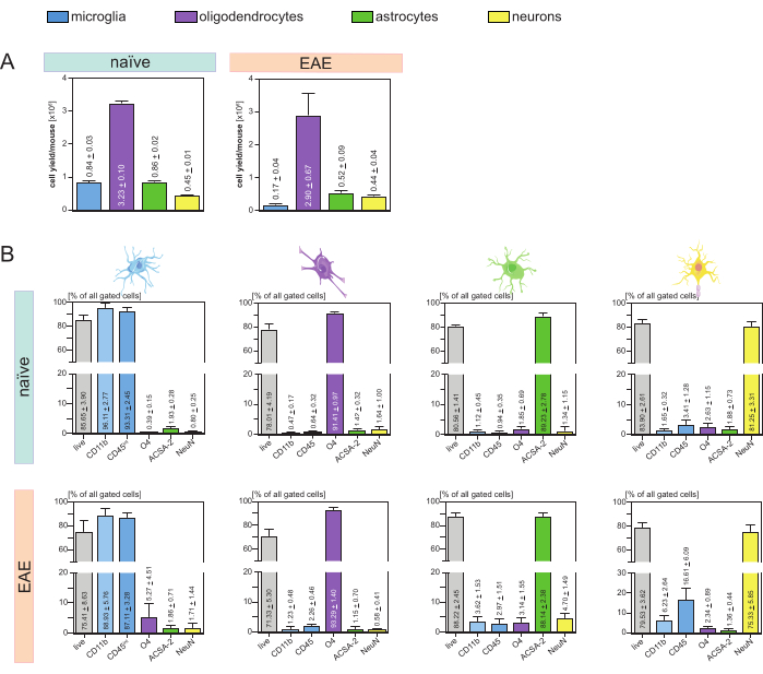
图 3:分离的中枢神经系统驻留细胞的细胞得率及基于流式细胞术的验证。(A) 在 naïve 小鼠和 EAE 小鼠中分离中枢神经系统驻留细胞后,每只小鼠每种细胞类型的细胞得率。柱状图展示了按照本方案操作后每只小鼠每种细胞类型的细胞得率。naïve 小鼠的数据来自五个生物学重复,EAE 小鼠的数据来自四个生物学重复。图中显示相应的均值 ± 标准误(SEM)。(B) 纯化后各细胞组分的纯度与活力分析。柱状图显示基于细胞类型特异性标志物表达水平所得的单细胞悬液的活力和纯度。NeuN 用作神经元的细胞类型特异性核标志物。每种细胞类型在健康小鼠和 EAE 小鼠中均采集并比较了五个生物学重复。图中显示相应的均值 ± 标准误(SEM)。缩写:Anti-ACSA-2 = 星形胶质细胞表面抗原-2;CD11b = 周期素依赖性激酶11B;CD45 = 受体型酪氨酸蛋白磷酸酶C;CNS = 中枢神经系统;EAE = 实验性自身免疫性脑脊髓炎;MACS = 磁珠激活细胞分选;NeuN = RNA结合蛋白fox-1同源物3;O4 = 少突胶质细胞标志物O4;SEM = 均值标准误。本图改编自49。请点击此处查看该图的放大版本。

图4分选小胶质细胞的设门策略(在分离CD11b+细胞后)。(A) 幼稚型小鼠和(B)EAE 小鼠中的门控策略。每个图面板的上排显示分选前的散点图,下排显示分选后的散点图。在选择活细胞(SSC-A / FSC-A)和单细胞(FSC-H / FSC-W)后,CD45阳性细胞群intCD11b+ 细胞被分选为小胶质细胞群体。 请点击此处以查看此图的放大版本。
讨论
目前,通过结合质谱和RNA测序技术对中枢神经系统(CNS)驻留细胞进行体外(ex vivo)定位的方法,能够在健康与疾病状态下实现极为精确的细胞图谱绘制,但该方法需要研究者具备较高的专业技术知识和相关领域经验50,51。此外,这些方法无法进行功能分析,且成本高昂。另一方面,微流控脑芯片系统可快速、经济地用于疾病机制研究和新治疗策略的筛选,但其在细胞生长和迁移方面存在限制52,53,54,55。未来,中枢神经系统类器官也可能成为研究细胞建模、细胞间连接以及疾病进程中相互作用的等效替代方案56,57,58,59。然而,目前荧光激活细胞分选和磁激活细胞分选仍是体外(ex vivo)制备高纯度且具活性的单细胞悬液最有效的方法35,60,61。尽管其他已建立的针对CNS驻留细胞类型分离的制备方案在磁分离及前期细胞解离步骤上与本方法相似,但这些方案通常要求针对每种细胞类型单独进行操作。相比之下,本方案将针对不同CNS驻留细胞类型的多种分离方法整合于一个逻辑框架中,使其能够从同一份CNS单细胞悬液中一次性同时完成各类细胞的分离(表1、表2)。因此,该方法支持从单一CNS细胞悬液中开展多组学分析,并最终有助于复杂神经网络的探索。尽管本方案并不要求必须合并来自多个动物的组织样本,但合并样本可确保获得足够数量的分离细胞以进行后续下游分析。若使用不同小鼠分别分离各类细胞,则将无法分析潜在的细胞间相互作用。此外,将针对不同CNS细胞类型的独立分离方法整合于同一CNS解离步骤之后,可通过使用同一份解离后的CNS细胞悬液完成所有后续磁分离步骤,从而节约材料成本。同时,也最大限度地减少了因使用不同小鼠而可能引入的技术偏差。
该方案的一个局限性可能是几乎 exclusively 使用雌性 C57BL/6J 小鼠。EAE 免疫方案最初是为雌性小鼠设计并建立的,因此本细胞分离方案也应用于雌性 C57BL/6J 小鼠。然而,在本方案的开发过程中也使用了未经处理的雄性小鼠,并未观察到对最终获得的细胞数量或纯度有任何影响。另一个限制涉及神经元的磁性细胞分离,因为目前尚无可用于神经元阳性筛选的特异性微珠。因此,本方案假设通过生物素标记并去除所有非神经元细胞,可以获得高纯度的单细胞悬液(表 2)。该假设通过使用 NeuN 作为神经元特异性核标记物,并将其纳入上述流式细胞术纯度检测方案中得以验证。另一项局限性涉及 EAE 小鼠中小胶质细胞的分离。在此情况下,与其他细胞类型相比,最终获得的细胞产量较低,这是由于在 MACS 方案之后增加了分选步骤。此外,有人认为,与其它细胞群体相比,分选过程可能增加了小胶质细胞所受的机械应力。不同的分选策略可能导致细胞得率存在差异。如果获得的细胞数量低于预期或需求,建议调整设门策略和/或优化活/死细胞的区分。
该实验方案中的一个关键步骤是去除碎片。必须非常缓慢且轻柔地铺加梯度液,以形成所需的三个独立相(图2A)。只有当最上两层中的髓鞘及其他碎片残留物被完全去除后(图2E),才能获得高纯度的单细胞悬液,并进一步减少污染。如果最终获得的细胞悬液纯度不足,除确保所有微珠正确使用外,该步骤通常是首先需要优化的部分。
在此类实验中,获得高纯度和高活力的细胞可能具有挑战性。以下是一些故障排除建议:
- 必须在无菌条件下操作,以防止不同微珠被污染,并确保其可重复使用,尤其是用于后续培养时。
- 强烈建议对每根试管进行标记,以避免混淆。
- 避免使用未冷却的试剂或缓冲液。整个实验过程中,所有细胞悬液均应置于冰上保存,以确保细胞具有较高的存活率。
- 应尽量缩短各操作步骤之间的时间间隔。本实验方案中未推荐任何可暂停实验的特定步骤。
- 严格遵守规定的孵育时间至关重要。
总之,本方案通过单次中枢神经系统样本即可同时分离出所有主要的中枢神经系统常驻细胞类型,从而能够从一份中枢神经系统细胞悬液中ex vivo分析复杂的神经元网络和神经炎症通路。因此,可在疾病进程的不同阶段(例如实验性自身免疫性脑脊髓炎中的神经炎症、神经退行性变和/或缓解期)对中枢神经系统常驻细胞进行研究。此外,还可对细胞间相互作用和生物化学通路进行单细胞水平的研究,并减少实验组内的个体差异。该方法还允许将分离得到的中枢神经系统细胞亚群进行单细胞培养,用于后续功能检测和验证。总体而言,本方案为临床前和临床研究方法提供了重要的技术进步。
披露
所有作者声明无利益冲突。
致谢
图示使用 Adobe Illustrator(版本 2023)和 Servier Medical Art (https://smart.servier.com)制作。Antonia Henes 得到 Jürgen Manchot Stiftung 的支持。
材料
| 姓名 | 公司 | 目录编号 | 评论 |
|---|---|---|---|
| 70 μm 细胞筛 | Corning, MA, USA | 352350 | 中枢神经系统组织解离 |
| ACSA-2 抗体,抗小鼠,PE-Vio 615(克隆 REA-969) | Miltenyi Biotec, Bergisch Gladbach, NRW, Germany | 130-116-244 | 流式细胞术,4 °C 保存 |
| 成年小鼠和大鼠脑组织解离试剂盒 | Miltenyi Biotec, Bergisch Gladbach, NRW, Germany | 130-107-677 | 组织解离,包含碎片和红细胞去除溶液;收到后立即分装酶 A 和酶 P,并于 -20 °C 保存;其余试剂盒组分于 4 °C 保存 |
| 抗 ACSA-2 MicroBead 试剂盒,小鼠 | Miltenyi Biotec, Bergisch Gladbach, NRW, Germany | 130-097-678 | 星形胶质细胞磁珠分选(MACS),4 °C 保存 |
| 抗小鼠 CD16/32 抗体 | BioLegend, London, UK | 101301 | 流式细胞术,4 °C 保存 |
| 抗 O4 MicroBeads,人、小鼠、大鼠 | Miltenyi Biotec, Bergisch Gladbach, NRW, Germany | 130-094-543 | 少突胶质细胞磁珠分选(MACS),4 °C 保存 |
| AstroMACS 分离缓冲液 | Miltenyi Biotec, Bergisch Gladbach, NRW, Germany | 130-091-221 | 星形胶质细胞磁珠分选(MACS),4 °C 保存 |
| 生物素抗体,PE(克隆 Bio3-18E7) | Miltenyi Biotec, Bergisch Gladbach, NRW, Germany | 130-113-853 | 流式细胞术,4 °C 保存 |
| BRAND Neubauer 计数板 | Thermo Fisher Scientific,Waltham, MA, USA | 10195580 | 细胞计数 |
| Brilliant Violet 510 抗小鼠 CD45 抗体(克隆 30-F11) | BioLegend, London, UK | 103137 | 流式细胞术,4 °C 保存 |
| CD11b MicroBeads,人、小鼠 | Miltenyi Biotec, Bergisch Gladbach, NRW, Germany | 130-049-601 | 小胶质细胞磁珠分选(MACS),4 °C 保存 |
| DNA酶 I,重组型,无RNase | Merck KGaA, Darmstadt, Germany | 4716728001 | 流式细胞术,-20°C 保存 |
| 含钙、镁、葡萄糖和丙酮酸盐的 D-PBS | Thermo Fisher Scientific,Waltham, MA, USA | 14287080 | 缓冲液,4 °C 保存 |
| 不含钙、镁的 D-PBS | Thermo Fisher Scientific,Waltham, MA, USA | 14190250 | 缓冲液,4 °C 保存 |
| eBioscience 可固定活力染料 eFluor 780 | Thermo Fisher Scientific,Waltham, MA, USA | 65-0865-14 | 流式细胞术,4 °C 保存 |
| eBioscience Foxp3/转录因子染色缓冲液套装 | Thermo Fisher Scientific,Waltham, MA, USA | 00-5523-00 | 流式细胞术,4°C 保存 |
| Falcon(15 mL) | Thermo Fisher Scientific,Waltham, MA, USA | 11507411 | 细胞管 |
| Falcon(50 mL) | Thermo Fisher Scientific,Waltham, MA, USA | 10788561 | 细胞管 |
| Falcon 带细胞筛 snap cap 的 5 mL 圆底聚苯乙烯试管 | Thermo Fisher Scientific,Waltham, MA, USA | 08-771-23 | 流式细胞术 |
| FcR 封闭试剂,小鼠 | Miltenyi Biotec, Bergisch Gladbach, NRW, Germany | 130-092-575 | 少突胶质细胞磁珠分选(MACS),4 °C 保存 |
| 雌性 C57BL/6J 小鼠 | Charles River Laboratories, Sulzfeld, Germany | 主动诱导实验性自身免疫性脑脊髓炎(EAE) | |
| 胎牛血清(FCS) | Merck KGaA, Darmstadt, Germany | F2442-50ML | 流式细胞术,-5 至 -20 °C 保存 |
| FITC 标记大鼠抗 CD11b 抗体 (克隆 M1/70) | BD Biosciences, San Jose, CA, USA | 553310 | 流式细胞术,4 °C 保存 |
| 弗氏完全佐剂(Freund’s Complete adjuvant) | Merck KGaA, Darmstadt, Germany | AR001 | 主动诱导实验性自身免疫性脑脊髓炎(EAE),4 °C 保存 |
| GentleMACS C 管 | Miltenyi Biotec, Bergisch Gladbach, NRW, Germany | 130-093-237 | 中枢神经系统组织解离 |
| 带加热模块的 GentleMACS Octo 组织解离仪 | Miltenyi Biotec, Bergisch Gladbach, NRW, Germany | 130-096-427 | 中枢神经系统组织解离 |
| Graphpad Prism 8.4.3 | Graphpad by Dotmatics | 图形分析 | |
| 异氟烷 | AbbVie, North Chicago, IL, USA | 主动诱导实验性自身免疫性脑脊髓炎(EAE),4 °C 保存 | |
| Kaluza 分析软件 V2.1.1 | Beckman Coulter, Indianapolis, IN, USA | 流式细胞术分析 | |
| LS 柱 | Miltenyi Biotec, Bergisch Gladbach, NRW, Germany | 130-042-401 | 磁珠分选(MACS) |
| MACS BSA 储存液 | Miltenyi Biotec, Bergisch Gladbach, NRW, Germany | 130-091-376 | PB 缓冲液 |
| MACS 多柱支架 | Miltenyi Biotec, Bergisch Gladbach, NRW, Germany | 130-042-303 | 磁珠分选(MACS) |
| MOG35–55 肽段 | Charité, Berlin, Germany;替代来源:Genosphere Biotechnologies(Paris, France)或 sb-Peptide(Saint Egrève, France) | 主动诱导实验性自身免疫性脑脊髓炎(EAE),-20 °C 保存 | |
| 结核分枝杆菌 H37 Ra 菌株 | Becton, Dickinson and Company (BD),Franklin Lakes, NJ, USA | 主动诱导实验性自身免疫性脑脊髓炎(EAE),4 °C 保存 | |
| 小鼠神经元分离试剂盒 | Miltenyi Biotec, Bergisch Gladbach, NRW, Germany | 130-115-390 | 神经元磁珠分选(MACS),4 °C 保存 |
| O4 抗体,抗人/小鼠/大鼠,APC(克隆 REA-576) | Miltenyi Biotec, Bergisch Gladbach, NRW, Germany | 130-119-897 | 流式细胞术,4 °C 保存 |
| 甘油中的百日咳毒素 | Hooke Laboratories Inc., Lawrence, MA, USA | BT-0105 | 主动诱导实验性自身免疫性脑脊髓炎(EAE);-20 °C 保存 |
| pluriStrainer Mini 100 μm | pluriSelect Life Science UG, Leipzig, Sachsen, Germany | 43-10100-40 | 流式细胞术 |
| QuadroMACS 分选仪 | Miltenyi Biotec, Bergisch Gladbach, NRW, Germany | 130-090-976 | 磁珠分选(MACS) |
| 重组 Alexa Fluor 647 标记抗 NeuN 抗体(克隆 EPR12763) | Abcam, Cambridge, UK | EPR12763 | 流式细胞术,-20 °C 保存 |
| 不锈钢脑组织切片模具,1 mm | Ted Pella, Redding, CA, USA | 15067 | 中枢神经系统组织解剖 |
| 台盼蓝溶液,0.4% | Thermo Fisher Scientific,Waltham, MA, USA | 15250061 | 细胞计数 |
| UltraPure 0.5 M EDTA,pH 8.0 | Thermo Fisher Scientific,Waltham, MA, USA | 15575020 | 流式细胞术,室温保存 |
参考文献
- Trapp, B. D., Nave, K. A. Multiple Sclerosis: An Immune or Neurodegenerative Disorder. Annu Rev Neurosci. 31 (1), 247-269 (2008).
- Stys, P. K., Zamponi, G. W., van Minnen, J., Geurts, J. J. Will the real multiple sclerosis please stand up. Nat Rev Neurosci. 13 (7), 507-514 (2012).
- Korn, T. Pathophysiology of multiple sclerosis. J Neurol. 255 (Suppl 6), 2-6 (2008).
- Ward, M., Goldman, M. D. Epidemiology and Pathophysiology of Multiple Sclerosis. CONTINUUM. 28 (4), 988-1005 (2022).
- Bittner, S., Afzali, A. M., Wiendl, H., Meuth, S. G. Myelin Oligodendrocyte Glycoprotein (MOG35-55) Induced Experimental Autoimmune Encephalomyelitis (EAE) in C57BL/6 Mice. J Vis Exp. (86), 51275(2014).
- Bittner, S., et al. The TASK1 channel inhibitor A293 shows efficacy in a mouse model of multiple sclerosis. Exp Neurol. 238 (2), 149-155 (2012).
- Göbel, K., et al. Plasma kallikrein modulates immune cell trafficking during neuroinflammation via PAR2 and bradykinin release. Proc Natl Acad Sci U S A. 116 (1), 271-276 (2019).
- Ballerini, C. Experimental Autoimmune Encephalomyelitis. Methods Mol Biol. 2285, 375-384 (2021).
- Birmpili, D., Charmarke Askar, I., Bigaut, K., Bagnard, D. The Translatability of Multiple Sclerosis Animal Models for Biomarkers Discovery and Their Clinical Use. Int J Mol Sci. 23 (19), 11532(2022).
- Tsatas, O., Ghasemlou, N. Isolation and RNA purification of macrophages/microglia from the adult mouse spinal cord. J Immunol Methods. 477, 112678(2020).
- Calvo, B., Rubio, F., Fernández, M., Tranque, P. Dissociation of neonatal and adult mice brain for simultaneous analysis of microglia, astrocytes and infiltrating lymphocytes by flow cytometry. IBRO Rep. 8, 36-47 (2020).
- Diaz-Amarilla, P., et al. Isolation and characterization of neurotoxic astrocytes derived from adult triple transgenic Alzheimer's disease mice. Neurochem Int. 159, 105403(2022).
- Galatro, T. F., Vainchtein, I. D., Brouwer, N., Boddeke, E. W. G. M., Eggen, B. J. L. Isolation of Microglia and Immune Infiltrates from Mouse and Primate Central Nervous System. Methods Mol Biol. 1559, 333-342 (2017).
- Altendorfer, B., et al. Transcriptomic Profiling Identifies CD8+ T Cells in the Brain of Aged and Alzheimer's Disease Transgenic Mice as Tissue-Resident Memory T Cells. J Immunol. 209 (7), 1272-1285 (2022).
- Lanfranco, M. F., Sepulveda, J., Kopetsky, G., Rebeck, G. W. Expression and secretion of apoE isoforms in astrocytes and microglia during inflammation. Glia. 69 (6), 1478-1493 (2021).
- Swire, M., Ffrench-Constant, C. Oligodendrocyte-Neuron Myelinating Coculture. Methods Mol Biol. 1936, 111-128 (2019).
- Park, J., Koito, H., Li, J., Han, A. Microfluidic compartmentalized co-culture platform for CNS axon myelination research. Biomed Microdevices. 11 (6), 1145-1153 (2009).
- Facci, L., Barbierato, M., Skaper, S. D. Astrocyte/Microglia Cocultures as a Model to Study Neuroinflammation. Methods Mol Biol. 1727, 127-137 (2018).
- Speicher, A. M., Wiendl, H., Meuth, S. G., Pawlowski, M. Generating microglia from human pluripotent stem cells: novel in vitro models for the study of neurodegeneration. Mol Neurodegener. 14 (1), 46(2019).
- Homayouni Moghadam, F., et al. Isolation and Culture of Embryonic Mouse Neural Stem Cells. J Vis Exp. (141), 58874(2018).
- Santos, R., et al. Differentiation of Inflammation-Responsive Astrocytes from Glial Progenitors Generated from Human Induced Pluripotent Stem Cells. Stem Cell Reports. 8 (6), 1757-1769 (2017).
- Tcw, J., et al. An Efficient Platform for Astrocyte Differentiation from Human Induced Pluripotent Stem Cells. Stem Cell Reports. 9 (2), 600-614 (2017).
- Miltenyi, S., Müller, W., Weichel, W., Radbruch, A. High gradient magnetic cell separation with MACS. Cytometry. 11 (2), 231-238 (1990).
- Huntemann, N., et al. An optimized and validated protocol for inducing chronic experimental autoimmune encephalomyelitis in C57BL/6J mice. J Neurosci Methods. 367, 109443(2022).
- Martin, E., El-Behi, M., Fontaine, B., Delarasse, C. Analysis of Microglia and Monocyte-derived Macrophages from the Central Nervous System by Flow Cytometry. J Vis Exp. (124), 55781(2017).
- Sarkar, S., et al. Rapid and Refined CD11b Magnetic Isolation of Primary Microglia with Enhanced Purity and Versatility. J Vis Exp. (122), 55364(2017).
- Rodríguez Murúa, S., Farez, M. F., Quintana, F. J. The Immune Response in Multiple Sclerosis. Annu Rev Pathol. 17, 121-139 (2021).
- Engelhardt, B., Ransohoff, R. M. Capture, crawl, cross: the T cell code to breach the blood-brain barriers. Trends Immunol. 33 (12), 579-589 (2012).
- Elia, G. Biotinylation reagents for the study of cell surface proteins. Proteomics. 8 (19), 4012-4024 (2008).
- Berl, S., et al. Enrichment and isolation of neurons from adult mouse brain for ex vivo analysis. J Neurosci Methods. 283, 15-22 (2017).
- Turvy, D. N., Blum, J. S. Biotin Labeling and Quantitation of Cell-Surface Proteins. Curr Protoc Immunol. 18 (7), (2001).
- Mao, S. Y. Biotinylation of Antibodies. Methods Mol Biol. 115, 39-41 (1999).
- Kantzer, C. G., et al. Anti-ACSA-2 defines a novel monoclonal antibody for prospective isolation of living neonatal and adult astrocytes. Glia. 65 (6), 990-1004 (2017).
- Batiuk, M. Y., et al. An immunoaffinity-based method for isolating ultrapure adult astrocytes based on ATP1B2 targeting by the ACSA-2 antibody. J Biol Chem. 292 (21), 8874-8891 (2017).
- Pan, J., Wan, J. Methodological comparison of FACS and MACS isolation of enriched microglia and astrocytes from mouse brain. J Immunol Methods. 486, 112834(2020).
- Donovan, J. A., Koretzky, G. A. CD45 and the immune response. J Am Soc Nephrol. 4 (4), 976-985 (1993).
- Hathcock, K. S., Hirano, H., Hodes, R. J. CD45 expression by murine B cells and T cells: Alteration of CD45 isoforms in subpopulations of activated B cells. Immunol Res. 12 (1), 21-36 (1993).
- Balogh, P., Tew, J. G., Szakal, A. K. Simultaneous blockade of Fc? receptors and indirect labeling of mouse lymphocytes by the selective detection of allotype-restricted epitopes on the kappa chain of rat monoclonal antibodies. Cytometry. 47 (2), 107-110 (2002).
- Becerril-García, M. A., et al. Langerhans Cells From Mice at Birth Express Endocytic- and Pattern Recognition-Receptors, Migrate to Draining Lymph Nodes Ferrying Antigen and Activate Neonatal T Cells in vivo. Front Immunol. 11, 744(2020).
- Dent, M. A., Segura-Anaya, E., Alva-Medina, J., Aranda-Anzaldo, A. NeuN/Fox-3 is an intrinsic component of the neuronal nuclear matrix. FEBS Lett. 584 (13), 2767-2771 (2010).
- Duan, W., et al. Novel Insights into NeuN: from Neuronal Marker to Splicing Regulator. Mol Neurobiol. 53 (3), 1637-1647 (2016).
- Monteiro, R., Sivasubramanian, M. K., Balasubramanian, P., Subramanian, M. Obesity-Induced Sympathoexcitation is Associated with Glial Senescence in the Brainstem. FASEB J. 34 (S1), 1-1 (2020).
- Li, S., Chang, L., Teissie, J. Electroporation protocols: mircroorganism, mammalian system, and nanodevice. , Humana Press, New York. (2020).
- Kettenmann, H., Hanisch, U. K., Noda, M., Verkhratsky, A. Physiology of Microglia. Physiol Rev. 91 (2), 461-553 (2011).
- Haage, V., et al. Comprehensive gene expression meta-analysis identifies signature genes that distinguish microglia from peripheral monocytes/macrophages in health and glioma. Acta Neuropathol Commun. 7 (1), 20(2019).
- Kosior, N., Petkau, T. L., Connolly, C., Lu, G., Leavitt, B. R. Isolating cells from adult murine brain for validation of cell-type specific cre-mediated deletion. J Neurosci Methods. 328, 108422(2019).
- Jurga, A. M., Paleczna, M., Kuter, K. Z. Overview of General and Discriminating Markers of Differential Microglia Phenotypes. Front Cell Neurosci. 14, 198(2020).
- Man, S., Ubogu, E. E., Ransohoff, R. M. Inflammatory Cell Migration into the Central Nervous System: A Few New Twists on an Old Tale. Brain Pathol. 17 (2), 243-250 (2007).
- Schroeter, C. B., et al. One Brain-All Cells: A Comprehensive Protocol to Isolate All Principal CNS-Resident Cell Types from Brain and Spinal Cord of Adult Healthy and EAE Mice. Cells. 10 (3), 651(2021).
- Sankowski, R., et al. Mapping microglia states in the human brain through the integration of high-dimensional techniques. Nate Neurosci. 22 (12), 2098-2110 (2019).
- Brennan, F. H., et al. Microglia coordinate cellular interactions during spinal cord repair in mice. Nat Commun. 13 (1), 4096(2022).
- Enright, H. A., et al. Functional and transcriptional characterization of complex neuronal co-cultures. Sci Rep. 10 (1), 11007(2020).
- Mofazzal Jahromi, M. A., et al. Microfluidic Brain-on-a-Chip: Perspectives for Mimicking Neural System Disorders. Mol Neurobiol. 56 (12), 8489-8512 (2019).
- Chin, E., Goh, E. Blood-brain barrier on a chip. Methods Cell Biol. 146, 159-182 (2018).
- Miccoli, B., Braeken, D., Li, Y. E. Brain-on-a-chip Devices for Drug Screening and Disease Modeling Applications. Curr Pharm Des. 24 (45), 5419-5436 (2019).
- Giandomenico, S. L., et al. Cerebral organoids at the air-liquid interface generate diverse nerve tracts with functional output. Nat Neurosci. 22 (4), 669-679 (2019).
- Pellegrini, L., et al. Human CNS barrier-forming organoids with cerebrospinal fluid production. Science. 369 (6500), eaaz5626(2020).
- Chhibber, T., et al. CNS organoids: an innovative tool for neurological disease modeling and drug neurotoxicity screening. Drug Discov Today. 25 (2), 456-465 (2020).
- Tang, X. Y., et al. Human organoids in basic research and clinical applications. Signal Transduct TargetTher. 7 (1), 168(2022).
- Sutermaster, B. A., Darling, E. M. Considerations for high-yield, high-throughput cell enrichment: fluorescence versus magnetic sorting. Sci Rep. 9 (1), 227(2019).
- Doughty, D., et al. Development of a novel purification protocol to isolate and identify brain microglia. Exp Biol Med. 247 (16), 1433-1446 (2022).
重印与许可
