本文表明,如果进行了适当的仪器O2 背景校正,0.5 mL高分辨率呼吸测量室所获得的结果与2.0 mL呼吸室的结果具有一致性。该方法所需样品量更少,对于样品来源有限或呼吸活性较低的研究具有优势。
本文内容
摘要
摘要
研究呼吸通量对于理解代谢过程之间复杂的相互作用至关重要。对于样本量有限且呼吸速率较低的细胞和组织研究,采用较小的实验体积可能更具优势。我们考察了在使用Oroboros进行高分辨率呼吸测定、样品浓度相同的情况下,0.5 mL反应室是否能获得与2.0 mL反应室一致的结果。背景氧2 在空气饱和条件下,0.5 mL chamber 中的通量比 2.0 mL chamber 高 4 倍,但结果可重复,允许进行准确的背景校正。0.5 mL chamber 的最佳搅拌速度为每分钟 550 至 750 转(rpm)。在 0.5 mL 和 2.0 mL chamber 中平行测定了活细胞、透化细胞和分离线粒体的呼吸通量。 使用底物-解偶联剂-抑制剂滴定法,进行14至20次滴定。O2 不同条件下的通量 在检测限范围内,腔室类型相同。0.5 mL 腔室所需样本量比2.0 mL腔室少近四倍,对于样本量有限或呼吸能力较低的研究具有显著优势。
引言
研究线粒体呼吸途径对于揭示各种疾病、衰老或应激反应背后的代谢机制至关重要,从而为治疗干预提供潜在靶点,并提升诊断能力1,2,3。高分辨率呼吸测定法(HRR)能够对多种样本类型和制备条件下氧化磷酸化(OXPHOS)中的线粒体通路控制和偶联控制进行详细分析4。实验腔室的体积是呼吸测定仪器的一项基本设计特征。Oroboros仪器先前确定的腔室体积为2.0 mL。该体积经过优化,可确保仪器背景氧(O2 )通量较低,并保证搅拌介质的均一性。这些是准确测量低氧条件和高氧条件下呼吸速率的关键质量标准,分别适用于线粒体氧动力学和透化肌纤维实验5,6,7,8,9。此外,多种多传感器插入装置10也需要占用2.0 mL腔室的空间(表 1)。
在本研究中,我们探讨了将高分辨率呼吸测定法(HRR)的腔室体积从2.0 mL减少至0.5 mL的可行性。在较小的腔室内进行呼吸测定实验时,是否可以使用较少的细胞数量或组织量,同时不降低检测的准确性?明确这一问题对于研究人体组织和液体活检样本、原代细胞以及生长缓慢的细胞培养物具有重要意义1同样,对于研究线粒体功能缺陷且呼吸活性较低的样品,减小反应室体积可能具有优势。11 以及固有容量较低的线粒体通路,例如大脑中的脂肪酸氧化12然而,由于O的伪影随腔室尺寸减小而 disproportionately 增加,分辨率可能会降低2 倒扩散与不准确的体积校准13,14因此,我们分析了Oroboros仪器的性能表现 使用小体积室(sV) 校准至最小实验体积为 0.5 mL,相较于传统体积腔室(cV) 校准至2.0 mL。特别地,我们分析了仪器背景O2 流速与最佳搅拌速度。
底物-解偶联剂-抑制剂滴定(SUIT)方案用于评估氧化磷酸化(OXPHOS)分析中的呼吸速率和呼吸状态。SUIT方案在每次呼吸测定实验中包含14至20次滴定,涉及的氧通量(O2 )范围从每秒每毫升几皮摩尔(pmol∙s-1∙mL-1)到数百皮摩尔(pmol∙s-1∙mL-1)不等。 偶联控制状态包括漏呼吸率 L,即呼吸中以耗散形式存在的组分,无法用于将腺苷-5'-二磷酸(ADP)磷酸化为腺苷-5'-三磷酸(ATP),其来源包括质子漏、质子滑移、阳离子循环和电子漏。OXPHOS容量 P 指在存在ADP的条件下,电子传递(以O2 消耗(氧化)来测量)与ATP合成(磷酸化)相偶联时线粒体的呼吸能力。解偶联状态下的电子传递容量 E 通过在存在线粒体底物的条件下滴加质子载体(protonophore)进行定量测定15。残余氧消耗 rox 则在加入复合体I和复合体III抑制剂鱼藤酮(rotenone)和抗霉素A(antimycin A)后测定。
我们在sV和cV室中测量了细胞和线粒体分离物的呼吸作用。血小板是巨核细胞的无核细胞质碎片,是易于获取的人类线粒体来源。因此,血小板被用作具有完整质膜的活细胞(或细胞颗粒)的模型。血细胞的呼吸作用可作为系统性线粒体疾病的生物标志物,并与代谢活跃器官的生物能量健康状况相关16,17,18,19,尽管这些发现尚不能普遍化20,21,22。人成纤维细胞被选为线粒体功能障碍的细胞模型1,23,24,25。质膜的完整性对细胞存活至关重要,但其选择性通透化可允许细胞质与周围介质之间的分子交换。因此,化学法通透化质膜的成纤维细胞被作为通透化细胞的示例进行研究,而分离的线粒体则用于评估线粒体功能,并可用于分离不同的线粒体亚群26,27。
方案
在人类血小板研究中,八名志愿者签署了书面知情同意书,同意参与本研究,该研究已获得瑞典伦理审查局批准(编号:2013/181)。两株来自男性新生儿的人类皮肤成纤维细胞系购自ATCC,并根据捷克共和国卫生部的伦理批准(项目编号:AZV MZ CR NU22-07-00474)进行使用。涉及小鼠的实验操作均依照奥地利动物实验法以及《欧洲实验和其他科学用途脊椎动物保护公约》进行(Tierversuchsgesetz 2012;指令 2010/63/EU;BMWFM-66.011/0128-WF/V/3b/2016)。实验中遵循3R原则,以尽量减少实验动物的使用数量。
1. 体积校准
- 将小体积腔室(sV)或经典体积腔室(cV)安装到Oroboros 上(图1A、B;表1)。
- 打开Oroboros 仪器,并连接至DatLab软件。
- 干燥腔室。
- 向sV 中加入0.54 mL H2O(有效实验体积0.50 mL,毛细管死体积0.04 mL),或向cV 中加入2.07 mL H2O(有效体积2.00 mL,毛细管死体积0.07 mL)。
注意:为避免液体在玻璃筒顶部与聚甲醛(POM)腔室支架之间流失,应将移液枪头插入足够深度,使其接触玻璃腔室壁。 - 启动搅拌器。
- 在关闭腔室前,润湿密封塞的O型圈,同时保持其毛细管部分干燥。松开体积校准环,将密封塞向下插入,直至液体充满密封塞毛细管,并在毛细管顶端的接收处形成一小滴液体(表1)。
- 拧紧体积校准环以固定其位置,确保每次插入密封塞时均可获得校准后的体积。
2. Oroboros 常规设置
- 开始 DatLab 软件
- 设置 实验温度 (37 °C)以及 搅拌转速 在 550 rpm (9.2 Hz 或每秒转数)在腔室中V 或 750 rpm (12.5 Hz) 在 Chamber c 中V. 设置 数据采样间隔 在 2 s 与 平滑处理 结束 80 s (40个数据点)在仪器O中2 背景测试或 40 s (20 个数据点)用于呼吸测定。
注意:磁力搅拌子(AlNiCo 材质,外包聚偏二氟乙烯 [PVDF]); 表1由腔室内搅拌器水平处强度为3.5 mT的脉冲旋转磁场驱动振荡。 - 进行空气校准28 在MiR05或后续实验所用的相同孵育介质中。
- 偶尔进行零氧校准,但切勿在开始实验前立即进行。
- 通过溶解 O 制备新鲜的 Na-连二亚硫酸盐储备液2-将Zero粉末溶于50 mM磷酸盐缓冲液(pH 8.0)中,配制成2.5 mM或10 mM浓度,用于sV 或 cV 分别
- 过量滴加连二亚硫酸钠以获得原始信号 R0 或零电流µA] 在 O2 浓度 0 µM14,28.
3. 仪器操作O2 背景测试
- 腔室 sV
- 空气校准后,关闭腔室。监测氧气信号约60分钟,直至O₂2 浓度从空气饱和状态下降至约 150 µM. 设置四个标记 J°1 到 J°4 在图 'O 上2 斜率负值
注意:执行此仪器操作时2 每天开始实验前进行背景测试。 - 可选:使用 Hamilton 注射器,滴加按步骤 2.4.1 制备的连二亚硫酸钠溶液,以达到所需的 O₂ 浓度。2 浓度 100 µM (图1C).
注意:在另一项方案中,连二亚硫酸钠被滴加到 使用滴定-注射微泵(TIP2k; 补充图1 和 补充图2) 与标准方法测定仪器本底O进行比较2 通量28.
- 空气校准后,关闭腔室。监测氧气信号约60分钟,直至O₂2 浓度从空气饱和状态下降至约 150 µM. 设置四个标记 J°1 到 J°4 在图 'O 上2 斜率负值
- 腔室 cV
- 空气校准后,关闭腔室,等待 O2 斜率,并设置 J°1 在图上标记“O2 斜率负值”28使用 Hamilton 注射器,按照 2.4.1 步骤制备的 Na-连二亚硫酸盐,以逐步滴定 0.7 µL 以匹配腔室的氧气环境V 并降低 O2 浓度至 150 µM.
- 添加另一步连二亚硫酸钠滴定(约 7 µL) 以降低 O2 浓度至 100 µM (图1D).
- 仪器 O2 背景数据分析
- 计算仪器的O2 DatLab 中的背景斜率28选择 通量/斜率 从顶部菜单。
- 打开 O2 背景校正 窗口。选择 活动文件 作为O的来源2 背景并选择 J° 标记 用于线性回归的计算。 应用 O2 背景校正。
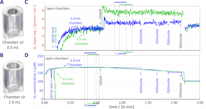
图1:小体积(sV)和经典体积(cV) 室和仪器 O2 背景检测 (A) 室V. (B) 室 cV. (C代表性轨迹图 斜率呈负值且 J°1 标记,sV (0.5 mL;绿色)和 cV (2.0 mL;蓝色) (D) 相应的氧浓度和R1标记。三次手动添加连二亚硫酸钠 滴定(约 0.7 µL,10 mM 储存液;cV)和最终 7.0 µL 2.5 mM 和 10 mM 的滴定 s中的储备V 和 cV 分别对腔室进行滴定,由此产生的峰信号已被消除。 请点击此处以查看此图的放大版本。
4. 将样品加入 Oroboros 反应室
注意:样本可通过两种方法加入腔室:部分体积置换法(本研究中的血小板和成纤维细胞)或完全体积置换法(本研究中的分离线粒体)。
- 部分体积置换
- 移除完全插入的塞子。用移液器吸取并弃去体积为 Vout 的 MiR05。对于血小板,Vout 为 60–70 µL;对于成纤维细胞,为 80–150 µL,具体数值根据储备细胞悬液中的细胞浓度计算确定。
- 用移液器将等体积 VJ.i 的储备细胞悬液(已知细胞浓度)加入氧电极室中,该体积与 Vout 相同(图2)。
- 根据实际体积 V'(0.54 mL 和 2.07 mL)计算实验中的细胞浓度,该体积大于有效实验体积 V(0.5 mL 和 2.0 mL)。
注:储备细胞悬液的浓度和实验细胞浓度应预先设计,以确保 VJ.i = Vout 的体积值可兼容较小的滴定误差。在血小板实验中,sV 室使用了一种原型塞子,其帽状部分的死体积为 0.05 mL。
- 完全体积置换
- 用虹吸法将室内的全部培养基吸出,并分别向 sV 或 cV 室中加入至少 0.54 mL 或 2.07 mL 含实验样品浓度的分离线粒体储备溶液。
- 关闭反应室,再用虹吸法将塞子接收腔中多余的线粒体储备液吸出。
注:完全体积置换可避免通过 Hamilton 注射器经塞子毛细管向密闭 反应室中滴加样品时可能引入的线粒体浓度变异。后一种方法适用于由高浓度储备悬液制备的小直径样品(例如分离的线粒体);然而,该方法会使脆弱细胞暴露于 Hamilton 注射器长 51 mm、内径 0.41 mm 的针头所产生的剪切力下,可能导致细胞质膜受损。通过移除塞子并采用部分或完全体积置换的方式将样品移入反应室,可避免此类损伤。本研究共分析了三种类型的线粒体制剂。样品制备方法详见 补充文件11,26,29,30。
- 活细胞:人血小板
- 将含有血小板的冻存管插入 37 °C 水浴中,缓慢融化至仅剩少量冰晶。
- 打开冻存管,缓慢逐滴加入 1 mL 预热培养基(DPBS + 10 mM EGTA,37 °C)。
- 轻轻混匀血小板,转移至 50 mL 离心管中,加入预热培养基(DPBS + 10 mM EGTA,37 °C)至终体积 10 mL。在室温(RT)下,使用摆动转子以 1000 g 离心 10 分钟(加速度 6,刹车 2)。
- 将血小板重悬于 0.25 mL DPBS + 10 mM EGTA(室温)中,通过部分体积置换加入反应室,使初始实验细胞浓度在 0.5 mL 室中为 157 · 106 至 210 · 106 x·mL-1,在 2.0 mL 室中为 164 · 106 至 220 · 106 x·mL-1(有关基本单位 x 的详细信息见31)。
注:一台 Oroboros 仪器在 A 和 B 位置分别装配 sV 和 cV 反应室,与另一台在 B 和 A 位置分别装配 sV 和 cV 反应室的 Oroboros 仪器并行使用。
- 透化细胞:人成纤维细胞
- 通过部分体积置换将细胞加入 Oroboros 反应室,名义细胞浓度为 0.75 · 106 x·mL-1 (两种反应室均适用),并计算实际浓度(步骤 4.1.3)。
注:并行使用了多台不同的 Oroboros 仪器,每台仪器分别装配 sV 或 cV 反应室。
- 通过部分体积置换将细胞加入 Oroboros 反应室,名义细胞浓度为 0.75 · 106 x·mL-1 (两种反应室均适用),并计算实际浓度(步骤 4.1.3)。
- 分离线粒体:小鼠心脏
- 将 15 µL 线粒体悬液加入 5985 µL 线粒体呼吸介质(MiR05)中,得到线粒体蛋白储备浓度范围为 0.013–0.026 mg∙mL-1。
- 通过完全体积置换将线粒体悬液加入 Oroboros 反应室中。
注:一台 Oroboros 仪器在 A 和 B 位置分别装配 sV 和 cV 反应室,与另一台在 B 和 A 位置分别装配 sV 和 cV 反应室的 Oroboros 仪器并行使用。
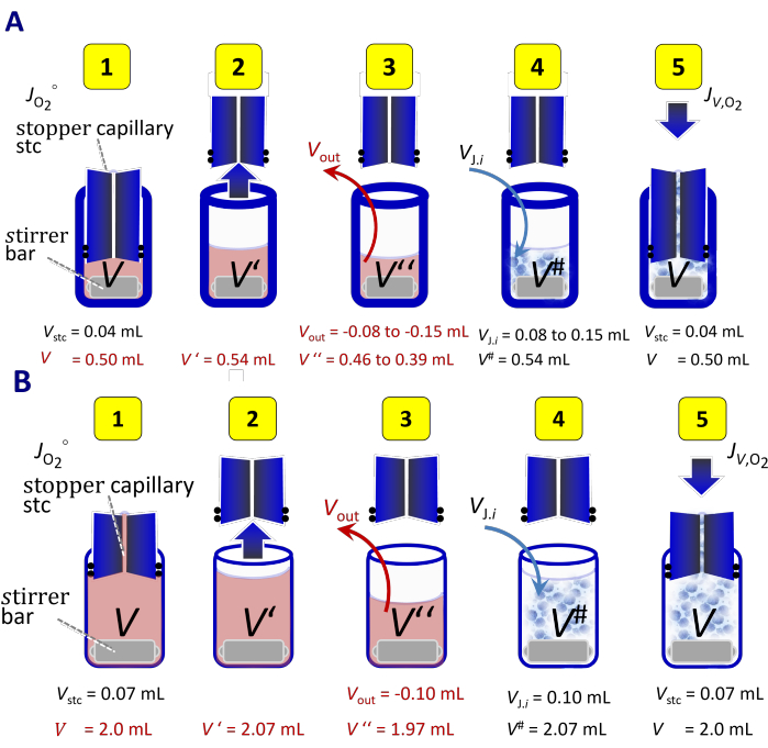
图2通过部分体积置换添加细胞悬液。 (A) 0.5 mL 反应室中的添加步骤V 以及(B2.0 mL 室 cV. (1) 带有呼吸介质体积的密闭室 V 并填充带塞毛细管 Vstc,用于测量仪器本底O2 通量 JO2°. (2塞子移除, Vstc 流入腔室,导致总体积 V'= V+Vstc. (3部分去除培养基 V出 = -VJ.i. 一个体积 V''= V'+V出 留在室中。4移液取体积子样本 VJ.i 将呼吸测定用储备细胞悬液加入腔室,终体积为 V# = V+Vstc 稀释后的细胞悬液。5) 带有填充塞的密闭腔室(死体积) Vstc用于测定体积特异性氧2 通量 JV,O2 在实验体积中 V此图已根据 图2A 细胞计数与HRR中的归一化41. 请点击此处以查看此图的放大版本。
5. SUIT 实验方案
- 使用 Hamilton 微量注射器将 补充表 1 中所列化学品的储备溶液滴加到腔室中,调整滴加体积,使 sV 和 cV 腔室中的实验浓度保持一致(参见图 6、图 8 和图 10 的图注)。
- 选择实验方案 SUIT-003_D01832,33(本研究中用于血小板)。
- 选择实验方案 SUIT-001_D0044,15,34,35 和 SUIT-002_D0074,15,35,36(本研究中用于成纤维细胞)。
- 选择实验方案 SUIT-002_D0054,15,35,37(本研究中用于分离的线粒体)。
6. SUIT 数据分析
- 在 DatLab 中设置标记,以定义所选图表的各个区段。
- 在顶部菜单中选择通量/斜率,打开通量/斜率窗口。应用滴定体积校正,以校正 SUIT 方案中每次滴定对悬浮样品造成的稀释效应。
- 从顶部菜单中选择标记,打开标记统计,显示经减去残余氧消耗(rox)进行基线校正后的标记数据点的中位数。
- 使用电子表格软件,通过反向回归分析25,28,计算 sV 和 cV 腔室中成对结果之间的截距和斜率。
- 使用软件 jamovi 2.3.1338,通过配对样本 t 检验比较 sV 和 cV 腔室中 rox 状态(最低 O2 通量)下的呼吸速率。
注意:由于 Oroboros 仪器中的两个腔室在功能上相互独立(除温度和 DatLab 记录间隔外),每个 腔室的测量可视为独立进行,类似于使用不同的 仪器。最终结果可报告为多个腔室的中位数或平均值,并附上其变异性信息。
结果
原始信号 R1 [µA氧气传感器(POS)的读数应不受“开放”室中与气相空气处于平衡状态的水相体积影响,这一点已得到证实。 补充表格 2在密闭腔室中,仪器背景O2 接近空气饱和的通量 J°1 [pmol∙s-1∙mL-1] 取决于体积 V (补充表格 2)。O2 传感器的消耗量 [pmol∙s-1] 可根据零点校正后的原始信号 R1-R0 测得的电子流进行预测 [µA],电荷数 4 e-/02 以及法拉第常数 F = 96 485.33 C∙mol-1. 1 µA 相当于电子流(e-10.36 pmol∙s-1. 除以电荷数可得到一个 O2 流15,39 2.591 pmol∙s-1·µA-1. 记录的 R1-R0 中位数为 2.09 µA 和 1.98 µA 分别在0.5 mL和2.0 mL的反应室中(补充表格 2;R0 = 0.006 ± 0.007 µA 在 sV 且 R0 = 0.004 ± 0.003 µA 在 c 中V 腔室),理论上对应于 O2 5.41 pmol的消耗速率∙s-1和 5.13 pmol∙s-1. O2 传感器的流速被转换为空气饱和条件下预期的体积特异性通量(J°R1) 为 5.41 pmol∙s-10.5 mL = 10.82 pmol∙s-1∙mL-1 和 5.13 pmol∙s-1/2.0 mL = 2.56 pmol∙s-1∙mL-1 在 sV 和 cV 室分别。在2.0 mL和0.5 mL chamber中使用相同类型的极谱式氧传感器时,其体积特异性背景O₂2 通量 - 由电流 R1-R0 计算得出J°R1)——理论上会因腔室体积的倒数关系而增加4倍。 J°空气 (在空气饱和条件下,此时反向扩散为零;比较 Table 2 和 补充表格 3) 针对 O 进行了计算2 空气饱和浓度由实验背景测试的线性回归确定(图3). J°空气 接近理论预期值 J°R1 两个腔室均(Table 2)。该 J°空气 / J°R1 比值为0.85 ± 0.04 和 0.92 ± 0.12 在 s 中V 和 cV 分别在2.0 mL室中进行的一系列独立测量(共52次)也与此一致,其中该比值为0.95 ± 0.12 (补充表格 4). J°空气 在0.5 mL室中比在2.0 mL室中高3.9倍。净O2 反向扩散 J°50,计算于 50 µM O2 来自实验背景测试的线性回归结果显示,在0.5 mL反应室中,该值仅高出三倍Table 2)。仪器背景氧的变异性2 两种不同腔室体积中的通量几乎相同,标准差(SD) 0.5 和 0.3 pmol∙s-1∙mL-1 用于 J°空气 0.9 和 0.8 pmol∙s-1∙mL-1 用于 J°50 在 sV 和 cV 室,分别(Table 2)。尽管仪器本底较高 O2 0.5 mL chamber 中的斜率,背景 O2 通量在 cO2 在两种培养室中均具有高度可重复性(图3D。这会导致残差值较低 <±1 pmol∙s-1∙mL-1两个腔室均(补充表格 5),符合Oroboros检测限的要求28.
我们量化了搅拌速度对POS在350 rpm(5.8 Hz)至950 rpm(15.8 Hz)范围内以100 rpm(1.7 Hz)为步长时校准原始信号R1的影响,实验在腔室处于“开放”配置下进行。在此配置中,O2 溶解在培养基中的气体与气相处于平衡状态;因此,原始信号(微安[µA]) 随搅拌速度的变化并不反映真实的差异 cO2. 氧传感器在默认转速750 rpm下,响应搅拌速度变化时相对于R1仅有微小偏差,且该偏差与腔室体积无关图4)。在“开放”构型中,O的噪声2 斜率,定义为信号稳定后每一步的标准差(SD),在两种腔室中均于850 rpm时最低,当搅拌速度向任一方向偏离该值时,斜率均增加(图5A。在550 rpm以下,O的噪声2 斜率超过了阈值 ±4 pmol∙s-1∙mL-1,定义最低的实用搅拌速度。由于技术限制,未能达到上限,因此将950 rpm定为最高的实用搅拌速度(图 5A)。在此范围内,O的偏差2 来自 R1 的信号 <2.5%。关闭腔室并未改变噪声与搅拌速度之间的关系图5B)。指数时间常数 τ,为2.53 ± 0.05 秒,550 转/分钟,2.50 ± 0.05 秒,750 转/分钟,适用于 0.5 mL 样品室(补充材料 图 3).
应用于血小板的 SUIT-003_D018 方案包含多达 14 步滴定操作。该方案旨在研究活细胞的偶联控制、ce 以及质膜通透性作为细胞活力的指标32。在两种类型的腔室中测得的氧通量(图6)在所有呼吸状态下均高度一致(图7)。需要间歇性重新充氧,以避免功能性缺氧或将实验中的 O2 浓度限制在特定范围内。重新充氧操作在鱼藤酮(Rot)滴定完成后通过将腔室打开至“开放”位置实现。关闭腔室后,2.0 mL 腔室中的 O2 通量在 4 分钟内即趋于稳定,而 0.5 mL 腔室则需要约 10 分钟才能稳定并设定新的基准值(图6 和 补充图 4)。在 O2 通量较低的 rox 状态下进行重新充氧,可避免在达到稳定所需的时间内 O2 浓度下降。在 sV 和 cV 腔室中,经鱼藤酮抑制后的 rox 值分别为 0.9 ± 0.6 和 2.0 ± 0.6 pmol∙s-1∙mL-1 ,对应的氧浓度分别为 98 ± 17 和 94 ± 17 µM O2 (均值 ± 标准差)。在 sV 和 cV 腔室中,重新充氧至 159 ± 8 和 147 ± 11 µM O2 后,rox 分别升高了 3.1 ± 0.9 和 0.9 ± 0.4 pmol∙s-1∙mL-1 。血小板中观察到的 rox 对氧的依赖性与人成纤维细胞和脐静脉内皮细胞的研究结果一致40。
在透化成纤维细胞的氧化磷酸化(OXPHOS)分析中,我们采用了两种SUIT方案,分别包含最多17步和20步的滴定步骤(SUIT-001和SUIT-002,分别35;图8);因此,滴定体积校正尤为重要,因为在SUIT-002结束时,sV和cV腔室的最大稀释度分别达到14%和10.5%。在0.5 mL和2.0 mL腔室中,成纤维细胞数量分别为0.35·106 x和1.44·106 x,最终获得相同的细胞浓度,即0.75·106 x∙mL-1。SUIT-001与SUIT-002的联合应用共检测了20种呼吸状态,所有状态在两种腔室类型之间均表现出高度相关性(SUIT-001的r2 = 0.950,SUIT-002的r2 = 0.959)。在两种方案中,复合物IV(Complex IV)的数据均表现出相对较高的离散度(图9)。在SUIT-001中,起始时体积特异性O2 通量较低,为18.7 ± 7.0 pmol∙s-1∙mL-1 (均值 ± 标准差),在“常规” 状态下无需重新供氧,允许O2 浓度下降至56 µM,直至实验方案中与CIV相关的部分(图8A;8Ama)。然而,在SUIT-002中,需在解偶联剂滴定前进行重新供氧,将O2 浓度从100 µM提升至180 µM(图8B;6Gp)。考虑到重新供氧后通量稳定所需时间较长,应避免在解偶联剂滴定过程中进行重新供氧。
采用简化的SUIT方案对分离的线粒体进行检测(图10)。线粒体蛋白浓度在呼吸测定实验后确定,因此无法预先设定反应腔内样品的准确浓度。体积特异性呼吸通量随线粒体浓度变化而变化。因此,数据以体积特异性和蛋白质量特异性通量两种形式呈现(图11)。两种表示方式均显示出在0.5 mL和2.0 mL反应腔中获得的测量结果之间具有高度相关性(图11)。
在使用抗霉素A抑制复合体III后测得的残余氧消耗量rox,可作为仪器在极低呼吸速率下性能的指标。在所有实验系列中,0.5 mL和2.0 mL检测腔之间的rox值具有可比性(血小板:t(5) = -0.34,p = 0.74;成纤维细胞:SUIT-001:t(10) = 20.00,p = 0.06;SUIT-002:t(10) = 11.00,p = 0.96;分离的线粒体:t(4) = -1.18,p = 0.27)。数据的离散程度主要对应于仪器的不确定度±1 pmol∙s-1∙mL-1 13,28,如图12所示。原始文件可在Zenodo数据库中开放获取:10.5281/zenodo.11634408。

图3仪器背景信号 O2 s的通量V (0.5 mL;绿色和黑色)以及 cV 腔室(2.0 mL;蓝色) O2 浓度范围从空气饱和(约 180 μM约 50 µM. 空心符号:O2 未加连二亚硫酸钠时测得的浓度。实心 符号:O2 由连二亚硫酸钠手动滴定控制浓度。黑色圆圈: O2 通过TIP2k使用连二亚硫酸钠滴定,在0.5 mL体系中控制浓度 腔室(N = 8)。仪器本底测试的结果汇总自(A系列 1 (sV: N = 2;cV: N = 2)在使用活细胞(ce)之前;(B系列 2(sV: N = 38; cV: N = 50)后使用透化细胞(pce);(C系列 3(sV: N = 10;cV: N = 10)后使用分离的线粒体(imt)。D) 叠加自各图面板的数据 A-C。 请点击此处以查看此图的放大版本。
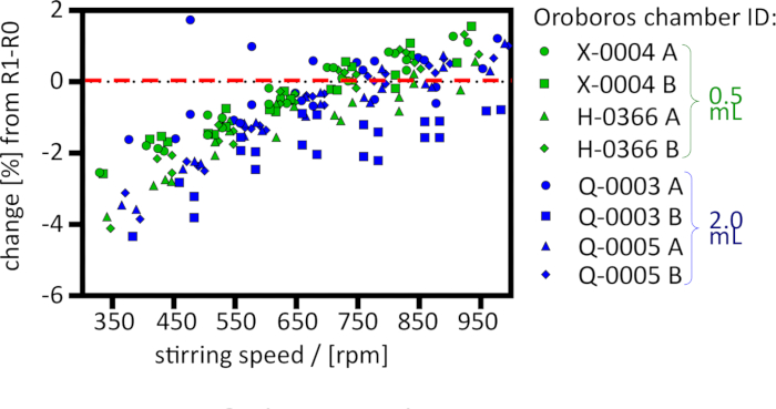
图4:搅拌转速对0.5 mL腔室(绿色)和2.0 mL腔室(蓝色)在空气饱和状态(“开放”腔室)下POS信号的影响。响应值 [%] 相对于750 rpm时经R0校正的空气标定信号R1-R0 [µA](sV:N = 4; cV:N = 4)。请点击此处查看该图的放大版本。
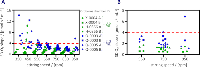
图5:O2 斜率噪声随搅拌转速的变化。 噪声以标准差表示,分别为(A)“开放”室;(B)封闭室(0.5 mL,绿色;2.0 mL,蓝色)。请点击此处查看该图的放大版本。
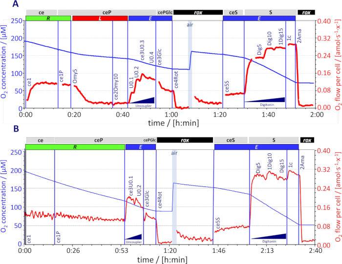
图6O2 0.5 mL腔室中冷冻保存的人血小板的消耗 O的代表性轨迹2 浓度 [µM](蓝线)和 O2 每细胞流量 [amol∙s-1∙x-1] 计算为 O2 通量 [pmol∙s-1∙mL-1] 除以细胞浓度 157∙106 x∙mL-1 (红色线条,已校正仪器背景O2 通量;由...引起的尖峰 滴定被消除)。O2 浓度保持在以上 50 µM 间歇性地 复氧(空气,开放室,蓝色阴影)。呼吸状态序列(滴定) 和速率;方案 SUIT-003_D018) ce1,常规 呼吸作用 R 活细胞 ce1P, 5 mM 丙酮酸作为外源底物。 ce2Omy10 (面板 A),Omy滴定以5 nM为步长;漏液 呼吸作用 L. ce3U0.3 (面板 A)和 ce3U0.1 (面板 B),解偶联剂滴定至 最佳CCCP浓度为0.3和 0.1 µM,分别;ET 容量 E. ce3Glc, 葡萄糖 11 mM E 被解偶联剂逐渐抑制。 ce4旋转鱼藤酮 0.5 µM 抑制复合体 I;残余氧消耗 rox. ce5S,琥珀酸 10 mM;刺激 E 在 细胞膜通透性细胞 1挖15 (面板 A)和 1挖10 (面板 B),皂苷 滴定在 5 µg·mL-115 和最佳浓度的步骤 10 µg·mL-1分别 用于完全的质膜透化;琥珀酸盐途径电子传递能力 SE. 1c细胞色素 c 10 µM线粒体外膜完整性检测 2Ama抗霉素A 2.5 µM 抑制复合体III; rox. 请点击此处以查看此图的放大版本。
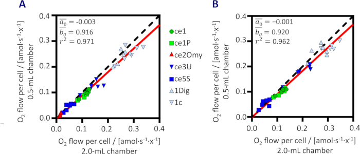
图7:0.5 mL和2.0 mL反应室中测定的冷冻保存人血小板呼吸的相关性 Rox校正的 O2 流程在方案 SUIT-003_D018 中进行评估(图6); N = 6. (A) 使用寡霉素 Omy;(B) 无 Omy 时的平均斜率  和截距
和截距  倒置最小二乘回归及决定系数 r2 根据所有数据计算得出。黑色虚线表示恒等线。红色线表示计算得到的回归线。O2 加入葡萄糖后处于该状态下的流被排除(参见 图6)。含寡霉素与不含寡霉素的实验方案分别展示于不同图中 A 和 B 以避免数据点重叠。 请点击此处以查看此图的放大版本。
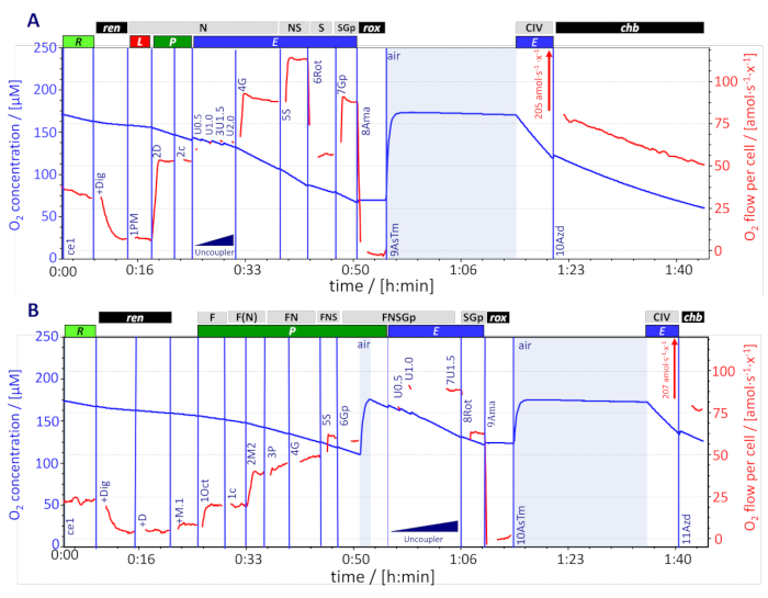
图8O2 0.5 mL chamber 中成纤维细胞的消耗量 代表性轨迹 cO2 (蓝色线条[μM]) 和 O2 每细胞流量计算为 O2 通量 [pmol∙s-1∙mL-1] 除以细胞浓度(0.75·106 x∙mL-1),并校正仪器背景O2 通量(红色线条,滴定引起的尖峰已被消除)。O2 浓度保持在以上 50 μM 通过间歇性再氧合(空气,“开放”室,蓝色阴影)进行。呼吸状态序列(滴定及速率)。A方案 SUIT-001_D004: ce1,常规 呼吸作用 R. +Dig,皂苷素 10 μg·mL-1 (细胞外聚合物转化为颗粒状细胞外聚合物),残余内源呼吸 仁. 1PM,丙酮酸钠 5 mM & 苹果酸 2 mM;N通路泄漏 呼吸作用 NL. 二维,ADP 2.5 mM;N通路氧化磷酸化能力 NP. 2c细胞色素 c 10 μM线粒体外膜完整性检测。 3U1.5,以确定最佳CCCP浓度的解偶联剂滴定 1.5 μM;NADH连接的电子传递链容量 NE. 4G,谷氨酸 10 mM;N(PGM)E. 5S, 琥珀酸 10 mM;NS途径电子传递能力 NSE. 6Rot鱼藤酮 0.5 μM;琥珀酸盐途径电子传递能力 SE. 7Gp,10 mM 甘油磷酸盐;SGpE. 8Ama抗霉素A 2.5 μM; rox. 9AsTm,抗坏血酸 2 mM & TMPD 0.5 mM;细胞色素c氧化酶(CIV)激活底物;反应室保持开放20分钟,以实现氧化还原平衡,同时避免氧气耗尽4;O2 关闭培养室后通量超出量程(205 amol·s-1·x-1). 10Azd,叠氮化物 100 mM;氧依赖性化学背景通量 中国台湾. (B方案 SUIT-002_D007: ce1, R. +Dig, 任. +D,刺激 任. +M.1,苹果酸 0.1 mM 和 10月1日,辛酰肉碱 0.5 mM;F通路氧化磷酸化能力 FP = J(10月1日)J(+M.1) 1c. 2M2,2 mM 苹果酸支持的回补性 N 途径;F(N)P. 3P 和 4G,向纤连蛋白的逐步活化P. 5S,FNSP. 6Gp,FNSGpP. 7U1.5,FNSGpE. 8Rot,SGpE. 9Ama, rox. 10AsTm O2 通量超出量程 207 amol·s-1·x-1. 11Azd, 中国台湾通常,复合体 IV 活性为反应速率之差, J(AsTm)-J(Azd)在紧密匹配的 O2 浓度。参见 图6 有关更多详细信息。 请点击此处以查看此图的放大版本。
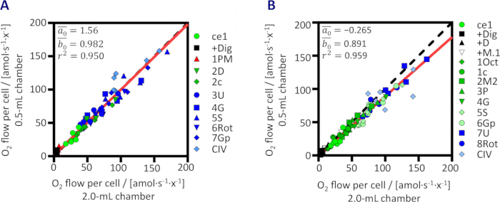
图9:0.5 mL和2.0 mL腔室中透化成纤维细胞呼吸作用的相关性。 经Rox校正的O2通量在(A) SUIT-001_D004和(B) SUIT-002_D007中的结果(N = 6)。所有数据经反向最小二乘回归分析得到的平均斜率 和截距 ,以及决定系数r2。黑色虚线表示恒等线。红色线表示计算得到的回归线。请点击此处查看该图的放大版本。
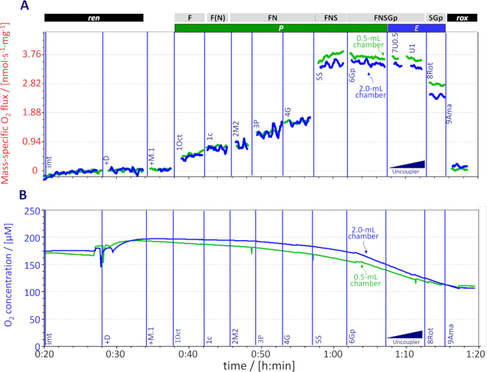
图10:0.5 mL(绿色)和2.0 mL(蓝色)腔室中分离的小鼠心脏线粒体的O2消耗。代表性曲线显示(A)线粒体蛋白浓度为0.023 mg∙mL-1时的单位质量O2通量(已校正仪器背景O2通量,滴定引起的尖峰已被消除)以及(B)O2浓度[μM]。按照SUIT-002_D005方案依次建立的呼吸状态(滴定及速率):imt,残余内源性呼吸 ren;+D,加入ADP(2.5 mM),刺激ren;+M.1,加入苹果酸(0.1 mM)和1Oct,辛酰肉碱(0.5 mM);F通路氧化磷酸化能力FP = J(1Oct)-J(+M.1);1c,细胞色素c(10 μM);检测线粒体外膜完整性;2M2,高浓度苹果酸(2 mM),支持补碳的N通路;F(N)P;3P,丙酮酸(5 mM);FNP;4G,谷氨酸(10 mM);FNP;5S,琥珀酸(10 mM);FNSP;6Gp,甘油磷酸(10 mM);FNSGpP;7U0.5,逐步加入解偶联剂至CCCP最适浓度(0.5 μM);FNSGpE;8Rot,鱼藤酮(0.5 μM)抑制复合体I(CI);SGpE;9Ama,抗霉素A(2.5 μM)抑制复合体III(CIII);残余氧消耗rox。请点击此处查看该图的放大版本。
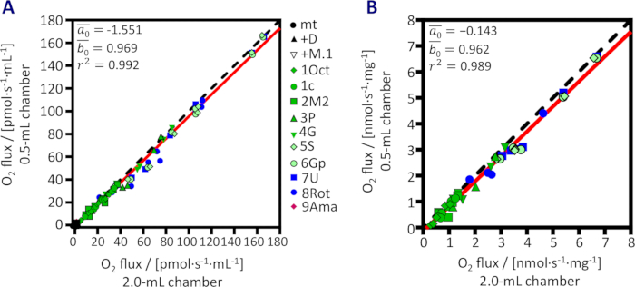
图11:在0.5 mL和2.0 mL腔室中测定的分离小鼠心脏线粒体呼吸的相关性。 按照图10所示方案测得的O2通量(N = 4)。(A) 体积特异性O2通量。(B) 经Rox校正后的O2通量除以线粒体蛋白浓度(0.013 - 0.026 mg∙mL-1)。均值斜率 和截距 由逆向最小二乘回归计算得出,并基于所有数据计算决定系数r2。黑色虚线表示恒等线。红色线表示计算得到的回归线。请点击此处查看该图的放大版本。
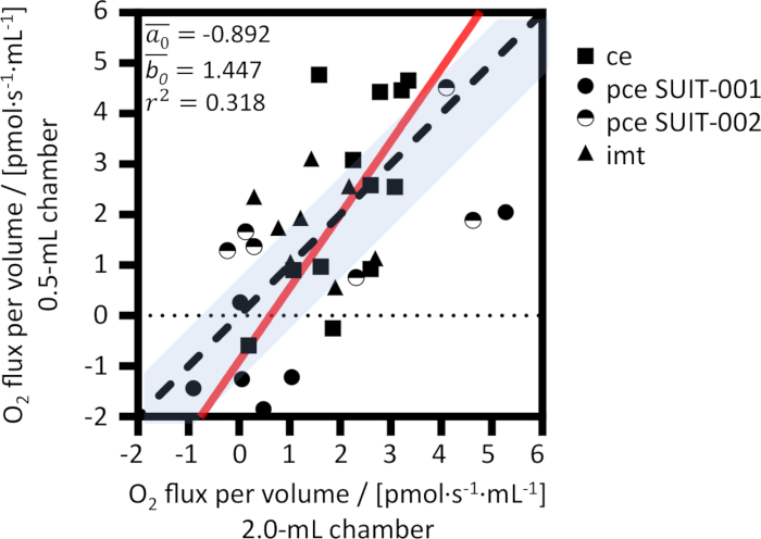
图12:0.5 mL和2.0 mL检测室中残余氧消耗速率 rox。在加入抗霉素A后测得的体积比通量,数据综合了活细胞(ce,方块)、透化细胞(pce,圆圈)和分离线粒体(imt,三角形)的实验结果。平均斜率 和截距 由反向最小二乘回归计算得出。虚线:恒等线。红线:计算得到的回归线。阴影区域:仪器分辨率范围。请点击此处查看该图的放大版本。
| 模块 | sV | cV |
| 实验腔室体积 V | 0.5 mL | 2.0 mL |
| 玻璃圆筒内径 | 12.0 mm | 16.0 mm |
| 玻璃圆筒外径 | 24.0 mm | 24.0 mm |
| 玻璃壁厚度 | 6.0 mm | 4.0 mm |
| PVDF涂层搅拌子 | 11.5 x 6.2 mm | 15.0 x 6.0 mm |
| 塞子毛细管直径 | 1.00 mm | 1.30 mm |
| 塞子毛细管横截面积 | 0.79 mm2 | 1.33 mm2 |
| 塞子毛细管长度 | 50.64 mm | 48.86 mm |
| 塞子毛细管体积 | 39.8 µL | 64.9 µL |
| 塞子毛细管死体积 Vstc(包含一小滴液体)a | 0.04 mL | 0.07 mL |
| 体积校准用体积 V´ | 0.54 mL | 2.07 mL |
a液滴体积应为5 µL,对应于小室cV,而小室sV则接近0 µL。
表1:采用聚醚醚酮(PEEK)密封垫的小体积(sV)和经典体积(cV)模块的尺寸。
| J°air / [pmol∙s-1∙mL-1] | J°50 / [pmol∙s-1∙mL-1] | |||
| 实验腔室体积 V | 0.5 mL | 2.0 mL | 0.5 mL | 2.0 mL |
| 平均值 | 9.22 | 2.37 | -3.17 | -1.17 |
| 中位数 | 9.25 | 2.36 | -3.08 | -1.04 |
| 标准差 | 0.49 | 0.34 | 0.93 | 0.83 |
| 最小值 | 8.20 | 1.79 | -5.11 | -3.72 |
| 最大值 | 10.41 | 3.42 | -0.82 | 0.67 |
表2:外推至空气饱和状态的J°air以及在50 μM O2浓度下计算的J°50。 第2系列(pce)和第3系列(imt)仪器本底测试的汇总数据,以及TIP2k 滴定前的数据(0.5 mL:N = 48;TIP2k:N = 8;2.0 mL:N = 60)。
补充文件 1:样品制备。 人血小板、人成纤维细胞以及从小鼠心脏中分离的线粒体。 请点击此处下载该文件。
补充图1:使用TIP2k进行仪器O2背景检测。 使用TIP2k在0.5 mL样品室中滴定Na-连二亚硫酸盐(2.5 mM)的代表性检测曲线。介质:MiR05,温度:37 °C,搅拌速度:750 rpm。滴定引起的信号尖峰已被消除。请点击此处下载该文件。
补充图2:仪器背景O2 通量 J°1 作为O的函数2 浓度 腔室 sV (0.5 mL;绿色, N = 48)和 Chamber cV (2.0 mL;蓝色, N 系列2(pce,圆圈)和系列3(imt,三角形)中n = 60,以及在0.5 mL样品池中进行TIP2k滴定前(黑圈, N = 8)。红线:中位数 ± SD 请点击此处下载此文件。
补充图3:指数时间常数τ随搅拌转速的变化关系。 在37 °C条件下,0.5 mL样品室的各个τ值(N = 2)。在0.5 mL样品室中,550 rpm时τ为2.53 ± 0.05 s,750 rpm时为2.50 ± 0.05 s,850 rpm时为2.47 ± 0.04 s。请点击此处下载该文件。
补充图4:O2 2.0 mL 室中冷冻保存的人类血小板的消耗 SUIT-003_D018 方案的代表性轨迹,包括寡霉素滴定(Omy)。通量 [pmol∙s-1∙mL-1] 已校正仪器本底信号2 通量除以细胞浓度(164∙106 x∙mL-1). 滴定引起的峰已被消除。 cO2 保持在以上 60 µM 通过间歇性再氧合(空气,“开放”室,蓝色阴影)进行。呼吸状态序列(滴定及速率): ce1,常规 呼吸作用 R 活细胞 ce1P,以丙酮酸作为外源底物。 ce2Omy10,Omy滴定以5 nM为步长;漏液 呼吸作用 L. ce3U2.0,解偶联剂滴定至最适浓度 2 µM;ET容量 E. ce3Glc,葡萄糖 11 mM; E 被解偶联剂逐渐抑制。 ce4Rot鱼藤酮 0.5 µM 抑制复合体 I;残余氧消耗 rox. ce5S,琥珀酸盐 10 mM;刺激 E 在细胞质膜具有通透性的细胞中。 1Dig20,皂苷滴定至最适浓度 20 µg·mL-1,用于完全的质膜透化;琥珀酸盐途径电子传递能力 SE. 1c细胞色素 c 10 µM线粒体外膜完整性检测。 2Ama抗霉素A 2.5 µM 抑制复合体III; rox. 请点击此处下载此文件。
补充表 1:底物-抑制剂-解偶联剂滴定(SUIT)方案中使用的化学品。 sV 和 cV 室所用试剂的缩写及储备液浓度。请点击此处下载该文件。
补充表 2:R1-R0 比较与 J°1 在0.5 mL和2.0 mL的腔室中。 第二系列(pce)和第三系列(imt)仪器本底测试的合并数据,以及TIP2k滴定前(0.5 mL)的数据 N = 48;TIP2k: N = 8;2.0 mL: N = 60)。数据仅来自实验系列2(pce)和系列3(imt),这两组实验在平均大气压为94.9 kPa(奥地利因斯布鲁克)的条件下进行,排除了在海平面条件下获得的系列1(ce)的数据。在0.5 mL和2.0 mL反应室中使用了相同类型但不同个体的POS传感器。因此,在比较s时必须考虑传感器之间的变异性V 和 cV 腔室。尽管 J°1 = 2.26 ± 0.41 pmol∙s-1∙mL-1 通过实验测得的2.0 mL室腔结果与计算值高度一致, 实验的 J°1 = 8.48 ± 0.53 pmol∙s-1∙mL-1 0.5 mL 检测室中的数值低于预期,原因如下:关闭检测室后,较高的背景 O2 0.5 mL chamber 中的通量降低 O2 标记处的浓度 J°1 设置为相近的时间间隔(图1)。氧气浓度在 J°1 在0.5 mL和2.0 mL反应室中,平均分别比空气饱和度低4.06%和1.72%(补充图2,补充表2)。在较低 cO2,减弱的O2 传感器消耗以及部分 O2 反向扩散降低实验测定值 J°1. 请点击此处下载此文件。
补充表3:获得校准值(R1)和J°1时的氧气浓度[µM]及其百分比差异。 系列2(pce)和系列3(imt)的仪器本底测试汇总数据,以及TIP2k滴定前的数据(0.5 mL:N = 48;TIP2k:N = 8;2.0 mL:N = 60)。请点击此处下载该文件。
补充表 4:测量值与预期值 J°1 在2.0 mL室中。 独立系列的仪器本底测试,使用O2k系列A(配备钛制塞子)13), V = 2.0 mL (37 °C,含O的NaCl溶液2 溶解度因子 0.92; N = 52). nO2/Qe = 106/(96485.33·4) = 2.591 pmol∙µC-1 = 2.591 pmol∙s-1∙µA-1. 基于以下假设:O2 反向扩散可忽略不计 cO2,1,这只在 cO2,1 ~ cO2*. 请点击此处下载此文件。
补充表5:线性回归残差(绝对值 [pmol O2∙s-1∙mL-1]) 针对每种仪器的 O 计算得出2 背景测试第2组(pce)和第3组(imt)仪器本底测试的汇总数据,以及TIP2k滴定前的测试结果。 请点击此处下载此文件。
讨论
呼吸测定实验中可获取的样品量通常有限,例如在活检组织、原代细胞或细胞数量较少的慢速生长细胞培养物的研究中1此外,由于线粒体损伤,呼吸活性受到抑制11 或在研究呼吸容量较低的线粒体通路时,例如脂肪酸氧化12. 在这些情况下,较低的呼吸测量室体积有助于提高 O₂ 流量检测的分辨率2 通量。然而,仪器的背景氧通量2 在较低的腔室体积下,通量会增加,若未能可靠测量并针对氧浓度进行适当校正,则可能引发误差2 浓度。仪器氧2 背景测试与背景校正对于小体积实验腔室尤为重要。理论值(J°R1)和实验(J°空气) O2 通量为使用4 e的极谱法测量的准确性提供了证据-/2 在 的 阴极 POS 反应。两者之间的差异 J°R1 以及 J°空气 为1.64 ± 0.43 和 0.22 ± 0.31 pmol∙s-1∙mL-1 在 sV 和 cV 分别表明测量的精确度处于2.0-mL chamber的检测限范围内±1 pmol∙s-1∙毫升-1)28. 在 2.0-mL 和 0.5-mL 实验 chamber 中使用相同类型的极谱氧传感器时,单位体积的背景 O₂ 通量2 通量随腔室体积成反比增加4倍,两种腔室体积下的变异程度几乎相同,标准差分别为0.5和0.3 pmol∙s-1∙mL-1 用于 J°空气 和 0.9 与 0.8 pmol∙s-1∙毫升-1 用于 J°50 在 sV 和 cV 室,分别(Table 2)。在最低呼吸活性(小体积 chamber 的设计目标)条件下,即使微小的背景效应也变得显著(补充 Table 3; 补充图 2)。因此,对仪器背景氧通量进行常规评估是必要的2 通量图1) 是进行适当校正所必需的28.
为了维持体系的均一性并保证稳定的氧信号,需要对介质进行高速且持续的搅拌。2.0 mL 搅拌腔的搅拌速度设定为 750 转/分钟(rpm),但 0.5 mL 搅拌腔的最佳搅拌速度尚不明确。由于玻璃腔室与搅拌子在几何结构上的差异(表 2),需对 0.5 mL 腔室的最佳搅拌速度进行独立评估。氧传感器对 550 rpm 至 950 rpm 之间的搅拌速度变化有响应,在 750 rpm 时,O2 信号 R1 仅有微小偏离,且不受腔室体积影响。在相同搅拌速度下,对 Chamber sV 计算得到的指数时间常数 τ 与 2.0 mL 腔室报道的 τ 值一致13。这些结果表明,两种腔室几何结构下的搅拌效率相当,因此 2.0 mL 腔室默认的 750 rpm 搅拌速度可直接应用于 0.5 mL 腔室。在“开放”和“封闭”两种腔室构型下,0.5 mL 腔室均表现出更高的信噪比,提示其具有更高的检测精度,这是 0.5 mL 腔室的一项优势(图 5),尽管个体间 POS 的差异影响尚不能排除。由于将搅拌速度从 750 rpm 降低至 550 rpm 时噪声并未增加,因此在 0.5 mL 腔室中将搅拌速度降至 550 rpm 可能有助于减小剪切应力,同时不损害信噪比。
几乎相同的O₂通量2 在应用适当的背景校正后,于三种实验模型中分别在 0.5-mL 和 2.0-mL 检测室中测定了耗氧速率。使用少量血小板所得的结果 µ直径为 m42,活细胞和透化纤维细胞以及分离的线粒体结果表明,在0.5-mL和2.0-mL chamber中可获得一致的呼吸速率 多种细胞类型和线粒体制备样品中均适用。然而,本研究尚未测试透化肌纤维。准确测量 rox 需要控制氧水平40 且具有高仪器分辨率1. 体积特异性氧2 通量 rox 可能低至检测限。当实验中使用较少的细胞数量或通常较少的样品量时,无需重新氧合。然而,如有必要,建议在氧通量较低的呼吸状态下进行重新氧合2 通量,以避免氧气的快速下降2 稳定 O₂ 所需时间段内的浓度2 通量。氧气2 复氧后的通量在0.5 mL chamber中恢复所需时间更长。
作为质量控制的一部分,在部分换液过程中对悬浮液中样品的稀释需特别注意。在完全插入的塞子被取出后,总体积 V' 呼吸介质在腔室中的体积由实验体积决定 V 以及封口毛细管体积 Vstc (Table 2)。因此,储备体积 VJ. i 在实际体积中被稀释 V',大于 V,导致稀释倍数 V/V' 0.5/0.54 = 0.926 和 2.0/2.07 = 0.966,分别对应 0.5 mL 和 2.0 mL 气室(图2)。在血小板实验中,使用了 Chamber 的原型塞子 sV 毛细管死体积为0.05 mL,对应的稀释因子为0.909。
在相同样品浓度(单位体积内的量)下,0.5-mL 比 2.0-mL 气室所需的活细胞、透化细胞和分离线粒体的用量少近四倍,经细胞数量或蛋白质量归一化后的呼吸性 O2 消耗量测定结果具有可比性。这表明 Chamber sV 是样品量有限的研究中一种有价值的工具。另一方面,在 Chamber sV 中使用相同量的样品可提高体积特异性 O2 通量,从而在呼吸活性较低时降低测量的不确定性。
本研究中的氧环境仅限于从空气饱和至30 µM的O2 范围。在该范围之外的氧水平下的适用性尚需进一步评估,以研究组织常氧或低氧状态下的线粒体功能43,以及高氧条件下的透化组织9。Oroboros NADH和Q- 模块的多种多传感器配置,以及pH、NO和TPP+-电极10,44,无法适配于sV腔室,仍需使用2.0-mL腔室。
披露
EL 是 Oroboros Instruments 的员工。EG 是 Oroboros Instruments 的创始人兼首席执行官。
在本项目中,Oroboros Instruments 使用了 LFGS。EÅF 和 EE 是位于瑞典隆德的 Abliva AB 公司的员工,该公司与 Oroboros Instruments 持有商业协议。
LR 声明不存在利益冲突。
致谢
我们感谢 Manuela Passrugger 在分离线粒体数据收集方面的贡献,Patrizia Guerth 在搅拌转速测试方面的技术支持,以及 Mateus Grings 对搅拌器测试时间常数的分析。我们感谢我们的合作伙伴奥地利 Kolsass 的 WGT Elektronik GmbH & Co KG 提供玻璃腔室、密封塞和磁场强度的技术参数信息。我们感谢 Luiza HD Cardoso、Alejandra Romero-Martinez、Ondrej Sobotka、Alba Timon-Gomez 和 Jaime Willis 提出的宝贵意见。本研究部分由欧盟“地平线2020”研究与创新计划资助,资助号为 859770(NextGen-O2k 项目)。同时感谢 COST 行动 CA15203 MitoEAGLE 项目提供的短期科学访问计划(LFGS、LR)的财政支持。
材料
| 姓名 | 公司 | 目录编号 | 评论 |
|---|---|---|---|
| Anaconda3 | Anaconda Software Distribution | https://www.anaconda.com/ | 软件 |
| ADP | Merck, Germany | 117105 | |
| 抗生素-抗真菌素 100倍浓缩液 | Biosera, France | XC-A4110 | 警告 H317 可能引起过敏性皮肤反应 |
| 抗霉素 A | Sigma-Aldrich, USA | A8674 | 警告 H300 吞咽致命,H400 短期(急性)对水生环境有害 |
| 抗坏血酸 | Sigma-Aldrich, USA | A7631 | |
| ATP | Sigma-Aldrich, USA | A 2383 | |
| 无脂肪酸牛血清白蛋白 | Sigma-Aldrich, USA | A 6003 | bovine serum albumin |
| CaCO3 | Sigma-Aldrich, USA | C 4830 | |
| CCCP | Sigma-Aldrich, USA | C2759 | 羰基氰化物间氯苯腙 警告 H301 + H311 + H331 吞咽、皮肤接触或吸入有毒 |
| Countess 自动细胞计数仪 | Thermo Fisher Scientific, USA | AMQAX100 | |
| 细胞色素 c | Sigma-Aldrich, USA | C7752 | |
| DatLab | Oroboros Instruments, Austria | Version 7.4 | 用于数据采集与分析的软件 |
| DatLab | Oroboros Instruments, Austria | Version 8.1 | 用于数据采集与分析的软件 |
| DC 蛋白检测试剂盒 | Bio-Rad, USA | 5000112 | 蛋白检测试剂盒;警告 H314 可引起严重皮肤灼伤和眼睛损伤 |
| 皂苷 | Sigma-Aldrich, USA | D5628 | 警告 H301 吞咽有毒 |
| 二硫苏糖醇 | Sigma-Aldrich, USA | D 0632 | 警告 H318 可引起严重眼睛损伤 |
| DMEM | Pan Biotech, Germany | P04-05551 | Dulbecco’s 改良 Eagle’s 培养基 |
| DMSO | Sigma-Aldrich, USA | D2650 | 二甲基亚砜 |
| DPBS | Lonza Bioscience, Switzerland | LONBE17-512F | 无钙 Dulbecco’s 磷酸盐缓冲液 |
| EGTA | Sigma-Aldrich, USA | E 4378 | |
| FBS | Thermo Fisher Scientific, USA | A5256701 | 胎牛血清 |
| 葡萄糖 | Sigma-Aldrich, USA | G7528 | |
| 谷氨酸 | Sigma-Aldrich, USA | G1626 | |
| 甘油磷酸 | Santa Cruz Biotechnology | sc-215789 | |
| Heraeus Multifuge 3s Large | Thermo Fisher Scientific, USA | HM3S | 离心机 |
| Hettich Rotina 380R | Andreas Hettich GmbH & Co. KG, Germany | 1701 | 离心机 |
| 咪唑 | Honeywell Fluka, Austria | 56750 | 警告 H302 吞咽有害,H314 可引起严重皮肤灼伤和眼睛损伤,H360D 可能损害未出生婴儿 |
| jamovi | The jamovi project | Version 2.3.13 | 软件 |
| K+-MES | Sigma-Aldrich, USA | M 8250 | |
| Leucosep 管 | Greiner Bio-One GmbH, Austria | Z642843-300EA | 带聚乙烯屏障的离心管 |
| 苹果酸 | Sigma-Aldrich, USA | M1000 | 警告 H319 可引起严重眼睛刺激。 |
| 甘露醇 | Sigma-Aldrich, USA | M 4125 | |
| MgCl2 | Scharlau, Spain | MA 0036 | |
| MiR05-试剂盒 | Oroboros Instruments, Austria | 60101-01 | 线粒体呼吸介质 |
| Mixer | Swelab Instruments, Sweden | 440S | 摇床 |
| Mr. Frosty 冷冻容器 | Thermo Fisher Scientific, USA | 5100-0001 | 冷冻容器 |
| Oroboros | Oroboros Instruments, Austria | NextGen-O2k 和 O2k 统称为 ‘Oroboros’ | |
| Oroboros Chamber cV | Oroboros Instruments, Austria | 23100-01 | 经典体积腔室 |
| Oroboros Chamber sV | Oroboros Instruments, Austria | 33100-01 | 小体积腔室 |
| Oroboros TIP2k-模块 | Oroboros Instruments, Austria | 11100-03 | 滴定-注射微泵 |
| O2-Zero 粉末 | Oroboros Instruments, Austria | 26600-02 | Na-连二亚硫酸盐 警告 H251 自热:可能起火,H302 吞咽有害,H319 可引起严重眼睛刺激 |
| 辛酰肉碱 | Biotrend - APExBIO Technology, Germany | B6371 | |
| 寡霉素 | Sigma-Aldrich, USA | O4876 | |
| 磷酸肌酸 | Sigma-Aldrich, USA | P 7936 | |
| 丙酮酸 | Sigma-Aldrich, USA | P2256 | 警告 H317 可能引起过敏性皮肤反应 |
| 鱼藤酮 | Sigma-Aldrich, USA | R8875 | 警告 H300 吞咽致命 |
| 皮肤成纤维细胞系 | ATCC, USA | ||
| 叠氮化钠 | Sigma-Aldrich, USA | S2002 | 警告 H300 + H310 + H330 吞咽、皮肤接触或吸入致命 |
| 枯草杆菌蛋白酶 A | Sigma-Aldrich, USA | P 5380 | 警告 H318 - 可引起严重眼睛损伤,H334 - 吸入可能引起过敏或哮喘症状或呼吸困难 |
| 琥珀酸 | Sigma-Aldrich, USA | S2378 | |
| 蔗糖 | Sigma-Aldrich, USA | S 7903 | |
| Swelab Alfa 血细胞计数仪 | Boule Diagnostics AB, Sweden | 1420041 | |
| 牛磺酸 | Sigma-Aldrich, USA | T 0625 | |
| Tecan Infinite TM F200 | Tecan, Switzerland | 分光光度计 | |
| TMPD | Sigma-Aldrich, USA | T3134 | N,N,N',N'-四甲基对苯二胺二盐酸盐 警告 H315 可引起皮肤刺激,H319 可引起严重眼睛刺激 |
| 胰蛋白酶 | Sigma-Aldrich, USA | T4799 | 警告 H334 吸入可能引起过敏或哮喘症状或呼吸困难 |
| Vacuette | Greiner Bio-One GmbH, Austria | 15452520 |
参考文献
- Zdrazilova, L., Hansikova, H., Gnaiger, E. Comparable respiratory activity in attached and suspended human fibroblasts. PLoS ONE. 17 (3), e0264496(2022).
- Schöpf, B., et al. OXPHOS remodeling in high-grade prostate cancer involves mtDNA mutations and increased succinate oxidation. Nat Commun. 11 (1), 1487(2020).
- Meszaros, A. T., et al. The predictive value of graft viability and bioenergetics testing towards the outcome in liver transplantation. Transplant International. 37, 12380(2024).
- Doerrier, C., Garcia-Souza, L. F., Krumschnabel, G., Wohlfarter, Y., Mészáros, A. T., Gnaiger, E. High-resolution fluorespirometry and OXPHOS protocols for human cells, permeabilized fibers from small biopsies of muscle, and isolated mitochondria. Methods Mol Biol. 1782, 31-70 (2018).
- Gnaiger, E., Steinlechner-Maran, R., Méndez, G., Eberl, T., Margreiter, R. Control of mitochondrial and cellular respiration by oxygen. J Bioenerg Biomembr. 27 (6), 583-596 (1995).
- Gnaiger, E., Méndez, G., Hand, S. C. High phosphorylation efficiency and depression of uncoupled respiration in mitochondria under hypoxia. Proc Natl Acad Sci. 97 (20), 11080-11085 (2000).
- Gnaiger, E. Oxygen Conformance of Cellular Respiration. Hypoxia. Advances in Experimental Medicine and Biology. Roach, R. C., Wagner, P. D., Hackett, P. H. , Springer. Springer, Boston, MA. (2003).
- Harrison, D. K., Fasching, M., Fontana-Ayoub, M., Gnaiger, E. Cytochrome redox states and respiratory control in mouse and beef heart mitochondria at steady-state levels of hypoxia. J Appl Physiol. 119 (10), 1210-1218 (2015).
- Pesta, D., Gnaiger, E. High-resolution respirometry: OXPHOS protocols for human cells and permeabilized fibers from small biopsies of human muscle. Methods Mol Biol. 810, 25-58 (2012).
- Komlódi, T., Cardoso, L. H. D., Doerrier, C., Moore, A. L., Rich, P. R., Gnaiger, E. Coupling and pathway control of coenzyme Q redox state and respiration in isolated mitochondria. Bioenerg Commun. 2021.3, (2021).
- Suomalainen, A., Nunnari, J. Mitochondria at the crossroads of health and disease. Cell. 187 (11), 2601-2627 (2024).
- Cardoso, L. H. D., et al. Fatty acid β-oxidation in brain mitochondria: Insights from high-resolution respirometry in mouse, rat and Drosophila brain, ischemia and aging models. Biochim Biophys Acta Mol Basis Dis. 1871, 167544(2025).
- Gnaiger, E. Bioenergetics at low oxygen: dependence of respiration and phosphorylation on oxygen and adenosine diphosphate supply. Respir Physiol. 128 (3), 277-297 (2001).
- Gnaiger, E. Polarographic Oxygen Sensors, the Oxygraph and High-Resolution Respirometry to Assess Mitochondrial Function. Mitochondrial dysfunction in drug-induced toxicity. Dykens, J. A., Will, Y. , John Wiley & Sons, Inc. Hoboken, NJ. 327-352 (2008).
- Gnaiger, E. Mitochondrial pathways and respiratory control. An Introduction to OXPHOS analysis. Bioenerg Commun. 2020.2, (2020).
- Diaz, E. C., Adams, S. H., Weber, J. L., Cotter, M., Børsheim, E. Elevated LDL-C, high blood pressure, and low peak VO₂ associate with platelet mitochondria function in children-The Arkansas Active Kids Study. Front Mol Biosci. 10, 1136975(2023).
- Ehinger, J. K., Morota, S., Hansson, M. J., Paul, G., Elmér, E. Mitochondrial dysfunction in blood cells from amyotrophic lateral sclerosis patients. J Neurol. 262 (6), 1493-1503 (2015).
- Heimler, S., et al. Platelet bioenergetics are associated with resting metabolic rate and exercise capacity in older adult women. Bioenerg Commun. 2022.2, (2022).
- Palacka, P., et al. Platelet mitochondrial bioenergetics reprogramming in patients with urothelial carcinoma. Int J Mol Sci. 23 (1), 388(2021).
- Fischer, C., et al. Mitochondrial respiration in response to iron deficiency anemia: Comparison of peripheral blood mononuclear cells and liver. Metabolites. 12 (3), 270(2022).
- JedIicka, J., et al. Impact of aging on mitochondrial respiration in various organs. Physiol Res. 71 (Suppl. 2), S227-S236 (2022).
- Westerlund, E., et al. Correlation of mitochondrial respiration in platelets, peripheral blood mononuclear cells and muscle fibers. Heliyon. 10 (5), e26745(2024).
- Hütter, E., Renner, K., Pfister, G., Stöckl, P., Jansen-Dürr, P., Gnaiger, E. Senescence-associated changes in respiration and oxidative phosphorylation in primary human fibroblasts. Biochem J. 380 (3), 919-928 (2004).
- Yépez, V. A., et al. OCR-Stats: Robust estimation and statistical testing of mitochondrial respiration activities using Seahorse XF Analyzer. PLoS One. 13 (7), e0199938(2018).
- Gnaiger, E. Bioenergetic cluster analysis - mitochondrial respiratory control in human fibroblasts. MitoFit Preprints. 2021.8, (2021).
- Palmer, J., Tandler, B., Hoppel, C. Biochemical properties of subsarcolemmal and interfibrillar mitochondria isolated from rat cardiac muscle. J Biol Chem. 252, 8731-8739 (1977).
- Lai, N. M., Kummitha, C., Rosca, M. G., Fujioka, H., Tandler, B., Hoppel, C. L. Isolation of mitochondrial subpopulations from skeletal muscle: Optimizing recovery and preserving integrity. Acta Physiol. 225 (2), e13182(2019).
- Baglivo, E., Cardoso, L. H., Cecatto, C., Gnaiger, E. Statistical analysis of instrumental reproducibility as internal quality control in high-resolution respirometry. Bioenerg Commun. 2022.8, (2022).
- Mela, L., Seitz, S. Isolation of mitochondria with emphasis on heart mitochondria from small amounts of tissue. Methods Enzymol. 55, 39-46 (1979).
- Cardoso, L. H. D., Doerrier, C., Gnaiger, E. Magnesium Green for fluorometric measurement of ATP production does not interfere with mitochondrial respiration. Bioenerg Commun. 2021.1, (2021).
- Gnaiger, E. The elementary unit — count and numbers in the International System of Units. BEC preprints. , (2025).
- Vernerova, A., et al. Mitochondrial respiration of platelets: Comparison of isolation methods. Biomedicines. 9 (12), 1859(2021).
- Oroboros Instruments SUIT-003 O2 ce-pce D018. , https://wiki.oroboros.at/index.php/SUIT-003_O2_ce-pce_D018 (2024).
- Oroboros Instruments SUIT-001 O2 ce-pce D004. , https://wiki.oroboros.at/index.php/SUIT-001_O2_ce-pce_D004 (2024).
- Timón-Gómez, A., et al. Bioenergetic profiles and respiratory control in mitochondrial physiology: Precision analysis of oxidative phosphorylation. Exp Physiol. , 1-33 (2025).
- Oroboros Instruments SUIT-002 O2 ce-pce D007. , https://wiki.oroboros.at/index.php/SUIT-002_O2_ce-pce_D007 (2024).
- Oroboros Instruments SUIT-002 O2 mt D005. , https://wiki.oroboros.at/index.php/SUIT-002_O2_mt_D005 (2024).
- The Jamovi Project jamovi (Version 2.3.13). , https://www.jamovi.org/ (2024).
- Gnaiger, E. Preface: A biologist´s view. Polarographic Oxygen Sensors. Aquatic and Physiological Applications. Gnaiger, E., Forstner, H. , Berlin, Heidelberg, New York. Springer. V-VII (1983).
- Hütter, E., Renner, K., Jansen-Dürr, P., Gnaiger, E. Biphasic oxygen kinetics of cellular respiration and linear oxygen dependence of antimycin A inhibited oxygen consumption. Mol Biol Rep. 29 (1/2), 83-87 (2002).
- Oroboros Instruments Cell count and normalization in HRR. , https://wiki.oroboros.at/index.php/Cell_count_and_normalization_in_HRR (2020).
- Williams, O., Sergent, S. R. Histology Platelets. , StatPearls Pubs. Treasure Island, FL. (2022).
- Donnelly, C., et al. The ABC of hypoxia - what is the norm. Bioenerg Commun. 2022.12.V2, (2022).
- Aguirre, E., Rodríguez-Juárez, F., Bellelli, A., Gnaiger, E., Cadenas, S. Kinetic model of the inhibition of respiration by endogenous nitric oxide in intact cells. Biochim Biophys Acta. 1797 (5), 557-565 (2010).
重印与许可
