方法文章

A Simple and Rapid Method for Simultaneous Isolation of Primary Islets and Primary Pancreatic Acinar Cells from Mice

DOI:

10.3791/68960

2026年1月9日

* These authors contributed equally

本文内容

摘要

Loading...
$$\rightleftharpoonup{xx}$$ $$\longleftharp{xx}$$, $$\longrightharp{xx}$$,

This study developed a simplified method to efficiently extract functional primary islets and acinar cells from the mouse pancreas, offering a valuable tool for studying intercellular communication in the pathogenesis of Acute Pancreatitis-Induced Diabetes.

摘要

Loading...
$$\rightleftharpoonup{xx}$$ $$\longleftharp{xx}$$, $$\longrightharp{xx}$$,

In the research on the pathogenesis of post-acute pancreatitis diabetes mellitus (PPDM-A), abnormal bidirectional communication between pancreatic acinar cells (PACs) and islet cells is a key focus. However, immortalized cell lines cannot replicate pathophysiological conditions, making the extraction of high-quality primary cells crucial. Current methods for extracting primary islets from mice mostly rely on in-vivo pancreatic perfusion via bile duct cannulation, which has a high technical barrier and is not conducive to operation by researchers without experience.

This study modified the method, eliminating the need for complex, in-vivo perfusion. SPF-grade C57BL/6J mice (6-8-week-old) were anesthetized and euthanized, followed by pancreas isolation. The pancreas was digested in vitro with collagenase P; primary islets were separated via Ficoll density gradient centrifugation, and acinar cells were obtained through cell sieve filtration and centrifugation. Cell viability and function were evaluated using calcein/propidium iodide (Calcein/PI) staining, glucose-stimulated insulin secretion assay, and amylase activity detection.

The results showed that the modified method was easy to operate: the yield per mouse was (120 ± 5) primary islets and 1.6-1.95 × 10⁷ acinar cells; the viability rates of islets and acinar cells were (97.52 ± 0.16)% and (96.55 ± 0.95)%, respectively. Moreover, the islets exhibited normal insulin secretion ability, and the acinar cells were sensitive to cerulein stimulation. This method is simple and reliable, providing a feasible framework for studying pancreatic exocrine-endocrine interactions and PPDM-A. However, it has limitations, such as an unvalidated application in rats.

引言

Loading...
$$\rightleftharpoonup{xx}$$ $$\longleftharp{xx}$$, $$\longrightharp{xx}$$,

Acute pancreatitis (AP) can disrupt pancreatic endocrine function through multiple pathways, such as pancreatic parenchymal injury and inflammatory cascades, thereby inducing post-acute pancreatitis diabetes mellitus (PPDM-A)1,2. Currently, the core pathogenesis of PPDM-A remains unclear, and abnormal bidirectional communication between the pancreatic endocrine and exocrine compartments is a key research focus3. Although existing immortalized β-cell lines (e.g., INS-1, MIN6) are commonly used tools for in-vitro diabetic cell models, their tumor-derived nature leads to significant differences from normal primary islet cells in terms of glucose responsiveness and cellular heterogeneity. As a result, they cannot truly replicate the pathophysiological state under the microenvironment of acute pancreatitis4,5. Therefore, obtaining high-quality primary islets and acinar cells has become the core technical basis for elucidating the mechanism of their interaction.

Current methods for isolating primary islets from mice primarily rely on duct cannulation technology, which involves precise localization and cannulation of the bile duct to inject collagenase solution into the pancreatic parenchyma for in vivo perfusion6,7. However, for isolating primary islets from neonatal mice, Huang et al.8 achieved excellent results using in-vitro digestion without in-vivo pancreatic perfusion. Based on our research team's practical experience, performing optimal pancreatic perfusion through bile duct cannulation is challenging to accomplish quickly and effectively without specialized training. Inspired by the method for isolating primary islets from neonatal mice, our team modified the extraction protocol to make it more straightforward and accessible for researchers without perfusion experience. This modified approach also provides a simpler alternative for isolating primary islets from young mice or those with delicate bile ducts. Furthermore, this method offers advantages in both the isolation efficiency (quantity) and purity (quality) of islets and acinar cells. It allows researchers to conduct experiments within the same pathological and physiological context, facilitating in-depth analysis of the interaction mechanism between islets and acinar cells.

The method requires strict control of pancreatic digestion time; mincing the pancreas before digestion is also a crucial step. Additionally, attention should be paid to the intensity of mechanical dispersion of pancreatic tissue after digestion. These factors all affect the quantity and quality of isolated islets and acinar cells. This method has not been validated in rats and cannot be scaled up for production at this time.

方案

Loading...
$$\rightleftharpoonup{xx}$$ $$\longleftharp{xx}$$, $$\longrightharp{xx}$$,

Procedures involving animals have been approved by the Ethics Committee or the Laboratory Animal Center Ethics Committee of Changhai Hospital (Approval No: CHEC (A.E)-2025-026). SPF-grade C57BL/6J mice (6-8 weeks old, weighing 18-25 g, male or female, n = 3) were maintained on a 12 h light/dark cycle with free access to food (maintenance diet) and water.

Waste sharps such as syringes and needles shall be collected in the laboratory's dedicated sharps containers and disposed of by qualified professional agencies. In accordance with the Guidelines for Ethical Conduct in the Care and Use of Nonhuman Animals in Research, mouse carcasses shall be placed in the laboratory's dedicated mouse carcass freezer and incinerated by professional agencies.

1. Reagent preparation

NOTE: Solutions like sterile density gradient medium shall be collected in the laboratory's "Chemical Waste Collection Drum". All chemical wastes are to be disposed of by qualified professional agencies as arranged by the laboratory.

  1. Preparation of collagenase P solution
    1. In a 50 mL centrifuge tube, sequentially weigh 25 mg of collagenase P, 42 mg of anhydrous calcium chloride, and 0.02 g of trypsin inhibitor. Add 45.5 mL of Hank's Balanced Salt Solution and 500 µL of HEPES solution, then vortex until completely dissolved. Sterilize the solution by filtration through a 0.22 µm filter, and transfer the filtered solution to a new sterile 50 mL centrifuge tube. This yields a 0.5 mg/mL collagenase P solution, which is used for subsequent pancreatic tissue digestion.
  2. Treatment of sterile density gradient solution
    1. Filter the sterile density gradient solution through a 0.22 µm filter for sterilization, then transfer it to a new sterile 50 mL centrifuge tube. Clearly label the solution information and store it at 4 °C for later use. Hereafter, it is referred to as "Ficoll".
  3. Preparation of stop buffer and complete medium
    1. Add 10% fetal bovine serum (FBS) and 1% penicillin-streptomycin solution to blank DMEM medium and RPMI-1640 medium, respectively, to prepare DMEM complete medium and RPMI-1640 complete medium (also the stop buffer for pancreatic digestion). Clearly label the solution information and store at 4 °C for later use.
  4. Preparation of glucose solutions with different concentrations
    1. In a 50 mL centrifuge tube, add 50 mL of Krebs-Ringer bicarbonate HEPES buffer (KRBH) and 0.25 g of Bovine Serum Albumin (BSA)-V to prepare a KRBH solution containing 0.5% BSA. Then, in 28.33 mL of the 0.5% BSA-containing KRBH solution, add 168 µL of glucose (1 M) and 1.5 mL of calcium chloride solution (50 mM) to prepare a 5.6 mM glucose solution. In 9.28 mL of the 0.5% BSA-containing KRBH solution, add 220 µL of glucose (1 M) and 500 µL of calcium chloride solution (50 mM) to prepare a 22 mM glucose solution.

2. Isolation of pancreatic islets and pancreatic acinar cells (PACs)

NOTE: All surgical instruments must undergo autoclave sterilization.

  1. Add Hank's Balanced Salt Solution and collagenase P solution to the 12-well plate. And add 3 mL of collagenase P solution to a 50 mL centrifuge tube for subsequent digestion.
    NOTE: Add 2 mL of Hank's Balanced Salt Solution to three wells and 2 mL of collagenase P solution to one well.
  2. Weigh and anesthetize the mouse with intraperitoneal injection of 1.25% tribromoethanol at a dose of 0.025 mL/g before surgery, check the anesthetic status by pinching the mouse's footpad: the depth of anesthesia is determined by a gradual decrease in respiratory rate and no response to footpad pinching. Once deep anesthesia is confirmed, euthanize the mice by cervical dislocation. Immerse the mice in 75% ethanol for 5 min for disinfection, then transfer them to a biosafety cabinet for pancreatic isolation.
    NOTE: Tribromoethanol (1.25%) is provided as a preprepared solution upon purchase; no manual preparation is required. Researchers must wear gloves and masks throughout the experiment. If skin contact occurs during the injection of 1.25% tribromoethanol, immediately wipe off residual reagent with anhydrous ethanol, rinse with running water for 5 min, and apply moisturizer to relieve dryness. No corrosive chemicals are involved in this study. Waste chemical reagents, such as 1.25% tribromoethanol, shall be collected in dedicated sealed containers labeled "Hazardous Chemical Waste".
  3. Make a "V"-shaped abdominal incision to open the mouse's abdominal cavity. The dark red lymphoid organ in the left hypochondriac region is the spleen, and the white organ attached to the spleen is the pancreas. Carefully perform blunt dissection of the pancreas along the lower edge of the stomach and the pancreaticoduodenal junction.
  4. Wash the isolated pancreatic tissue in Hank's Balanced Salt Solution and remove residual mesenteric attachments, the spleen, and peripancreatic adipose tissue.
  5. Move the preprocessed pancreas to a 12-well plate containing 2 mL of collagenase P solution. Hold the pancreas in place with tweezers using the left hand, and use a 1 mL syringe with the right hand to inject collagenase P solution into the pancreatic parenchyma until the pancreatic tissue appears translucent and edematous.
    NOTE: The volume of collagenase P solution for perfusion is 600-800 µL.
  6. Cut the pancreas into 1-2 mm³ tissue pieces quickly with surgical scissors.
    NOTE: The size of the tissue pieces is approximately equal to the inner diameter of a standard 200 µL pipette tip.
  7. Cut off the distal 1-1.5 cm of a 1 mL pipette tip (to enlarge the opening) and use this modified tip to transfer the pancreatic tissue pieces together with 2 mL of collagenase P solution into a 50 mL centrifuge tube pre-loaded with 3 mL of collagenase P solution.
  8. Incubate the centrifuge tube in a 37 °C water bath for 12 min, gently shaking the tube every 5-6 min during this period.
    NOTE: The purpose of shaking the centrifuge tube is to ensure full contact between the pancreatic tissue and collagenase P solution.
  9. Add 10 mL of RPMI-1640 complete medium to terminate the digestive effect of collagenase P solution.
  10. Pipette the pellet repeatedly with a 5 mL Pasteur pipette (15-20 up-and-down strokes) until no obvious large tissue clumps remain.
  11. Add 10 mL of RPMI-1640 complete medium, then centrifuge at 180 × g for 2 min at 4 °C, and discard the supernatant.
  12. Resuspend the pellet with 20 mL of RPMI-1640 complete medium. Filter the suspension through a 40-mesh sieve, then centrifuge at 180 × g for 2 min at 4 °C.
  13. Discard the supernatant gently, add 20 mL of Ficoll solution to resuspend the pellet, and slowly add 15 mL of RPMI-1640 complete medium.
    NOTE: At this point, a clear liquid interface can be observed.
  14. Centrifuge gently at 640 × g for 20 min at 25 °C.
    ​NOTE: Avoid violent shaking during centrifugation to prevent interface disruption.
  15. Carefully remove the centrifuge tube, and aspirate the upper red medium layer using a 5 mL Pasteur pipette. Then, carefully aspirate the liquid containing the islets at the liquid layer interface and transfer it to a new 50 mL centrifuge tube. Add 20 mL of RPMI-1640 complete medium, centrifuge at 180 × g for 2 min at 4 °C, and discard the supernatant.
  16. Resuspend the pellet with 20 mL of RPMI-1640 complete medium, centrifuge at 180 × g for 2 min at 4 °C, and discard the supernatant.
  17. Resuspend the pellet with 10 mL of RPMI-1640 complete medium and transfer it to a 60 mm cell culture dish.
  18. Under an inverted biological microscope (100x magnification), manually pick islets using a 20 µL pipette. Transfer the selected islets to a 24-well cell culture plate preloaded with 500 µL of RPMI-1640 complete medium per well.
  19. Discard the Ficoll solution layer, resuspend the pellet (from step 2.15) with 10 mL of DMEM complete medium, filter through a 100 µm cell strainer, centrifuge at 180 × g for 2 min at 4 °C, and discard the supernatant.
  20. Resuspend the pellet with 10 mL of DMEM complete medium, centrifuge at 180 × g for 2 min at 4 °C, and discard the supernatant. Then, resuspend the pellet with 5 mL of DMEM complete medium for subsequent experiments.

3. Viability detection of pancreatic islets and acinar cells

  1. Transfer the isolated islets and an appropriate number of acinar cells to 12-well/24-well cell culture plates containing 1 mL of RPMI-1640 complete medium and 1 mL of DMEM complete medium, respectively. Place the plates in a 37 °C, 5% CO₂ cell incubator for 15 min to equilibrate.
  2. Centrifuge the 12-well/24-well cell culture plates at 180 × g for 30 s at 25 °C. Meanwhile, prepare the staining reagent according to the instructions of the Calcein/Propidium Iodide (Calcein/PI) Cell Viability and Cytotoxicity Assay Kit (protect from light during preparation).
  3. Gently discard the medium, wash the islets and acinar cells once with phosphate-buffered saline (PBS), then centrifuge under the same conditions as step 3.2.
  4. Discard the PBS, and resuspend the islets and acinar cells with the prepared staining reagent. Wrap the culture plate with aluminum foil (to protect from light) and incubate in a 37 °C, 5% CO₂ cell incubator for 30 min.
  5. Under a light-protected environment, capture images using a fluorescence microscope (100x magnification) and perform cell viability analysis via ImageJ.

4. Acinar amylase assay

  1. Seed the acinar cells in a 12-well plate at a density of 1.5 × 10⁶ cells/well with 1 mL of medium. Set up the control (Ctrl) and cerulein-stimulated cells (10 nM, 20 nM, 50 nM; labeled as C10, C20, C50). After 30 min of stimulation, collect supernatants for amylase activity detection according to the manufacturer's instructions.

5. Glucose-stimulated insulin release assay

  1. Seed 10 islets per well in a 24-well plate. Stabilize the islets in complete RPMI-1640 medium for 2 h, then discard the medium.
  2. Incubate the islets in 1 mL of 5.6 mM glucose solution for 1 h. Collect the supernatant afterward.
  3. Wash the islets with KRBH buffer. Next, incubate the islets in 22 mM glucose solution for 1 h and collect the supernatant again.
  4. Detect the insulin concentrations in the two supernatants (from the low-glucose and high-glucose conditions) using a Mouse INS (Insulin) ELISA Kit. Calculate the Glucose-Stimulated Insulin Index (GSI):
    GSI = Insulin concentration in high-glucose medium / Insulin concentration in low-glucose medium

结果

Loading...
$$\rightleftharpoonup{xx}$$ $$\longleftharp{xx}$$, $$\longrightharp{xx}$$,

After Ficoll solution density gradient centrifugation, islets were observed near the interface between the transparent colorless liquid layer and the medium, with PACs present as sediment at the bottom of the tube (Figure 1).

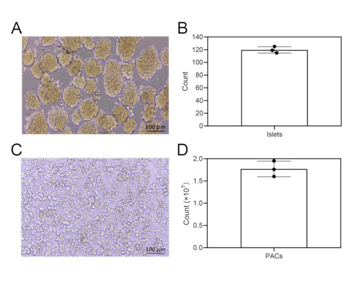
Under an optical microscope, islets were typically round or oval and golden-brown in color, with a stable yield of (120 ± 5) islets per mouse (Figure 2A,B). Freshly isolated PACs were spherical and mainly distributed in clusters (3-8 acinar cells per cluster). The apical ends of the acinar cells were darker in color, with clearly visible zymogen granules; no granules or vesicular structures were observed around the cells, and the solution background was clear. The yield of acinar cells ranged from 1.6 to 1.95 × 10⁷ cells per mouse [(1.77 × 10⁷) ± (1.75 × 10⁶)] (Figure 2C,D).

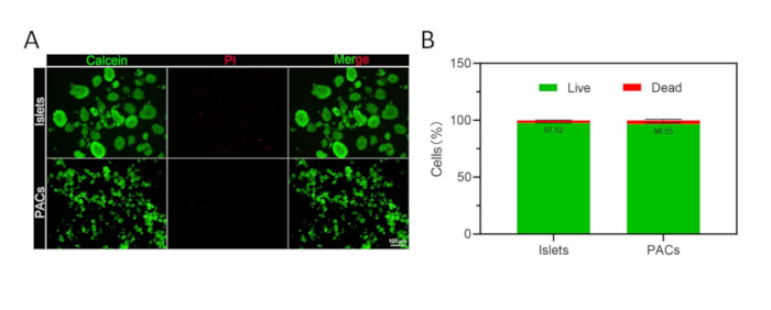
Calcein/PI staining results of islets and acinar cells are shown in Figure 3A: Calcein-stained cells (green) accounted for the majority, while PI-stained cells (red) were rare. Quantitative analysis of the live/dead staining images was performed using ImageJ. The results showed that the viability rates of the isolated islets and acinar cells were (97.52 ± 0.16)% and (96.55 ± 0.95)%, respectively (Figure 3B).

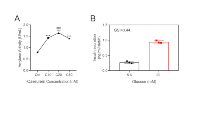
The basal amylase activity of the isolated pancreatic acinar cells was (0.79 ± 0.01) U/mL. After stimulation with caerulein at concentrations of 10 nM, 20 nM, and 50 nM, the amylase activities of the acinar cells were (1.45 ± 0.03) U/mL, (1.65 ± 0.05) U/mL, and (1.39 ± 0.02) U/mL, respectively. One-way ANOVA results indicated that compared with the control (Ctrl) group, all cerulein-stimulated groups showed significant differences in amylase activity (all P < 0.001). Additionally, a significant difference in amylase activity was observed between the 20 nM and the 10 nM cerulein groups (P < 0.001) (Figure 4A).

When stimulated with 5.6 mM glucose solution, the insulin secretion of the isolated islets was (0.27 ± 0.04) ng/mL/islet/h. When stimulated with 22 mM glucose solution, the insulin secretion of the isolated islets was (0.94 ± 0.04) ng/mL/islet/h, with a GSI of 3.44. The results indicate that the isolated islets exhibit a typical and effective insulin secretion response under stimulation with glucose solutions of different concentrations (Figure 4B).

Members of our research team observed that the quantity and quality of isolated islets and acinar cells are closely related to digestion time, which requires strict control during operation and is less affected by different operators. Meanwhile, fluctuations in yield and viability are also closely related to the complete dissection of the pancreas and the mechanical separation of pancreatic tissue, which requires attention during operation.

Islet isolation process, centrifuge tube with layers, cell separation, lab experiment setup.
Figure 1: Results of Ficoll solution density gradient centrifugation. The islet layer is located at the interface between the colorless transparent liquid layer and the culture medium. The precipitate at the bottom of the centrifuge tube is PACs. Abbreviation: PACs: pancreatic acinar cells. Please click here to view a larger version of this figure.

Cellular morphology microscopy of pancreatic islets and PACs with bar charts of cell counts.
Figure 2: Morphological and quantitative characteristics of islets and PACs. (A) Morphology of islets isolated from mouse pancreatic tissue. Scale bar = 100 µm. (B) Quantitative analysis of the number of islets isolated from mouse pancreatic tissue (n = 3). (C) Morphology of PACs isolated from mouse pancreatic tissue. Scale bar = 100 µm. (D) Quantitative analysis of the number of PACs isolated from mouse pancreatic tissue (n = 3). All data were expressed as mean ± SD. Abbreviation: PACs: pancreatic acinar cells. Please click here to view a larger version of this figure.

Live-dead cell analysis of islets and PACs; microscopy image with calcein and PI stain, bar graph.
Figure 3: Viability assessment of islets and PACs. (A) Calcein/propidium iodide fluorescence staining for evaluating the viability of mouse islets and PACs. Green: live cells. Red: dead cells. Scale bar = 100 µm. (B) Quantitative analysis of the viability of islets and PACs (n = 3). All data were expressed as mean ± SD. Abbreviations: PACs: pancreatic acinar cells; PI = propidium iodide. Please click here to view a larger version of this figure.

Amylase activity vs caerulein concentration; insulin secretion vs glucose; experimental graphs.
Figure 4: Assessment of amylase activity of PACs and insulin secretion capacity of islets. (A) Amylase activity of PACs under stimulation with different concentrations of cerulein (Ctrl: Control group; C10, C20, and C50: The concentrations of cerulein were 10 nM, 20 nM, and 50 nM (n = 3). (B) Quantitative analysis of glucose-stimulated insulin secretion concentrations (n = 3). All data were expressed as mean ± SD. ***P < 0.001. Abbreviations: GSI = Glucose-stimulated insulin index; PACs: pancreatic acinar cells. Please click here to view a larger version of this figure.

讨论

Loading...
$$\rightleftharpoonup{xx}$$ $$\longleftharp{xx}$$, $$\longrightharp{xx}$$,

In recent years, post-pancreatitis diabetes mellitus (PPDM) has received widespread attention1,9,10. Investigating the molecular mechanisms by which PACs affect islet hormone secretion in the context of AP is of great significance.

The pathogenesis of AP is mainly associated with the excessive activation of pancreatic enzymes in acinar cells, which leads to autodigestion of pancreatic tissue11,12. Although cell lines such as AR42J, 266-6 (acinar cell lines) and MIN6, INS-1 (β-cell lines) are currently available, these in-vitro cell models cannot fully replicate the physiological complexity of primary acinar cells and islets4,5. Therefore, isolating primary acinar cells and islets is crucial for in-depth studies on the interaction between the exocrine and endocrine compartments of the pancreas. Currently, methods for islet isolation are well-established; however, most fail to meet the research needs for studying the interaction between islets and pancreatic acinar cells6,7,8.

This experimental protocol optimizes the pancreatic perfusion procedure, lowering the technical barrier, thereby enabling researchers without specialized training in bile duct cannulation to conduct experiments. Meanwhile, it ensures the quantity and quality of isolated islets and acinar cells, providing a simple and rapid method for the simultaneous isolation of primary mouse islets and primary pancreatic acinar cells.

Although this method simplifies the islet isolation process, key steps still require strict control to ensure high yields and effective separation of islets and pancreatic acinar cells. These steps include adequate pancreatic perfusion, proper tissue disruption (ensuring thorough mincing), pancreatic digestion (precise control of digestion time and manual shaking during digestion), and mechanical pipetting (controlling the number and intensity of pipetting strokes). Before islet selection, resuspended islets should be stabilized in a cell incubator for approximately 10 min to facilitate more efficient islet picking.

It is important to note that during pancreatic perfusion, better results are achieved when clearer fluid vesicles are injected; the entire pancreatic tissue should be fully perfused, but the perfusion time should be controlled within 1-2 min. If low cell viability is detected, adjust the contact time between pancreatic tissue and collagenase P (including perfusion time and water bath digestion time). Additionally, pay attention to mechanical damage during isolation-for example, avoid excessive force when dispersing pancreatic tissue. Aseptic technique must be maintained throughout the isolation of islets and acinar cells to prevent contamination. Prepared reagents should be filtered through a 0.22 µm filter. The isolation process should be performed in a biosafety cabinet, and all instruments and consumables used during isolation must be sterile. The two prepared complete media also contain 1% penicillin-streptomycin solution.

For mouse anesthesia: fix the mouse's neck skin with the left thumb and index finger, and support its abdomen with the ring and little fingers (maintaining a head-down, abdomen-up posture to expose the abdominal cavity). Hold the syringe with the right hand, insert it at a 30° angle into the skin of the mouse's lower left abdomen (1 cm from the groin and 0.5 cm from the midline), inject the anesthetic slowly, and press the injection site with a sterile cotton swab for 10 s after needle withdrawal to prevent drug leakage. Only euthanize the mouse by cervical dislocation once it is fully anesthetized.

If pricked by a contaminated needle (exposed to mouse blood or tissue) or bitten by a mouse, immediately squeeze the area around the wound at the nearest sink (squeeze from the proximal to distal end of the wound to expel a small amount of blood), rinse the wound continuously with running water for 15 min, then disinfect it with 75% ethanol or 0.5% povidone-iodine.

Results of acinar cell amylase activity assays showed that the isolated pancreatic acinar cells had a low level of basal activation and were sensitive to cerulein stimulation: amylase activity increased gradually with rising cerulein concentration, reached the highest level at 20 nM cerulein, and decreased slightly when the cerulein concentration was 50 nM. The results of the glucose-stimulated insulin secretion assay showed that the isolated islets exhibited a typical and effective insulin secretion response under stimulation with glucose solutions of different concentrations. These results indicate that the acinar cells and islets extracted by this protocol are suitable for subsequent in-vitro experiments.

The islet and acinar cell isolation procedure of this experimental protocol is easy to operate. Experimental results show that the islets and acinar cells isolated via this protocol exhibit excellent quantity and viability. Additionally, multiple members of our team have validated this protocol using multiple mice; the validation results indicate that it has good reproducibility, reliable data, and minimal fluctuations in cell yield. However, this protocol also has certain limitations. Although it has low dependence on the operator's technical skills, it still requires the operator to have basic mouse anatomy knowledge to completely dissect the mouse pancreas-this is a prerequisite for the entire isolation process. If isolating pancreatic tissues from multiple mice simultaneously, the amount of collagenase P solution and other reagents must be adjusted accordingly. Additionally, simultaneous isolation from more than two mice is not recommended: the time difference between processing pancreatic tissues of different mice during dissection, perfusion, and mincing will affect the contact time between pancreatic tissue and collagenase P, thereby impairing the efficiency and viability of cell isolation. Thus, its large-scale application may be limited. Furthermore, this protocol has not been validated in rats. If isolating islets and acinar cells from rats, the dosage of some reagents and the digestion time may need further adjustment.

In conclusion, this experimental protocol provides a simple and rapid method for the simultaneous isolation of primary mouse islets and primary pancreatic acinar cells. It is more suitable for inexperienced researchers to perform islet and acinar cell isolation, while also offering a practical experimental framework for in-vitro studies on pancreatic exocrine-endocrine interactions.

披露

Loading...
$$\rightleftharpoonup{xx}$$ $$\longleftharp{xx}$$, $$\longrightharp{xx}$$,

The authors have no conflicts of interest to disclose.

致谢

Loading...
$$\rightleftharpoonup{xx}$$ $$\longleftharp{xx}$$, $$\longrightharp{xx}$$,

X.T., H.C., and J.L. contributed equally to this work. The work was supported by the National Natural Science Foundation of China (grant nos. 82370658, 82170657, 82370655, and 82400760) and the Natural Science Foundation of Zhejiang Province (grant no. LGF22H030014). The authors thank bioRender (www.biorender.com) for their help in creating the images.

材料

本文使用的材料清单
姓名公司目录编号评论
0.22 & mu;M滤波器MillexSLGPR33RB过滤制备好的胶原蛋白酶P溶液
0.25 米氯化钙(无菌)碧昂泰姆ST365激活胰岛素分泌的钙依赖信号通路
1毫升注射器杰瑞10084692516405用于胶原酶P灌注到胰腺组织。
1.25% 三溴乙醇溶液北京约西达生物技术有限公司JT0781麻醉
1.5毫升离心管埃彭多夫30121589收集细胞上清液进行离心和储存。
1x 汉克平衡盐溶液生物锐利BL561A准备胶原酶P溶液并冲洗胰腺组织
12孔细胞培养板萨斯泰特83.3921把胰腺组织和培养中提取的肾上腺素拿出来
15毫升离心管北京拉布吉科技有限公司CT-002-15A实验期间保持溶液
24孔细胞培养板萨斯泰特83.3922养殖开采的岛屿
40-mesh 筛子魏丹仪器滤胰腺组织
5毫升巴斯德移液管生物锐利BS-XG-03L输液和物理破坏胰腺组织
50毫升离心管北京拉布吉科技有限公司CT-002-50A实验期间储存溶液、组织封存和离心
60毫米细胞培养碟北京拉布吉科技有限公司12211用于存放含有小岛的培养基容器,便于采摘
75%乙醇溶液海诺特把老鼠浸泡消毒。
Adobe Photoshop 2023Adobe 公司生成图像。
贝克曼阿勒格拉 X-12/X-12R 离心机贝克曼阿莱格拉 X-12/X-12R离心机
牛血清白蛋白(BSA)-V太阳生物A8020保持胰岛的正常生理功能,并利用(它/这个)制备不同浓度的葡萄糖溶液。
C57BL/6J小鼠,SPF级,6–8周大,体重18岁;25克海军医科大学实验动物中心及;
钙化素/PI细胞活性与细胞毒性检测套件碧昂泰姆C2015L通过同时对活细胞和死亡细胞进行钙化素-AM(Calcein AM)和碘化丙苣(PI)双荧光染色,检测动物细胞的细胞活性和细胞毒性。
氯化钙(无水)桑贡生物技术10043-52-4准备胶原酶P溶液
细胞过滤器(100 & mu;m)生物锐利BS-100-XBS滤化腺皮细胞
胶原酶P西格玛11213857001消化胰腺组织
D-(+)-葡萄糖溶液(20%,无菌)碧昂泰姆ST491刺激胰岛上的胰岛分泌。
DMEM 基础(1x)(Dulbecco'S改良版鹰式中介)吉布科C11995500BT培养腺窝细胞
胎儿牛血清(FBS)生物西部S140B-500准备培养基
Ficoll-Paque PREMIUM 无菌溶液西提瓦17544203密度梯度分层的密度梯度介质
GraphPad 棱镜 8.0.1GraphPad 软件有限责任公司生成图像并进行统计分析。
HEPES溶液(1 M)生物锐利BL1061A准备胶原酶P溶液
高速/冷藏离心机西格玛3K15离心机
图片J美国国立卫生研究院光学与计算仪器实验室(LOCI)分析细胞荧光图像并计算细胞存活能力。
倒置生物显微镜徕卡观察细胞形态并选择小岛。
倒置荧光显微镜奥林匹斯IX73观察细胞荧光信号并捕捉荧光图像。
克雷布斯-林格碳酸氢盐HEPES缓冲液利阿金CZ0103保持胰岛的正常生理功能,并利用(它/这个)制备不同浓度的葡萄糖溶液。
鼠用INS(胰岛素)ELISA套件武汉精品生物技术有限公司EM0260检测胰岛的胰岛分泌水平。
眼科产钳(10厘米,带钩弯)青衣医疗器械协助解剖
眼用产钳(10厘米,直带牙齿)青衣医疗器械协助切除并突然分离胰腺组织,并提取胰腺组织
青霉素链霉素溶液吉布科15140-122准备培养基以防止污染
RPMI-1640 中型,KeyGEN 生物技术KGL1501-500终止胶原酶P消化液并培养胰岛
来自Glycine max(大豆)的胰蛋白酶抑制剂西格玛T6522准备胶原酶P溶液
急迫剪刀(10厘米,直尖)青衣医疗器械解剖辅助,将胰腺组织切成小块
水浴OAICLABOW-HF保持消化温度。
&α;-淀粉酶和β;-淀粉酶活性检测试剂盒ElabscienceE-BC-K006-M检测腺苷细胞在不同条件下分泌的淀粉酶水平

参考文献

Loading...
$$\rightleftharpoonup{xx}$$ $$\longleftharp{xx}$$, $$\longrightharp{xx}$$,
  1. Manrai, M., et al. Diabetes mellitus as a consequence of acute severe pancreatitis: unraveling the mystery. World J Diabetes. 14 (8), 1212-1225 (2023).
  2. García-Compeán, D., et al. Post-acute pancreatitis diabetes: a complication waiting for more recognition and understanding. World J Gastroenterol. 29 (28), 4405-4415 (2023).
  3. Shen, H. N., Yang, C. C., Chang, Y. H., Lu, C. L., Li, C. Y. Risk of diabetes mellitus after first-attack acute pancreatitis: a national population-based study. Am J Gastroenterol. 110 (12), 1698-1706 (2015).
  4. Hohmeier, H. E., et al. Isolation of INS-1-derived cell lines with robust ATP-sensitive K⁺ channel-dependent and -independent glucose-stimulated insulin secretion. Diabetes. 49 (3), 424-430 (2000).
  5. Hohmeier, H. E., Newgard, C. B. Cell lines derived from pancreatic islets. Mol Cell Endocrinol. 228 (1-2), 121-128 (2004).
  6. Neuman, J. C., Truchan, N. A., Joseph, J. W., Kimple, M. E. A method for mouse pancreatic islet isolation and intracellular cAMP determination. J Vis Exp. (88), e50374(2014).
  7. Villarreal, D., et al. A simple high-efficiency protocol for pancreatic islet isolation from mice. J Vis Exp. (150), e57048(2019).
  8. Huang, C., Gu, G. Effective isolation of functional islets from neonatal mouse pancreas. J Vis Exp. (119), e55160(2017).
  9. Petrov, M. S., Yadav, D. Global epidemiology and holistic prevention of pancreatitis. Nat Rev Gastroenterol Hepatol. 16 (3), 175-184 (2019).
  10. Xiao, A. Y., et al. Global incidence and mortality of pancreatic diseases: a systematic review, meta-analysis, and meta-regression of population-based cohort studies. Lancet Gastroenterol Hepatol. 1 (1), 45-55 (2016).
  11. Sendler, M., Weiss, F. U., Golchert, J., et al. Cathepsin B-mediated activation of trypsinogen in endocytosing macrophages increases severity of pancreatitis in mice. Gastroenterology. 154 (3), 704-718.e10 (2018).
  12. Modenbach, J. M., Möller, C., Asgarbeik, S., et al. Biochemical analyses of cystatin-C dimers and cathepsin-B reveals a trypsin-driven feedback mechanism in acute pancreatitis. Nat Commun. 16 (1), 1702(2025).

重印与许可

申请许可以重复使用本 JoVE 文章的文本或图表

申请许可

标签

Ficoll P PI

相关文章