Las interacciones neurona-glial en la neurodegeneración no se comprenden bien debido a herramientas y métodos inadecuados. Aquí, describimos protocolos optimizados para obtener neuronas inducidas, células precursoras de oligodendrocitos y oligodendrocitos a partir de células madre pluripotentes humanas y proporcionamos ejemplos de los valores de estos métodos para comprender las contribuciones específicas del tipo de célula en la enfermedad de Alzheimer.
Se requiere una suscripción a JoVE para ver este contenido. Inicie sesión o comience su prueba gratuita.
Artículo de método
Generación de neuronas humanas y oligodendrocitos a partir de células madre pluripotentes para modelar las interacciones neuronal-oligodendrocitos
* These authors contributed equally
En este artículo
Aviso de erratum
Important: There has been an erratum issued for this article. View Erratum Notice
Resumen
Resumen
En la enfermedad de Alzheimer (EA) y otros trastornos neurodegenerativos, la insuficiencia oligodendroglial es una característica patológica temprana común, pero la forma en que contribuye al desarrollo y progresión de la enfermedad, particularmente en la materia gris del cerebro, sigue siendo en gran parte desconocida. La disfunción de las células del linaje de oligodendrocitos se caracteriza por deficiencias en la mielinización y alteración de la auto-renovación de las células precursoras de oligodendrocitos (OPC). Estos dos defectos son causados, al menos en parte, por la interrupción de las interacciones entre la neurona y los oligodendrocitos a lo largo de la acumulación de patología. Las OPC dan lugar a oligodendrocitos mielinizantes durante el desarrollo del SNC. En la corteza cerebral madura, las OPC son las principales células proliferativas (que comprenden ~ 5% del total de células cerebrales) y controlan la formación de nueva mielina de una manera dependiente de la actividad neuronal. Tales comunicaciones neuronómicas a oligodendrocitos están significativamente poco estudiadas, especialmente en el contexto de afecciones neurodegenerativas como la EA, debido a la falta de herramientas adecuadas. En los últimos años, nuestro grupo y otros han hecho progresos significativos para mejorar los protocolos actualmente disponibles para generar neuronas funcionales y oligodendrocitos individualmente a partir de células madre pluripotentes humanas. En este manuscrito, describimos nuestros procedimientos optimizados, incluido el establecimiento de un sistema de cocultivo para modelar las conexiones neurona-oligodendrocitos. Nuestros resultados ilustrativos sugieren una contribución inesperada de OPC / oligodendrocitos a la amiloidosis cerebral y la integridad de la sinapsis y destacan la utilidad de esta metodología para la investigación de la EA. Este enfoque reduccionista es una herramienta poderosa para diseccionar las interacciones heterocelulares específicas de la complejidad inherente dentro del cerebro. Se espera que los protocolos que describimos aquí faciliten futuros estudios sobre defectos oligodendrogliales en la patogénesis de la neurodegeneración.
Introducción
Las células del linaje de oligodendrocitos, incluidas las células precursoras de oligodendrocitos (OPC), los oligodendrocitos mielinizantes y los tipos de transición intermedios, constituyen un grupo importante de células cerebrales humanas1 que participan activamente en muchas funciones críticas para el correcto funcionamiento y mantenimiento de nuestro sistema nervioso central a lo largo del desarrollo neuronal y el envejecimiento 2,3,4 . Mientras que los oligodendrocitos son bien conocidos por producir mielina para facilitar la transmisión de la actividad neuronal y apoyar la salud axonal en la sustancia blanca, los OPC son abundantes (~ 5%) en la materia gris donde la mielinización es escasa y realizan funciones de señalización dependientes de la actividad para gobernar el comportamiento de aprendizaje y la formación de la memoria 5,6,7,8 . La forma en que las células oligodendrogliales funcionan y la disfunción en la patogénesis de la enfermedad de Alzheimer (EA) y otras afecciones neurodegenerativas asociadas a la edad han sido poco estudiadas9. Las insuficiencias de un sistema modelo apropiado y las deficiencias en el conocimiento general para guiar un camino experimental hacia adelante son las principales razones de esta brecha.
A la luz de los últimos avances en la obtención de células cerebrales humanas a partir de células madre pluripotentes, incluidas las células madre embrionarias (ES) y las células madre pluripotentes inducidas (iPS), tales modelos celulares junto con las herramientas modernas de edición de genes han surgido como herramientas robustas para manejar el intrincado nexo de las interacciones celulares en el cerebro, y son capaces de demostrar manifestaciones de enfermedades específicas del ser humano10, 11. Teniendo en cuenta que los tipos individuales de células cerebrales pueden exhibir efectos distintos e incluso conflictivos frente a las mismas condiciones promotoras de la EA12,13, esta metodología de células madre ofrece de manera única información específica del tipo de célula que anteriormente se había pasado por alto utilizando modelos establecidos in vivo o in vitro que solo proporcionan lecturas agregadas de colecciones de tipos de células cerebrales. En la última década, se han desarrollado un buen número de protocolos confiables para generar neuronas humanas a partir de la transdiferenciación de células ES/iPS o la conversión directa de otros tipos de células diferenciadas terminalmente (por ejemplo, fibroblastos)14,15. En particular, la aplicación de factores de transcripción neurogénicos clave (por ejemplo, neurogenina 2, Ngn2)16 a células madre pluripotentes humanas puede generar una población homogénea de tipos de células neuronales bien caracterizadas para cultivos puros sin necesidad de cocultivo con células gliales12,17,18. Para los oligodendrocitos humanos inducidos, hay algunos protocolos publicados que pueden generar células funcionales muy parecidas a sus contrapartes primarias, con una amplia gama de eficiencia y demanda en tiempo y recursos 19,20,21,22,23,24,25,26,27,28 . Hasta la fecha, ninguno de estos protocolos se ha aplicado para investigar cómo las células oligodendrogliales responden y afectan la patogénesis de la EA.
Aquí, describimos nuestros protocolos mejorados para cultivos únicos y mixtos de neuronas inducidas humanas (iNs) y OPC / oligodendrocitos (iOPC / iOL). El protocolo iN descrito aquí se basa en el enfoqueNgn2 16 ampliamente utilizado, y tiene la característica adicional de estar libre de glía. Los iNs resultantes son homogéneos y se asemejan mucho a las neuronas excitadoras de la capa cortical 2/3, con morfología piramidal característica, patrón de expresión génica y características electrofisiológicas17,18 (Figura 1). Para superar algunas de las barreras fundamentales en la diferenciación dirigida de células madre pluripotentes, hemos desarrollado un método simple y eficaz de pretratamiento con dosis bajas de dimetilsulfóxido (DMSO)29,30, y hemos reportado una mayor propensión de las células ES/iPS humanas a transdiferenciarse en iOPCs e iOLs31, basado en un protocolo ampliamente adaptado por Douvaras y Fossati 32 . Hemos simplificado aún más el protocolo e incorporado un compuesto robusto que promueve la diferenciación, clemastine 7,33,34, para acelerar el proceso de maduración oligodendroglial. Como resultado (Figura 2), las iOPC se pueden generar en 2 semanas (~95% positivo para el marcador O4) y las iOLs en cuatro semanas (expresando marcadores maduros MBP y PLP1). Curiosamente, encontramos que las iOPC solas secretan una cantidad notable de β amiloide (Aβ), consistente con los datos transcriptómicos independientes que muestran la abundante expresión de la proteína precursora amiloide (APP) y la proteasa de procesamiento β-secretasa (BACE1) en células de linaje oligodendrocitos35,36. Además, nuestro sistema de cocultivo iN-iOPC promueve el envoltorio de axones mediante procesos iOL positivos para MBP y proporciona un apoyo significativo para la formación de sinapsis (Figura 3). Por lo tanto, los protocolos que hemos detallado a continuación tienen ventajas técnicas y biológicas sobre los métodos de cocultivo neurona-oligodendroglia previamente catalogados, y prometen modelar mejor la neurodegeneración en la EA.
Acceso restringido. Inicie sesión o comience una prueba gratuita para ver este contenido.
Protocolo
1. Inducción de neuronas humanas a partir de células madre pluripotentes humanas
- Preparación de lentivirus (~ 5 días, protocolo detallado como se describió anteriormente16)
- Placa ~1 millón de células HEK293T cada matraz T75, para tenerlas ~40% confluentes al realizar la transfección. Transfectarlos con plásmidos que expresan Ngn2 inducible por tetraciclina y gen resistente a la puromicina (PuroR; bajo el mismo control del promotor TetO), rtTA y los tres plásmidos auxiliares pRSV-REV, pMDLg/pRRE y VSV-G (12 μg de ADN vectorial lentiviral y 6 μg de cada uno de los ADN plásmidos auxiliares). Preparar al menos tres frascos por preparación de lentivirus. Use PEI para la transfección siguiendo las instrucciones del fabricante. Cambie el soporte después de 16 h y deséchelo.
- La cosecha liberó partículas virales recolectando medios de cultivo todos los días y reemplazándolos con medios nuevos durante 3 días. Agrupe los medios recolectados que contienen partículas virales para su purificación. Filtrar el virus a través de un filtro de 0,22 μm y centrifugar a 49.000 x g durante 90 min. Resuspender el pellet en el volumen apropiado de PBS-glucosa (~150 μL).
- Inducción neuronal (~5 días)
NOTA: Este protocolo de inducción (Figura 1A; diagrama de flujo) es altamente efectivo para células iPS y ES de pluripotencia validada (que puede ensayarse mediante tinción inmunohistoquímica de marcadores de pluripotencia bien caracterizados; Figura 1B).- Utilice células madre embrionarias humanas H1 disponibles comercialmente al paso de 52 (ver Tabla de materiales). Cultivar las células en placas de 6 pocillos recubiertas con solución de matriz extracelular (~0,5 mg de solución de matriz por placa de 6 pocillos; ver Tabla de materiales) utilizando medio de mantenimiento de células madre embrionarias (ver Tabla de materiales) e incubar las placas a 37 °C con 5% de CO2.
- En el día -2, separe las células madre embrionarias (80% confluentes) con 1 ml de solución de desprendimiento celular (consulte la Tabla de materiales) e incube a temperatura ambiente durante 10 minutos. Transfiera las células a un tubo; lavar el pozo con 2 mL de medio y combinar en el mismo tubo. Centrifugar a 300 x g durante 5 min, resuspender el pellet en medios y colocar las células en placas de 6 pocillos recubiertas con matriz a la densidad de siembra de 1 x 10⁵ células por pocillo.
- El día -1, agregue lentivirus que expresen Ngn2 más PuroR y rtTA junto con polebreno (8 μg/ml) a las células madre embrionarias en un medio de mantenimiento de células madre embrionarias frescas (ver Tabla de materiales). La cantidad exacta de virus debe determinarse por títulos reales o la titulación. Por lo general, agregamos 5 μL cada virus por pocillo en una placa de 6 pocillos.
- El día 0, añadir doxiciclina (2 μg/ml, para activar la expresión de Ngn2) en medio DMEM-F12 con suplemento N2 sin morfógenos.
- El día 1, agregue puromicina en medio fresco de DMEM-F12 más N2 y doxiciclina, a la concentración final de 1 μg / ml de medio. Seleccione las células transducidas en Puromicina durante al menos 24 h. Puede ser necesaria una mayor concentración de puromicina (hasta 5 μg/ml) y un período de selección más largo (hasta 48 h) para eliminar adecuadamente las células subtransducidas si el título del virus es bajo.
- El día 2, separar las neuronas diferenciadoras con una solución de desprendimiento celular (ver Tabla de materiales) y volver a colocarlas en placas de 24 pocillos (entre 80,000-200,000 células / pocillo) recubiertas con solución de matriz (ver Tabla de materiales), y mantenerlas en medio NBA / B27 sin doxiciclina. La densidad de siembra es crítica.
- En esta etapa, las neuronas separadas pueden congelarse en un medio de congelación comercial especializado (consulte la Tabla de materiales) y almacenarse en nitrógeno líquido hasta por 3 meses. Las neuronas puras pueden ser plateadas que representan la típica muerte celular de ~15%-20% después del deshielo, cultivadas solas o cocultivadas con otros tipos de células cerebrales (ver paso 3.2.3. para el cocultivo con OPC).
- Cultive iNs puros en las placas recubiertas con soluciones basadas en matriz extracelular según las instrucciones del fabricante (consulte la Tabla de materiales). La morfología piramidal característica debe ser evidente en el día 4 (y el día 6; Figura 1C). La formación de sinapsis se puede detectar ya en el día 14 al 16 y es prominente en el día 24 mediante tinción inmunohistoquímica con marcadores pre y postsinápticos estándar. (Figura 1D; etiquetado con el marcador presináptico Synapsin 1 y el marcador dendrítico Map2).
2. Inducción de células precursoras de oligodendrocitos humanos (OPC) a partir de células madre pluripotentes y maduración de oligodendrocitos
- Generación de células progenitoras neurales (NPC): protocolo monocapa (~7 días). Consulte la Figura 2A para el diagrama de flujo.
- Cultivar células madre embrionarias humanas H1 como se describió anteriormente (ver paso 1.2.1.) y transdiferenciarlas en células progenitoras neurales (NPC) mediante un enfoque establecido llamado SMADi dual, con inhibidores de moléculas pequeñas para múltiples vías de señalización. Aquí utilizamos un kit comercial ampliamente aceptado y seguimos el protocolo monocapa proporcionado por el fabricante (ver Tabla de materiales).
- En el día -1, placa 0.5–1 x 10 6 células por pocillo en una placa de 6 pocillos recubierta por una solución de matriz reducida con factor de crecimiento (ver Tabla de materiales; ~0.5 mg de solución de matriz por placa de6 pocillos) con medio de mantenimiento de células madre embrionarias (ver Tabla de materiales). Esta solución de matriz reducida de factor de crecimiento se utiliza para recubrir todas las placas que se utilizarán en los siguientes pasos.
- El día 0, tratar las células durante 24 h con medio de mantenimiento de células madre embrionarias (ver Tabla de materiales) complementado con DMSO al 2%.
- En el día 1-6, cambie el medio completo con un medio de inducción neural tibio (37 °C) que contenga los inhibidores de SMAD del kit comercial (consulte la Tabla de materiales). Si las células se dividen y alcanzan la confluencia antes del día 7, pasarlas a la densidad de siembra de 0,5–1 x 10⁶, como se describió anteriormente en el paso 2.1.2.
- El día 7, los NPC de paso usan una solución de desprendimiento celular (ver Tabla de materiales) y una placa a una densidad de siembra de 1–2 x 10⁵ células/pocillo de una placa de 24 pocillos.
- Evaluar la eficiencia de diferenciación mediante tinción inmunohistoquímica (IHC) para detectar la ausencia de marcador de pluripotencia, OCT4, por ejemplo, y la presencia de marcadores NPC como PAX6, Nestin y Sox1.
- En esta etapa, los NPC separados pueden congelarse en los medios de congelación NPC comerciales especializados (consulte la Tabla de materiales) y almacenarse en nitrógeno líquido hasta por 3 meses. Después de congelar y descongelar por una vez, los NPC aún conservan la multipotencia para dar lugar a neuronas, astrocitos y OPC con protocolos confiables.
- Generación de células precursoras de oligodendrocitos (OPC) (~7 días). Consulte la Figura 2A para ver el diagrama de flujo.
- El día 7, pase NPC usando solución de desprendimiento celular (ver Tabla de materiales) y plaquelos a una densidad de siembra de 1–2 x 10⁵ células por pocillo en una placa de 24 pocillos en medio de inducción neural caliente (37 °C) más inhibidores de SMAD del kit comercial (ver Tabla de materiales).
- El día 8, preparar una solución de DMSO al 1% en el medio de diferenciación OPC y tratar los NPC plateados durante 24 h. El medio de diferenciación OPC está compuesto por: medio DMEM/F12, suplemento de N2 al 1%, suplemento de B27 al 1%, bFGF a 20 ng/ml, SAG a 1 μM, PDGF-AA a 10 ng/ml (ver Tabla de materiales).
- El día 9, reemplace los medios con un nuevo medio de diferenciación OPC sin DMSO. Alimente las células cada dos días hasta el día 15. Si las células alcanzan la confluencia antes del día 15, pasarlas a la densidad de siembra de 1–2 x 10⁵ células por pocillo, como se describe en el paso 2.2.1.
- El día 14, OPC en placa en medio de diferenciación OPC a una densidad de 1–2 x 10⁵ células/pocillo en una placa de 24 pocillos.
- En esta etapa (Día 15), las células de prueba para detectar la presencia de marcadores específicos de OPC mediante tinción IHC o qPCR (p. ej., O4, Olig1/2, CSPG4/Ng2, NKX2.2, PDGFRa; Figura 2B) y por la ausencia de marcadores NPC (Pax6 o Nestin; Figura 2D). Por lo general, detectamos la inmunorreactividad O4 en más del 95% de las células en el día 15. De particular relevancia para la enfermedad de Alzheimer, la expresión de APP (proteína precursora amiloide), BACE1 (la proteasa de procesamiento β-secreatasa 1) y péptido amiloide-β (Aβ) es abundante en OPC (Figura 2F).
- Maduración de oligodendrocitos (OL) (~7–20 días)
- El día 15, reemplace los medios con medio de maduración OL: medio neurobasal-A, suplemento de B27 al 2%, cAMP de 1 μM, triyodotironina T3 de 200 ng / ml y clemastina de 1 μM (consulte la Tabla de materiales). Cambie el medio cada dos días o todos los días, si es necesario.
- Cuando las células alcanzan el 90% de confluencia, dividir en una proporción de 1:3 hasta 2 pasajes o hasta que la división celular disminuya sustancialmente. Si los OPC se dividen demasiado rápido y alcanzan la confluencia en menos de 3 días, agregue Ara-C (consulte la Tabla de materiales) a una concentración de 2-5 μM durante 1-3 días. La proliferación activa indica una menor eficiencia de maduración.
- Examinar la eficiencia de la maduración oligodendroglial evaluando la expresión de marcadores OL, por ejemplo, CLDN11, PLP1, MBP por qPCR, tinción IHC o immunoblotting. La morfología característica de estructuras altamente complejas (Figura 2C) y la expresión de marcadores OL (Figura 2E) deben detectarse fácilmente antes del día 28.
3. Cocultivo de neuronas humanas inducidas (iNs) y células precursoras de oligodendrocitos (iOPC)
- Recubrimiento iOPC (~3 días)
- IOPC en placa en el día 14 a una densidad de 1 x 10⁵ células por pocillo en una placa de 24 pocillos (como se describe anteriormente en el paso 2.2.4.) en medio de diferenciación OPC (como se describe en el paso 2.2.2.).
- Creación de la cocultura iN-iOPC
- El día 15, separar las neuronas humanas inducidas en el paso del día 2 después de la selección de puromicina (como se describe en el paso 1.2.6.) con solución de desprendimiento celular (ver Tabla de materiales).
- Añadir neuronas a las OPC cultivadas, colocando en placa la densidad de siembra de 2 x 10⁵ células por pocillo en la placa de 24 pocillos con OPC en crecimiento (a partir del paso 3.1.1). Use el medio de cocultivo que contiene medio neurobasal-A, suplemento de B27 al 2% y triyodotironina T3 de 100 ng / ml. Cambie el medio al día siguiente y luego cada dos días después. Si los OPC proliferan demasiado rápido y alcanzan la confluencia en menos de 3 días, agregue Ara-C a una concentración de 2-5 μM. Una imagen representativa de las iNs e iOPC cultivadas en cocultivo después de 7 días de neuronas se muestra en la Figura 3A.
- Utilice neuronas congeladas preparadas como se describe anteriormente en el paso 1.2.7 para el cocultivo con OPC. Las neuronas de congelación y descongelación de placas a una densidad mayor de 3 x 10⁵ células por pocillo.
- Después del día 14-16 en cocultivos, la formación de sinapsis en iNs se puede observar mediante tinción IHQ de marcadores pre y postsinápticos, y para el día 21 los puntos sinápticos deben ser abundantes (Figura 3C) y las actividades neuronales se pueden registrar de manera confiable.
- A partir del día 21, pruebe las células para marcadores específicos de OL (por ejemplo, MBP y PLP1). Para el día 28, normalmente observamos el fenómeno de envainamiento de los axones iN mediante procesos iOL, marcados por tinción IHC para marcadores específicos (Figura 3B; neurofilamento NF para axones iN y MBP para procesos iOPC).
Acceso restringido. Inicie sesión o comience una prueba gratuita para ver este contenido.
Resultados
Generación directa de neuronas humanas inducidas a partir de células madre pluripotentes humanas
Es muy importante que las células madre pluripotentes humanas iniciales exhiban un alto grado de pluripotencia para la generación exitosa de iNs o iOPCs/iOLs. Por lo tanto, las células deben teñirse para marcadores específicos, como Oct4 y SOX2, antes de iniciar cualquiera de los protocolos de inducción descritos en el presente manuscrito (Figura 1A). Se utilizaron células H1 ...
Acceso restringido. Inicie sesión o comience una prueba gratuita para ver este contenido.
Discusión
Además del apoyo físico y metabólico para estabilizar las estructuras sinapsis y facilitar la conducción de señales saltatorias por mielinización, las células del linaje de oligodendrocitos pueden dar forma al patrón de actividad neuronal a través de conversaciones cruzadas rápidas y dinámicas con las neuronas 5,6,7. Mientras que en la patología de la EA las respuestas oligodendrogliales se consideraron inicialmente como meramen...
Acceso restringido. Inicie sesión o comience una prueba gratuita para ver este contenido.
Divulgaciones
Los autores no tienen nada que revelar.
Agradecimientos
Este trabajo fue apoyado por las subvenciones de los Institutos Nacionales de Salud (R00 AG054616 a Y.A.H. y T32 GM136566 a K.C.), la Escuela de Medicina de la Universidad de Stanford y una beca Siebel (otorgada a SC). Y.A.H. es profesor traslacional de GFL del Centro de Neurociencia Traslacional del Instituto Brown de Ciencias Traslacionales.
Acceso restringido. Inicie sesión o comience una prueba gratuita para ver este contenido.
Materiales
| Nombre | Empresa | Número de catálogo | Comentarios |
|---|---|---|---|
| Accutase | STEMCELL Technologies | 7920 | |
| B27 suplemento | ThermoFisher | 17504044 | |
| bFGF | ThermoFisher | PHG 0266 | |
| cAMP | MilliporeSigma | A9501 | |
| Clemastine | MilliporeSigma | SML0445 | |
| DMEM/F12 medio | STEMCELL Technologies | 36254 | |
| DMSO | ThermoFisher | D12345 | |
| Doxiciclina | MilliporeSigma | D3072 | |
| Suero fetal bovino | ScienCell | 10 | |
| H1 células madre embrionarias humanas | WiCell | WA01 | |
| Matrigel | Corning | 354234 | |
| mTeSR plus | STEMCELL Technologies | 5825 | |
| Suplemento de N2 | ThermoFisher 17502001 | ||
| Neurobasal A medio | ThermoFisher | 10888-022 | |
| Aminoácidos no esenciales | ThermoFisher | 11140-050 | |
| PDGF-AA | R& D Systems | 221-AA-010 | |
| PEI | VWR | 71002-812 | |
| pMDLg/pRRE | Addgene | 12251 | |
| Polybrene | MilliporeSigma | TR-1003-G | |
| pRSV-REV | Addgene | 12253 | |
| Puromicina | ThermoFisher | A1113803 | |
| Inhibidor de ROCK Y-27632 | STEMCELL Technologies | 72302 | |
| SAG | Tocris | 4366 | |
| STEMdiff Progenitor Neural Progenitor Medios de congelación | STEMCELL Technologies | 5838 | |
| STEMdiff SMADi Kit de inducción neuronal | STEMCELL Technologies | 8581 | |
| T3 triyodotironina | MilliporeSigma | T6397 | |
| Tempo-iOlogo: OPC humanas derivadas de iPSC | Tempo BioScience | SKU102 | |
| TetO-NG2-Puro | Addgene | 52047 | |
| VSV-G | Addgene | 12259 |
Referencias
- Pelvig, D. P., Pakkenberg, H., Stark, A. K., Pakkenberg, B. Neocortical glial cell numbers in human brains. Neurobiology of Aging. 29 (11), 1754-1762 (2008).
- Barres, B. A. The mystery and magic of glia: a perspective on their roles in health and disease. Neuron. 60 (3), 430-440 (2008).
- De Strooper, B., Karran, E. The cellular phase of Alzheimer's disease. Cell. 164 (4), 603-615 (2016).
- Monje, M. Myelin plasticity and nervous system function. Annual Review of Neuroscience. 41, 61-76 (2018).
- Hughes, E. G., Orthmann-Murphy, J. L., Langseth, A. J., Bergles, D. E. Myelin remodeling through experience-dependent oligodendrogenesis in the adult somatosensory cortex. Nature Neuroscience. 21 (5), 696-706 (2018).
- Gibson, E. M., et al. Neuronal activity promotes oligodendrogenesis and adaptive myelination in the mammalian brain. Science. 344 (6183), 1252304(2014).
- Pan, S., Mayoral, S. R., Choi, H. S., Chan, J. R., Kheirbek, M. A. Preservation of a remote fear memory requires new myelin formation. Nature Neuroscience. 23 (4), 487-499 (2020).
- Thornton, M. A., Hughes, E. G. Neuron-oligodendroglia interactions: Activity-dependent regulation of cellular signaling. Neuroscience Letters. 727, 134916(2020).
- Ettle, B., Schlachetzki, J. C. M., Winkler, J. Oligodendroglia and myelin in neurodegenerative diseases: more than just bystanders. Molecular Neurobiology. 53 (5), 3046-3062 (2016).
- Essayan-Perez, S., Zhou, B., Nabet, A. M., Wernig, M., Huang, Y. A. Modeling Alzheimer's disease with human iPS cells: advancements, lessons, and applications. Neurobiology of Disease. 130, 104503(2019).
- Li, L., et al. GFAP mutations in astrocytes impair oligodendrocyte progenitor proliferation and myelination in an hiPSC model of Alexander disease. Cell Stem Cell. 23 (2), 239-251 (2018).
- Lin, Y. T., et al. APOE4 causes widespread molecular and cellular alterations associated with Alzheimer's disease phenotypes in human iPSC-derived brain cell types. Neuron. 98 (6), 1294(2018).
- TCW, J., et al. Cholesterol and matrisome pathways dysregulated in human APOE ε4 glia. bioRxiv. , (2019).
- Ang, C. E., Wernig, M. Induced neuronal reprogramming. Journal of Comparitive Neurology. 522 (12), 2877-2886 (2014).
- Penney, J., Ralvenius, W. T., Tsai, L. H. Modeling Alzheimer's disease with iPSC-derived brain cells. Molecular Psychiatry. 25 (1), 148-167 (2020).
- Zhang, Y., et al. Rapid single-step induction of functional neurons from human pluripotent stem cells. Neuron. 78 (5), 785-798 (2013).
- Huang, Y. A., Zhou, B., Nabet, A. M., Wernig, M., Sudhof, T. C. Differential signaling mediated by ApoE2, ApoE3, and ApoE4 in human neurons parallels Alzheimer's Disease risk. Journal of Neuroscience. 39 (37), 7408-7427 (2019).
- Huang, Y. A., Zhou, B., Wernig, M., Sudhof, T. C. ApoE2, ApoE3, and ApoE4 Differentially Stimulate APP Transcription and Abeta Secretion. Cell. 168 (3), 427-441 (2017).
- Yang, N., et al. Generation of oligodendroglial cells by direct lineage conversion. Nature Biotechnology. 31 (5), 434-439 (2013).
- Douvaras, P., et al. Efficient generation of myelinating oligodendrocytes from primary progressive multiple sclerosis patients by induced pluripotent stem cells. Stem Cell Reports. 3 (2), 250-259 (2014).
- Lee, E. H., Park, C. H. Comparison of reprogramming methods for generation of induced-oligodendrocyte precursor cells. Biomolecules & Therapeutics (Seoul). 25 (4), 362-366 (2017).
- Ehrlich, M., et al. Rapid and efficient generation of oligodendrocytes from human induced pluripotent stem cells using transcription factors. Proceedings of the National Academy of Sciences of the United States of America. 114 (11), 2243-2252 (2017).
- Rodrigues, G. M. C., et al. Defined and scalable differentiation of human oligodendrocyte precursors from pluripotent stem cells in a 3D culture system. Stem Cell Reports. 8 (6), 1770-1783 (2017).
- Hu, B. Y., Du, Z. W., Li, X. J., Ayala, M., Zhang, S. C. Human oligodendrocytes from embryonic stem cells: conserved SHH signaling networks and divergent FGF effects. Development. 136 (9), 1443-1452 (2009).
- Izrael, M., et al. Human oligodendrocytes derived from embryonic stem cells: Effect of noggin on phenotypic differentiation in vitro and on myelination in vivo. Molecular and Cellular Neuroscience. 34 (3), 310-323 (2007).
- Yamashita, T., et al. Differentiation of oligodendrocyte progenitor cells from dissociated monolayer and feeder-free cultured pluripotent stem cells. PLoS One. 12 (2), 0171947(2017).
- Wang, S., et al. Human iPSC-derived oligodendrocyte progenitor cells can myelinate and rescue a mouse model of congenital hypomyelination. Cell Stem Cell. 12 (2), 252-264 (2013).
- Chanoumidou, K., Mozafari, S., Baron-Van Evercooren, A., Kuhlmann, T. Stem cell derived oligodendrocytes to study myelin diseases. Glia. 68 (4), 705-720 (2020).
- Chetty, S., et al. A simple tool to improve pluripotent stem cell differentiation. Nature Methods. 10 (6), 553-556 (2013).
- Li, J., et al. A transient DMSO treatment increases the differentiation potential of human pluripotent stem cells through the Rb family. PLoS One. 13 (12), 0208110(2018).
- Sambo, D., Li, J., Brickler, T., Chetty, S. Transient treatment of human pluripotent stem cells with DMSO to promote differentiation. Journal of Visualized Experiments: JoVE. (149), (2019).
- Douvaras, P., Fossati, V. Generation and isolation of oligodendrocyte progenitor cells from human pluripotent stem cells. Nature Protocols. 10 (8), 1143-1154 (2015).
- Mei, F., et al. Micropillar arrays as a high-throughput screening platform for therapeutics in multiple sclerosis. Nature Medicine. 20 (8), 954-960 (2014).
- Madhavan, M., et al. Induction of myelinating oligodendrocytes in human cortical spheroids. Nature Methods. 15 (9), 700-706 (2018).
- Zhang, Y., et al. Purification and characterization of progenitor and mature human astrocytes reveals transcriptional and functional differences with mouse. Neuron. 89 (1), 37-53 (2016).
- Grubman, A., et al. A single-cell atlas of entorhinal cortex from individuals with Alzheimer's disease reveals cell-type-specific gene expression regulation. Nature Neuroscience. 22 (12), 2087-2097 (2019).
- Goldman, S. A., Kuypers, N. J. How to make an oligodendrocyte. Development. 142 (23), 3983-3995 (2015).
- Behrendt, G., et al. Dynamic changes in myelin aberrations and oligodendrocyte generation in chronic amyloidosis in mice and men. Glia. 61 (2), 273-286 (2013).
- Patzke, C., et al. Neuromodulator signaling bidirectionally controls vesicle numbers in human synapses. Cell. 179 (2), 498-513 (2019).
- Piao, J., et al. Human embryonic stem cell-derived oligodendrocyte progenitors remyelinate the brain and rescue behavioral deficits following radiation. Cell Stem Cell. 16 (2), 198-210 (2015).
- Keirstead, H. S., et al. Human embryonic stem cell-derived oligodendrocyte progenitor cell transplants remyelinate and restore locomotion after spinal cord injury. Journal of Neuroscience. 25 (19), 4694-4705 (2005).
- Kim, D. S., et al. Rapid generation of OPC-like cells from human pluripotent stem cells for treating spinal cord injury. Experimental & Molecular Medicine. 49 (7), 361(2017).
Acceso restringido. Inicie sesión o comience una prueba gratuita para ver este contenido.
Reimpresiones y permisos
Erratum
Formal Correction: Erratum: Generation of Human Neurons and Oligodendrocytes from Pluripotent Stem Cells for Modeling Neuron-Oligodendrocyte Interactions
Posted by JoVE Editors on 12/29/2020. Citeable Link.
An erratum was issued for: Generation of Human Neurons and Oligodendrocytes from Pluripotent Stem Cells for Modeling Neuron-Oligodendrocyte Interactions. The Representative Results section has been updated.
Figure 3 was updated from:
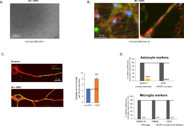
Figure 3: Co-culture of iNs and iOPCs. (A) Representative bright field image of co-cultured iNs and iOPCs at Day 7, showing a proper density for further maturation. (B) Representative immunofluorescence image of iNs and iOPCs co-cultured for 28 days. Axonal marker neurofilament NF is shown in green and oligodendrocytic marker MBP in red. Right, a segment of iN axon ensheathed by iOL process (MBP+). (C) Synapse formation assayed in 4-week-old co-cultures. Cells were stained for Synapsin 1 (Syn1, green) and MAP2 (red), and synaptic puncta were quantified by confocal analysis of density along the dendritic segments as described17,18. (D) In our co-cultures of iNs and iOPCs (7 days of co-culturing), the expression of astrocyte markers, ALDHL1 and GFAP, is minimal (top), and the expression of microglia markers, TMEM119, TREM2, and CD33, is not detected (N.D.) by qPCR. The contamination from these two glial cell types is thus excluded. Please click here to view a larger version of this figure.
to:
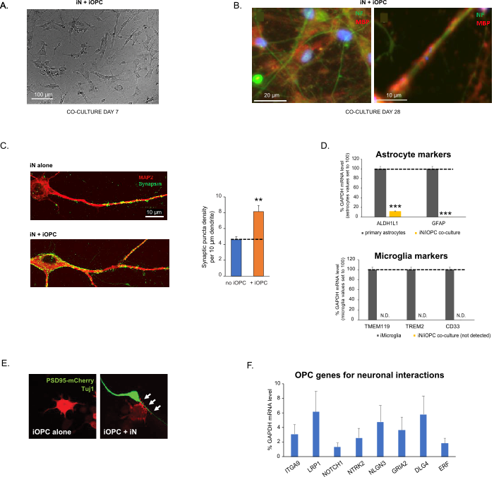
Figure 3: Co-culture of iNs and iOPCs. (A) Representative bright field image of co-cultured iNs and iOPCs at Day 7, showing a proper density for further maturation. (B) Representative immunofluorescence image of iNs and iOPCs co-cultured for 28 days. Axonal marker neurofilament NF is shown in green and oligodendrocytic marker MBP in red. Right, a segment of iN axon ensheathed by iOL process (MBP+). (C) Synapse formation assayed in 4-week-old co-cultures. Cells were stained for Synapsin 1 (Syn1, green) and MAP2 (red), and synaptic puncta were quantified by confocal analysis of density along the dendritic segments as described17,18. (D) In our co-cultures of iNs and iOPCs (7 days of co-culturing), the expression of astrocyte markers, ALDHL1 and GFAP, is minimal (top), and the expression of microglia markers, TMEM119, TREM2, and CD33, is not detected (N.D.) by qPCR. The contamination from these two glial cell types is thus excluded. (E) Coculturing iOPC with iN leads to the formation of neuron-OPC synapses. The fluorescence-tagged post-synaptic marker PSD95-mCherry is expressed only in OPCs, and display a diffuse pattern in single cultures (left) but aggregate to form puncta in cocultures (right, indicated by arrows; Tuj1, neuronal marker). (F) The expression of well-characterized oligodendroglial genes that can sense and respond to neuronal activities in the pure cultures of iOPCs at Day 14. Please click here to view a larger version of this figure.
The fourth paragraph was updated from:
Co-culturing of iNs and iOPCs
This protocol is optimized specifically for co-culturing iNs and iOPCs and allow our real-time monitoring of the inter-cellular communications between these two cell types along the course of neural development. The ideal plating densities for both cell types need to be decided with a series of cell number titration to achieve proper differentiation (Figure 3A). After 4 weeks in co-cultures, the iOPCs are expected to be adequately differentiated into OLs that are positive for specific markers such as MBP and extend processes to ensheath axons (Figure 3B). The co-culture system can robustly boost up the number of synapses, indicating that the iOPCs provide a neuronal support through physical contacts or release of trophic factors (Figure 3C). We can maintain the co-cultures in acceptable health condition for up to 6 weeks and observe that the synapse number and other neuronal attributes plateau around the fifth week. Of note, astrocytes and microglia are not present in our preparations and their absence can be documented by checking the expression of specific markers (Figure 3D).
to:
Co-culturing of iNs and iOPCs
This protocol is optimized specifically for co-culturing iNs and iOPCs and allow our real-time monitoring of the inter-cellular communications between these two cell types along the course of neural development. The ideal plating densities for both cell types need to be decided with a series of cell number titration to achieve proper differentiation (Figure 3A). After 4 weeks in co-cultures, the iOPCs are expected to be adequately differentiated into OLs that are positive for specific markers such as MBP and extend processes to ensheath axons (Figure 3B). The co-culture system can robustly boost up the number of synapses, indicating that the iOPCs provide a neuronal support through physical contacts or release of trophic factors (Figure 3C). We can maintain the co-cultures in acceptable health condition for up to 6 weeks and observe that the synapse number and other neuronal attributes plateau around the fifth week. Of note, astrocytes and microglia are not present in our preparations and their absence can be documented by checking the expression of specific markers (Figure 3D). The iOPCs express a good number of well-characterized genes that can potentially respond to and mediate the activity-dependent signals from neighboring neurons, in a paracrine (e.g. neurotrophins and metabolites) and/or a synaptic manner (Figure 3E and 3F).
