Este artículo describe los protocolos para la preparación de muestras, la reducción de datos y el análisis de datos en estudios de eco de espín de neutrones (NSE) de membranas lipídicas. El etiquetado juicioso de los lípidos con deuterio permite el acceso a diferentes dinámicas de membrana en escalas de longitud y tiempo mesoscópicas, sobre las cuales ocurren procesos biológicos vitales.
Se requiere una suscripción a JoVE para ver este contenido. Inicie sesión o comience su prueba gratuita.
Artículo de método
Espectroscopía de eco de espín de neutrones como una sonda única para la dinámica de la membrana lipídica y las interacciones membrana-proteína
En este artículo
Aviso de erratum
Important: There has been an erratum issued for this article. View Erratum Notice
Resumen
Resumen
Las bicapas lipídicas forman la matriz principal de las membranas celulares y son la plataforma principal para el intercambio de nutrientes, las interacciones proteína-membrana y la brotación viral, entre otros procesos celulares vitales. Para una actividad biológica eficiente, las membranas celulares deben ser lo suficientemente rígidas como para mantener la integridad de la célula y sus compartimentos, pero lo suficientemente fluidas como para permitir que los componentes de la membrana, como las proteínas y los dominios funcionales, se difundan e interactúen. Este delicado equilibrio de las propiedades de la membrana elástica y fluida, y su impacto en la función biológica, requieren una mejor comprensión de la dinámica colectiva de la membrana en escalas de tiempo y duración mesoscópicas de procesos biológicos clave, por ejemplo, deformaciones de membrana y eventos de unión a proteínas. Entre las técnicas que pueden sondear eficazmente este rango dinámico se encuentra la espectroscopia de eco de espín de neutrones (NSE). Combinado con el etiquetado de deuterio, NSE se puede utilizar para acceder directamente a las fluctuaciones de flexión y espesor, así como a la dinámica mesoscópica de características de membrana seleccionadas. Este documento proporciona una breve descripción de la técnica NSE y describe los procedimientos para realizar experimentos de NSE en membranas liposomales, incluidos los detalles de la preparación de muestras y los esquemas de deuteración, junto con instrucciones para la recopilación y reducción de datos. El documento también presenta métodos de análisis de datos utilizados para extraer parámetros clave de la membrana, como el módulo de rigidez de flexión, el módulo de compresibilidad del área y la viscosidad en el plano. Para ilustrar la importancia biológica de los estudios de NSE, se discuten ejemplos seleccionados de fenómenos de membrana sondeado por NSE, a saber, el efecto de los aditivos en la rigidez de flexión de la membrana, el impacto de la formación de dominios en las fluctuaciones de la membrana y la firma dinámica de las interacciones membrana-proteína.
Introducción
La comprensión de las membranas celulares y su función ha evolucionado notablemente en las últimas décadas. La visión anterior de las membranas celulares como bicapas lipídicas pasivas que definen los límites celulares y albergan proteínas de membrana1 se ha transformado gradualmente en un modelo dinámico en el que las bicapas lipídicas desempeñan un papel importante en la regulación de los procesos biológicos vitales, incluida la señalización celular, el intercambio molecular y la función de las proteínas, por nombrar algunos2,3,4,....
Acceso restringido. Inicie sesión o comience una prueba gratuita para ver este contenido.
Protocolo
1. Esquema de deuteración requerido para el experimento
- Para las mediciones de fluctuación de flexión, realice liposomas completamente protiated en D2O (D 99.9%) o D2O-buffer (por ejemplo, tampón de fosfato preparado con D2O en lugar de H2O). Utilice DMPC (C36H72NO8P) y DSPC (C44H88NO8P) totalmente protiados con
 133,4 mg, donde XDMPC y XDSPC son las fracciones molares de DMPC y DSPC, aquí establecidas en 0,7 y ....
Acceso restringido. Inicie sesión o comience una prueba gratuita para ver este contenido.
Resultados
Los estudios de NSE que acceden a las fluctuaciones de flexión se realizan típicamente en un rango Q de ~ (0.04 - 0.2) Å-1. Este rango Q corresponde a escalas de longitud intermedia entre el grosor de la membrana y el radio liposomal, donde domina la dinámica de flexión. La medición en un rango Q extendido puede dar acceso a modos dinámicos adicionales, incluida la difusión liposomal y la dinámica intramembrana. Para obtener más detalles sobre el cruce en la dinámica de membranas a la que accede NSE, consulte .......
Acceso restringido. Inicie sesión o comience una prueba gratuita para ver este contenido.
Discusión
NSE es una técnica poderosa y única en la medición de la dinámica mesoscópica de las membranas lipídicas bajo diversas condiciones. La utilización efectiva de NSE depende de la calidad de la muestra, el contraste de neutrones y el rango de dinámica accesible que se puede sondear para una muestra determinada. Por lo tanto, se requieren varios pasos críticos para realizar experimentos exitosos de NSE y recopilar datos de alta calidad. Un paso clave para garantizar el uso efectivo del tiempo de haz de neutrones durante un e.......
Acceso restringido. Inicie sesión o comience una prueba gratuita para ver este contenido.
Divulgaciones
Los autores declaran que no hay conflictos de intereses y no tienen nada que revelar.
Agradecimientos
R. Ashkar gracias M. Nagao, L.-R. Stingaciu y P. Zolnierczuk por muchas discusiones útiles y por su frecuente asistencia con experimentos de NSE en sus respectivas líneas de haz. Los autores reconocen el uso de espectrómetros de eco de espín de neutrones en NIST y ORNL. El espectrómetro NSE en el NIST cuenta con el apoyo del Centro de Dispersión de Neutrones de Alta Resolución, una asociación entre el Instituto Nacional de Estándares y Tecnología y la Fundación Nacional de Ciencias bajo el acuerdo no. DMR-1508249. El espectrómetro NSE en la fuente de neutrones de espalación de ORNL cuenta con el apoyo de la División de Instalaciones de Usuarios Científicos, Oficina de....
Acceso restringido. Inicie sesión o comience una prueba gratuita para ver este contenido.
Materiales
| Nombre | Empresa | Número de catálogo | Comentarios |
|---|---|---|---|
| Cloroformo (grado biotecnológico) | Sigma Aldrich | 496189 | grado biotecnológico, ≥ 99,8%, contiene 0,5-1,0% de etanol como estabilizador |
| Baño de agua circulante | Julabo | SE-12 | Circulador de calefacción con bomba inteligente, ajustes de temperatura programables y conexión de sensor externo para medición y control |
| Óxido de deuterio | Cambridge Isotopes Laboratories | DLM-4 | Agua deuterada; Agua pesada (D2O) (D, 99,9 %) |
| Semimicrobalanza digital | Mettler Toledo | MS105 | Semimicrobalanza con 120 g de capacidad, legibilidad de 0,01 mg, célula de pesaje de alta resolución, puertas ergonómicas y aplicación de control de pipetas |
| Etanol (grado de biología molecular) | Sigma Aldrich | E7023 | Etanol de 200 grados para aplicaciones de biología molecular |
| Pipetas de vidrio | VWR | 36360-536 Vidrio de | cal y soda desechable Pipetas Pasteur |
| Viales de vidrio | Thermo Scientific | B7990-1 | Viales de vidrio de borosilicato con tapones de tabique de PTFE/silicona |
| Congelador | Fisher Scientific | IU2886D | Congelador de temperatura ultrabaja (de -86 a -50 C) para el almacenamiento a largo plazo de lípidos y proteínas |
| Lípidos (protaiados o perdeuterados) | Avanti Polar Los lípidos | varían según el lípido | Los lípidos se pueden comprar a Avanti en forma de polvo o en una solución de cloroformo con las cantidades y esquemas de deuteración requeridos. |
| Purificador de agua Millipore | Millipore Sigma | ZRQSVP3US | Direct-Q® 3 Sistema de purificación de agua UV que suministra agua pura y ultrapura con una lámpara UV incorporada para reducir los niveles de compuestos orgánicos para productos biológicos. aplicaciones |
| Mini extrusora Avanti | Polar Lipids | 610020 | El mini-extrusora incluye miniextrusora, bloque calefactor, 2 jeringas GasTight y 2 juntas tóricas, membranas de policarbonato y soportes de filtro |
| Accesorios de conexión rápida | Grainger | 2YDA1 y 2YDA7 | Accesorios de tubo de botón para aplicaciones de circulación de agua QuickConnect, por ejemplo, extrusión de vesículas de alta temperatura |
| Bomba de | jeringaSyringePump.com | New Era-1000 | Bomba de jeringa totalmente programable para infusión y extracción; programa hasta 41 fases de bombeo con velocidades de bombeo, volúmenes dispensados y ciclos de extrusión ajustables |
| Baño ultrasónico | Fisher Scientific | CPX2800 | Baño ultrasónico con control de temperatura con funcionalidad programable para aplicaciones de desgasificación y ultrasonidos |
| Horno de vacío | Thermo Scientific | 3608 | Horno de vacío de 0.7 pies cúbicos con termostato de límite alto incorporado que protege contra el sobrecalentamiento |
| Mezclador de vórtice | Fisher Scientific | 02-215-414 | Control analógico de velocidad variable que permite el arranque a bajas revoluciones para una agitación suave o una mezcla a alta velocidad para un vórtice vigoroso de las muestras |
Referencias
- Singer, S. J., Nicolson, G. L. The fluid mosaic model of the structure of cell membranes. Science. 175 (4023), 720-731 (1972).
- Andersen, O. S., Koeppe, R. E. Bilayer thickness and membrane protein function: an energetic perspective. Annual ....
Acceso restringido. Inicie sesión o comience una prueba gratuita para ver este contenido.
Reimpresiones y permisos
Erratum
Formal Correction: Erratum: Neutron Spin Echo Spectroscopy as a Unique Probe for Lipid Membrane Dynamics and Membrane-Protein Interactions
Posted by JoVE Editors on 8/06/2021. Citeable Link.
An erratum was issued for: Neutron Spin Echo Spectroscopy as a Unique Probe for Lipid Membrane Dynamics and Membrane-Protein Interactions. The Introduction, Protocol, and Representative Results sections have been updated.
In the Introduction, the fith pargraph was updated from:
Besides direct access to the length and time scale of membrane dynamics, NSE has the inherent capabilities of neutron isotope sensitivity52. Specifically, the ability of neutrons to interact differently with the isotopes of hydrogen, the most abundant element in biological systems, results in a different neutron scattering length density,34 or NSLD (the equivalent of the optical index of refraction50), when protium is substituted by deuterium. This enables an approach known as contrast variation, which is commonly used to highlight specific membrane features or conceal others — the latter scenario is referred to as contrast matching. A frequent application of contrast variation/matching is the substitution of water (NSLD = -0.56 × 10⁻⁶ Å-2) by heavy water or D2O (NSLD = 6.4 × 10⁻⁶ Å-2) to amplify the neutron signal from protiated lipid membranes (NSLD ~ 2 × 10⁻⁶ Å-2). This approach is highly effective in studies of membrane structure because the penetration of D2O into the headgroup region of the membrane allows accurate determination of the membrane thicknesses (see Figure 2A, left panel) and of the location of different lipid subgroups when more sophisticated models are applied53,54. This paper highlights some examples on the use of contrast variation for studies of collective dynamics in biomimetic membranes and select membrane features.
to:
Besides direct access to the length and time scale of membrane dynamics, NSE has the inherent capabilities of neutron isotope sensitivity52. Specifically, the ability of neutrons to interact differently with the isotopes of hydrogen, the most abundant element in biological systems, results in a different neutron scattering length density,34 or NSLD (the equivalent of the optical index of refraction50), when protium is substituted by deuterium. This enables an approach known as contrast variation, which is commonly used to highlight specific membrane features or conceal others — the latter scenario is referred to as contrast matching. A frequent application of contrast variation/matching is the substitution of water (NSLD = -0.56 × 10⁻⁶ Å-2) by heavy water or D2O (NSLD = 6.4 × 10⁻⁶ Å-2) to amplify the neutron signal from protiated lipid membranes (NSLD ~ 0 × 10-6 Å-2). This approach is highly effective in studies of membrane structure because the penetration of D2O into the headgroup region of the membrane allows accurate determination of the membrane thicknesses (see Figure 2A, left panel) and of the location of different lipid subgroups when more sophisticated models are applied53,54. This paper highlights some examples on the use of contrast variation for studies of collective dynamics in biomimetic membranes and select membrane features.
In the Protocol, step 1.1 was updated from:
For bending fluctuation measurements, make fully protiated liposomes in D2O (D 99.9%) or D2O-buffer (e.g., phosphate buffer prepared with D2O instead of H2O). Use fully protiated DMPC (C36H72NO8P) and DSPC (C44H88NO8P) with 133.4 mg, where XDMPC and XDSPC are the mole fractions of DMPC and DSPC, here set to 0.7 and 0.3, respectively, and MwDMPC and MwDSPC are the molar weights given by 677.9 g/mol and 790.1 g/mol, respectively. Similarly, mDSPC = 66.6 mg. This deuteration scheme increases the scattering contrast between the membrane (NSLD ~ 2 × 10⁻⁶ Å-2) and the deuterated buffer (NSLD ~ 6.4 × 10⁻⁶ Å-2) and amplifies the signal from membrane undulations (see Figure 2A left panel).
to:
For bending fluctuation measurements, make fully protiated liposomes in D2O (D 99.9%) or D2O-buffer (e.g., phosphate buffer prepared with D2O instead of H2O). Use fully protiated DMPC (C36H72NO8P) and DSPC (C44H88NO8P) with 133.4 mg, where XDMPC and XDSPC are the mole fractions of DMPC and DSPC, here set to 0.7 and 0.3, respectively, and MwDMPC and MwDSPC are the molar weights given by 677.9 g/mol and 790.1 g/mol, respectively. Similarly, mDSPC = 66.6 mg. This deuteration scheme increases the scattering contrast between the membrane (NSLD ~ 0 × 10-6 Å-2) and the deuterated buffer (NSLD ~ 6.4 × 10⁻⁶ Å-2) and amplifies the signal from membrane undulations (see Figure 2A left panel).
In the Representative Results, the fist pagargaph was updted from:
NSE studies accessing bending fluctuations are typically performed over a Q-range of ~ (0.04 - 0.2) Å-1. This Q-range corresponds to intermediate length scales between the membrane thickness and the liposomal radius, where bending dynamics dominate. Measurement over an extended Q-range can give access to additional dynamic modes, including liposomal diffusion and intramembrane dynamics. For more details on the cross-over in membrane dynamics accessed by NSE, check these relevant publications25,71. It is important to emphasize that NSE signals are proportional to:  , where Icoh and Iinc are, respectively, the coherent and incoherent scattering intensity from the sample. Therefore, it is advisable to prepare NSE liposomal samples in deuterated buffers (i.e., buffers prepared with D2O instead of H2O) to minimize the incoherent scattering signal, mainly contributed by the hydrogen content of the sample. However, in some cases intermediate deuteration schemes (i.e., using mixtures of D2O and H2O) might be necessary to obtain optimal contrast conditions. Typically, NSE measurements of membrane bending fluctuations are performed on fully protiated liposomes in deuterated buffer, referred to as fully contrasted liposomes in Figure 5. This deuteration scheme results in a large NSLD difference between the membrane core (~2 × 10⁻⁶ Å-2) and its deuterated fluid environment (~6.4 × 10⁻⁶ Å-2), which significantly enhances the scattering signal from the liposomal membranes and improves the measurement statistics of bending dynamics. This contrast scheme (Figure 2A left panel) is frequently utilized in studies of bending rigidity of lipid membranes with single38,72 and multiple39,66 lipid components and in studies of membrane softening/stiffening by biological inclusions (e.g., cholesterol, drug molecules, peptides/proteins)36,37,73,74,75, and synthetic additives (e.g., nanoparticles)76,77.
to:
NSE studies accessing bending fluctuations are typically performed over a Q-range of ~ (0.04 - 0.2) Å-1. This Q-range corresponds to intermediate length scales between the membrane thickness and the liposomal radius, where bending dynamics dominate. Measurement over an extended Q-range can give access to additional dynamic modes, including liposomal diffusion and intramembrane dynamics. For more details on the cross-over in membrane dynamics accessed by NSE, check these relevant publications25,71. It is important to emphasize that NSE signals are proportional to: , where Icoh and Iinc are, respectively, the coherent and incoherent scattering intensity from the sample. Therefore, it is advisable to prepare NSE liposomal samples in deuterated buffers (i.e., buffers prepared with D2O instead of H2O) to minimize the incoherent scattering signal, mainly contributed by the hydrogen content of the sample. However, in some cases intermediate deuteration schemes (i.e., using mixtures of D2O and H2O) might be necessary to obtain optimal contrast conditions. Typically, NSE measurements of membrane bending fluctuations are performed on fully protiated liposomes in deuterated buffer, referred to as fully contrasted liposomes in Figure 5. This deuteration scheme results in a large NSLD difference between the membrane core (~0 × 10-6 Å-2) and its deuterated fluid environment (~6.4 × 10⁻⁶ Å-2), which significantly enhances the scattering signal from the liposomal membranes and improves the measurement statistics of bending dynamics. This contrast scheme (Figure 2A left panel) is frequently utilized in studies of bending rigidity of lipid membranes with single38,72 and multiple39,66 lipid components and in studies of membrane softening/stiffening by biological inclusions (e.g., cholesterol, drug molecules, peptides/proteins)36,37,73,74,75, and synthetic additives (e.g., nanoparticles)76,77.
In the Representative Reults, Figure 2 was updated from:
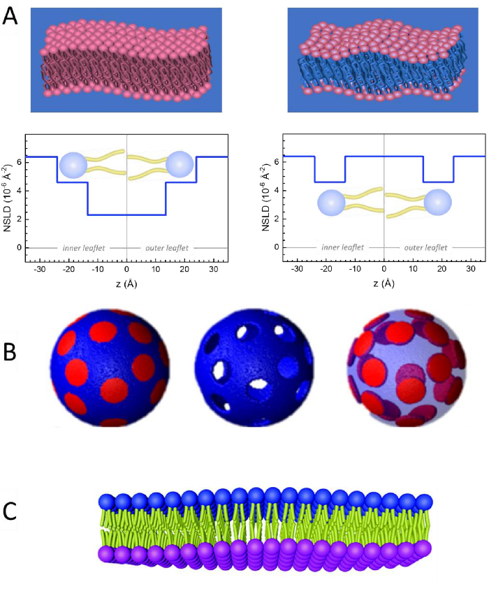
Figure 2: Examples of possible deuteration schemes in NSE experiments on lipid membranes. (A) Left: Fully contrasted membranes, e.g., protiated membranes in deuterated buffer, showing the NSLD profile along the normal to the membrane surface. The difference in the NSLD between the headgroup (~2 × 10⁻² Å-2) and tail region (~4.5 × 10⁻⁶ Å-2) of the membrane is due to the headgroup hydration with deuterated buffer. Right: Tail-contrast matched membranes such that the hydrocarbon tail region of the membrane has the same NSLD as the buffer, as shown in the corresponding NSLD profile along the membrane normal. (B) Domain-forming membranes with two neutron contrast schemes where the domains (center) or the matrix (left) are contrast-matched to the buffer, enabling selective studies of matrix or domain dynamics, respectively. This figure has been modified from Nickels et al., JACS 201541. (C) Asymmetric membranes prepared by cyclodextrin exchange between protiated and deuterated lipid vesicles, resulting in the deuteration of one membrane leaflet while keeping the other leaflet protiated. This allows studies of the bending dynamics of the protiated leaflet and provides insights into the mechanical coupling between opposing leaflets in asymmetric membranes. This figure has been modified from Rickeard et al., Nanoscale 202040. Please click here to view a larger version of this figure.
to:
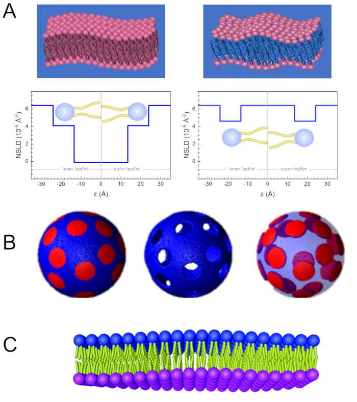
Figure 2: Examples of possible deuteration schemes in NSE experiments on lipid membranes. (A) Left: Fully contrasted membranes, e.g., protiated membranes in deuterated buffer, showing the NSLD profile along the normal to the membrane surface. The difference in the NSLD between the tail region (~0 × 10-2 Å-2) and headgroup region (~4.5 × 10⁻⁶ Å-2) of the membrane is due to the headgroup hydration with deuterated buffer. Right: Tail-contrast matched membranes such that the hydrocarbon tail region of the membrane has the same NSLD as the buffer, as shown in the corresponding NSLD profile along the membrane normal. (B) Domain-forming membranes with two neutron contrast schemes where the domains (center) or the matrix (left) are contrast-matched to the buffer, enabling selective studies of matrix or domain dynamics, respectively. This figure has been modified from Nickels et al., JACS 201541. (C) Asymmetric membranes prepared by cyclodextrin exchange between protiated and deuterated lipid vesicles, resulting in the deuteration of one membrane leaflet while keeping the other leaflet protiated. This allows studies of the bending dynamics of the protiated leaflet and provides insights into the mechanical coupling between opposing leaflets in asymmetric membranes. This figure has been modified from Rickeard et al., Nanoscale 202040. Please click here to view a larger version of this figure.
