A subscription to JoVE is required to view this content. Sign in or start your free trial.

Method Article

Concurrent Electroencephalographic and Local Field Potential Recordings in an Anesthetized Rat

1.3K views

⸱

July 8th, 2025

In This Article

Abstract

Source: Kang, S., et al. Concurrent Recording of Co-localized Electroencephalography and Local Field Potential in Rodent. J. Vis. Exp. (2017).

This video demonstrates the procedure for concurrent recordings of co-localized electroencephalography (EEG) and local field potentials (LFPs) in an anesthetized rat. The process involves preparing the rat and attaching stimulating electrodes to its whisker pads. An EEG spider electrode is placed on the rat's skull for surface EEG measurements. At the same time, a multi-channel microelectrode is inserted into a specific brain region through a burr hole in the skull to record neural activity from a localized brain area.
Protocol

All procedures involving animal models have been reviewed by the local institutional animal care committee and the JoVE veterinary review board.

1. Co-localized EEG/LFP Setup

  1. Clean and dry the skull surrounding the burr hole using a cotton swab.
  2. Carefully place the conductive EEG paste on one flat side of an EEG spider electrode. Leave a small hole clear of the EEG paste on the spider electrode to allow a multi-laminar microelectrode to pass through the hole without contacting the paste and the spider electrode. This prevents electrical contact between the EEG electrode and the microelectrode.

Access restricted. Please log in or start a trial to view this content.

Results

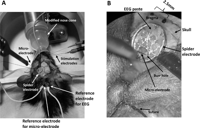
Micro-electrode setup showing EEG and stimulation electrodes in rat skull for neuroscience study.
Figure 1: General setup for concurrent EEG/LFP recording. (A) The setup consists of a modified nose cone for ease of whisker pad stimulation under isoflurane anesthesia, two stimulating electrodes inserted into the whisker pad, a spider el.......

Access restricted. Please log in or start a trial to view this content.

Disclosures

No conflicts of interest declared.

Materials

List of materials used in this article
NameCompanyCatalog NumberComments
Female Lister Hood ratsCharles Rivers
Spider electrodeUnimed Electrode Supplies LtdSCS24-426
EEG paste: Ten20Unimed Electrode Supplies Ltd10-20-S
Stereotaxic holder with dual micromanipulator arms: Dual Manipulator Stereotaxic Frame with 18° Ear BarsWPI (World Precision Instruments)502603
Stainless steel stimulating electrodesPlasticsOneE363/1/SPC
Isolated current stimulatorMade in House
16-channel micro-electrode, 100 μm spacing, area of each site 177 μm2NeuroNexusA1x16-10mm-100-177-A16
16-channel acute headstageTucker David Technologies Inc., TDTRA16AC-Z
Pre-Amplifier: Z-Series 64-Channel Neuro-Digitizing PreampTDTPZ5-64
Passive signal splitter: 32-Channel Splitter Box for PZ5TDTS-BOX_PZ5
Data acquisition unit: RZ2 BioAmp Processor. Z-Series 4-DSP ultra high performance processorTDTRZ2-4
Isoflurane National Vet Services Limited 50878
Small animal isoflurane anaesthetic systemWPI EZ-B800A
Thermostatic heating pad: Rat Blanket System 230VHarvard Apparatus UK 50-7221-F
Ophthalmic ointment: Optixcare eye lubeViovet 203865
Faraday cage Newport Corporation VIS-FDC-3600
Vibration isolation workstation: Vision IsoStationNewport Corporation M-VIS3660-RG4-325A
Oximeter Control Unit and sensor:
MouseOxPlus, Starr Life Sciences
Corp.
WPI O15001
Transparent soft nose cone: Microflex Non-Rebreathing Unit with a Rat NoseconeWPI EZ-103A

Explore More Articles

ElectroencephalographyConcurrent RecordingStereotaxic FrameBurr HoleMicroelectrode InsertionEEG Spider ElectrodeWhisker Pad StimulationData Acquisition