This video demonstrates the procedure for implanting a cannula into the cisterna magna of an anesthetized mouse. The technique involves guiding a needle-mounted cannula through the dura into the inverted triangular cisterna magna and securing it for targeted delivery into the cerebrospinal fluid.
A subscription to JoVE is required to view this content. Sign in or start your free trial.
Method Article
Cannula Implantation into the Cisterna Magna in a Mouse Model
3K views
⸱
August 29th, 2025
In This Article
Abstract
Protocol
All procedures involving animal models have been reviewed by the local institutional animal care committee and the JoVE veterinary review board.
- Surgical procedure
NOTE: The chronic cisterna magna (CM) cannulation allows the animals to recover from the surgical procedure. CM injections are made the day following cannula implantation and, importantly, can be performed in either anesthetized or awake animals. Since this is a recovery surgery, procedures should be carried out under sterile conditions.- Weigh the mouse (C57BL/6JRj, both sexes, 8 weeks) and anesthetize it with a mixture of ketamine and xylazine (100 mg/kg; 10 mg/kg, respectively) via intraperitoneal (i.p.) injection. If necessary, redose the mouse with a half dose of ketamine (50 mg/kg) during the surgical procedure. Alternatively, for chronic cannulation, anesthetize the mouse by placing it into an isoflurane induction chamber at 2.5 - 3% isoflurane, at circa 1 L/min oxygen (O2). In this case, use a nose cone to sustain isoflurane anesthesia at 1.5 - 2% throughout the surgery.
- When toe pinch reflexes cease and the respiration becomes slow and steady, place the animal in a stereotaxic frame over a heating pad.
- Apply ophthalmic ointment. Repeat during surgery whenever necessary.
- Shave the head and neck of the mouse, remove fur, and sterilize exposed skin first with an alcohol swab and then two times with chlorhexidine (0.5%) or iodine solution (2%). Repeat the sterilization two more times.
NOTE: Change the surgical drape to remove debris and hair after shaving. Then, drape the animal to protect the sterile field. - For chronic cannulation, administer 0.5 - 1 ml lidocaine/bupivacaine (1 mg/ml and 0.25 mg/ml, respectively) subcutaneously (s.c.) at the incision site. Administer buprenorphine (0.05 mg/kg; s.c.) for post-surgical analgesia.
- Fix the mouse in the stereotaxic frame. After ensuring fixation, either intraaural or by the zygomatic arch, tilt the head slightly so that it forms an angle of 120° to the body (Figure 1E).
- Find the part of the skull protruding immediately above the neck muscles - the occipital crest. Lift the overlying skin using a pair of tweezers, and cut an almond-shaped piece of skin of approximately 1 cm along the midline. Use cotton swabs or eye spears to control any resultant bleeding.
- Using the occipital crest as a reference point, pull apart the superficial connective tissue to expose the neck muscles below.
- Separate the muscles at the midline by carefully running the forceps down the middle of the incision site in the anterior-to-posterior axis. With a pair of curved forceps in each hand, join the tips in the middle near the bottom of the skull and pull the muscles aside.
NOTE: This should expose the CM, which appears as a tiny inverted triangle, outlined by the cerebellum above and medulla below, behind the translucent dural membrane (Figure 1B and 1C). - Using a surgical eye spear or cotton swab, wipe the dural membrane covering the CM.
- Insertion of the cannula into the CM
- Remove the cannula from the artificial cerebrospinal fluid (aCSF)-filled syringe, keeping the 30G needle attached to the rear end of the tubing.
- Attach the 30G needle to a distilled water-filled 100 µL syringe connected to a syringe pump.
- Insert an air bubble of approximately 1 cm into the cannula by withdrawing air with the aid of a syringe pump.
- Using a syringe pump, withdraw 12 μL of the desired cerebrospinal fluid (CSF) tracer into the cannula.
- Grasp the cannula, filled with the CSF tracer, near the tube-covered needle with a pair of curved tweezers held in the dominant hand. Rest the middle finger of the non-dominant hand on the ear bar of the opposite side and hold it steady for later use as a rest for the cannula.
- Insert the cannula at an angle of 45° relative to the mouse head, passing into the center of the CM, identified by its triangular aspect seen through the dura. Avoid any penetration of the cerebellum or medulla. Ensure that the needle is only inserted to a depth of 1 - 2 mm, such as to the point where the bevel is entirely under the dura. Release the tweezers holding the cannula, and let the cannula rest on the non-dominant hand.
NOTE: The beveled end of the dental needles will require the application of some force to pierce the dural membrane covering the CM. - If necessary, dry off any CSF leak upon penetration using a surgical eye spear or cotton swabs.
- Apply 2-3 drops of cyanoacrylate glue onto the dural membrane surrounding the cannula. Add a drop of glue accelerator to cure the glue immediately. Cover the skull and needle with a mixture of dental cement (approximately 0.5 mg) and cyanoacrylate glue (3 - 5 drops). Immediately after, apply a drop of glue accelerator to cure.
Access restricted. Please log in or start a trial to view this content.
Results
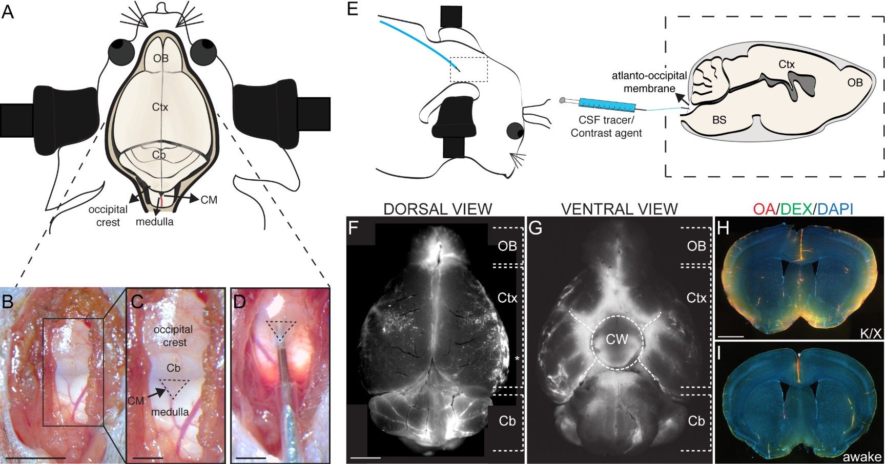
Figure 1: Injection of tracers into the cisterna magna. (A) Schematic overview of the m...
Access restricted. Please log in or start a trial to view this content.
Materials
| Name | Company | Catalog Number | Comments |
|---|---|---|---|
| SOPIRA carpule 30G 0.3 x 12 mm | Kulzer | AA001 | |
| Chlorhexidine 0.5% (chlorhexidine digluconate) | Meda AS | http://www.meda.dk/behandlingsomrader/desinfektion/desinfektion-af-hud/klorhexidin-sprit-medic-05/ | |
| Alcohol swab 70% isopropyl alcohol 30 x 60 mm | Vitrex Medical A/S | 520213 | |
| Viskoese Oejendraeber Ophtha | Ophtha | 145250 | |
| Wooden applicator, double cotton bud (Ø approximately, 4 - 5 mm, length approximately, 12 mm) | Heinz Herenz | 1032018 | |
| Eye spears | Medicom | A18005 | |
| Kimtech Science precision wipes tissue wipers | Kimberly Clark Professional | 5511 | |
| Loctite super glue precision 5 g | Loctite | http://www.loctite-consumer.dk/da/produkter/superglue-liquid.html | |
| Insta-Set CA accelerator | Bob Smith Industries | BSI-152 | |
| Dental cement powder | A-M Systems | 525000 | |
| Phosphate-buffered saline (PBS; 0.01 M; pH 7.4) | Sigma Aldrich | P3813 | |
| Ovalbumin, Alexa Fluor 647 conjugate | Thermo Fisher Scientific | O34784 | |
| DAPI (4′,6-diamidino-2-phenylindole) solution (1 mg/mL) | Thermo Fisher Scientific | 62248 | |
| Dextran, fluorescein, 3000 MW, anionic | Thermo Fisher Scientific | D3305 | |
| Ketaminol Vet 100 mg/mL (ketamine) | Intervet International BV | 511519 | |
| Rompin Vet 20 mg/mL (xylazine) | KVP Pharma + Veterinär Produkte GmbH | 148999 | |
| Xylocain 20 mg/mL (lidocain) | AstraZeneca | 158543 | |
| Marcain 2.5 mg/mL (bupivacain) | AstraZeneca | 123918 | |
| Bupaq Vet 0.3 mg/mL (buprenorphine) | Richter Pharma AG | 185159 |
