Method Article

Preparing a Single-Cell Suspension of Immune Cells from Murine Brain Tissue

1.2K views

July 8th, 2025

In This Article

Abstract

Source: DiSano, K. D., et al. Isolating Central Nervous System Tissues and Associated Meninges for the Downstream Analysis of Immune Cells. J. Vis. Exp. (2020).

The video demonstrates the preparation of a single-cell suspension of immune cells from a murine brain tissue. It involves enzymatic digestion to release cells from the brain tissue, followed by density gradient centrifugation to isolate the immune cells based on their densities to obtain the single-cell suspension.
Protocol

All procedures involving animal samples have been reviewed and approved by the appropriate animal ethical review committee.

1. Preparing single-cell suspensions of brain and spinal cord tissue

  1. Transfer the brain or spinal cord tissue with media to the top of the 100 mm Petri dish by pouring the tissue and media from the tube. Using forceps, move the tissue to the bottom of the Petri dish. Finely mince the brain or spinal cord with a sterile razor blade. Using the razor blade, move the minced tissue to the bottom of the plate by scraping to gather the tissue.
    NOTE: For the enzymatic digestion protocol below, up to two spinal cords may be pooled together for processing. Brains should be processed individually.
  2. Using a 5 mL serological pipette, add 3 mL of RPMI (Roswell Park Memorial Institute medium (supplemented with 10% fetal calf serum (FCS) to the Petri dish. Using a 10 mL serological pipette, pipette up and down to resuspend the tissue in the media and transfer to a 15 mL conical tube.
    NOTE: If the downstream application is single-cell or bulk RNA sequencing analysis or cell culture, FCS lots should be tested to ensure cells are not activated prior to analysis. Alternatively, cells can be processed using 1x PBS with 0.04% BSA instead of RPMI with 10% FCS.
  3. Using a 10 mL serological pipette, wash the Petri dish with an additional 2 mL of media to collect any residual tissue and transfer to a conical tube for a 5 mL total volume. Keep the tubes on ice between processing steps.
  4. Using a pipette, resuspend the collagenase I powder in Hank's Balanced Salt Solution (HBSS) media to obtain the desired concentration (i.e., 100 mg/mL). Add collagenase type I to the conical tube containing the minced tissue sample to obtain the desired final concentration (i.e., 50 µL for 1 mg/mL).
    NOTE: Higher concentrations of collagenase will increase cell yields but can cleave cell surface markers. Therefore, collagenase I lots should be titrated to determine the optimal concentration needed to obtain the highest number of viable cells with all required cell surface markers intact. For example, collagenase I was tested at final concentrations of 0.5 mg/mL, 1 mg/mL, and 2 mg/mL on single brain or spinal cord samples. Cell viability was determined using the trypan blue exclusion method and cell surface markers CD45, CD19, and CD4 were assessed by flow cytometry. A 1 mg/mL concentration of collagenase I yielded the highest live cell count while retaining all cell surface markers of interest. Thus, this concentration was used for further experiments examining these cell types.
  5. Gently resuspend DNase I powder using 0.15 M sodium chloride to the desired stock concentration. Add the resuspended DNase I to the conical tube containing the minced tissue sample to obtain a final concentration of 20 U/mL.
    NOTE: DNase I lots vary by units of activity per mL. The concentration to be added to the tissue sample will change based on the stock vial's units of activity per milliliter. The final desired concentration per sample is 20 U/mL.
  6. Place the tubes in a tube rack in a 37 °C water bath and incubate for 40 min. Invert the tubes every 15 min to thoroughly mix the tissue with the enzymes. After incubation, add 500 µL of 0.1 M EDTA (pH = 7.2) to each tube for a final concentration of 0.01 M EDTA and incubate for an additional 5 min to inactivate the collagenase.
  7. Using a 10 mL serological pipette, add 9 mL of RPMI supplemented with 10% FCS to each tube to bring the volume of each tube to ~14.5 mL. Centrifuge at 450 x g for 5 min at 4 ˚C. Using a Pasteur pipette with a vacuum, aspirate the supernatant, being careful not to touch the cell pellet.
  8. Using a 5 mL serological pipette, add 3 mL of 100% stock isotonic density gradient solution to the tube containing the cell pellet. Using a 10 mL serological pipette, add additional RPMI 10% FCS media to bring the final volume to 10 mL and resuspend the cell pellet to create a 30% stock isotonic density gradient solution layer.
    NOTE: Prepare the 100% stock isotonic density gradient medium in advance, aliquot, and store at 4 °C for up to 3 months. To prepare the 100% stock isotonic density gradient solution, dilute the density gradient media (Table of Materials) with density gradient media dilution buffer. Prepare the density gradient media dilution buffer (80.0 g/L NaCl, 3.0 g/L KCl; 0.73 g/L Na2HPO4, 0.20 g/L KH2PO4; 20.0 g/L glucose) and filter sterilize using a vacuum filter system. Make the 100% stock isotonic density gradient solution by mixing 1 part of the density gradient dilution buffer and 9 parts density gradient media. Mix well.
  9. Invert and mix each tube well prior to adding the 70% stock isotonic density gradient solution underlay. Insert a 1 mL serological pipette containing 1 mL of 70% stock isotonic density gradient solution into the bottom of the tube. Slowly underlay 1 mL of the solution, being careful not to make bubbles. Slowly remove the serological pipet from the tube, being careful not to disturb the gradient.
    NOTE: For the 70% underlay, the 100% stock isotonic density gradient solution should be diluted to 70% using RPMI media (i.e., 7 mL of 100% stock isotonic density gradient solution and 3 mL of RPMI media mixed well). Additionally, creating a clean, undisturbed 70% underlay is essential for the removal of myelin debris and for obtaining pure single-cell suspensions at the gradient interface following centrifugation.
  10. Centrifuge at 800 x g for 30 min at 4 °C with no brake. Aspirate the supernatant, including the myelin debris layer until 2–3 mL remains in the tube, being careful not to disturb the cell layer. Harvest the cell layer between the 30/70% density gradient using a 1 mL pipette and transfer to a new 15 mL conical tube.
  11. Using a 10 mL serological pipette, add RPMI 10% FCS media to bring the final volume to 15 mL. Centrifuge 450 x g for 5 min at 4 °C.
    NOTE: During this step, cell layers from two tubes can be pooled if needed. Do not pool more than two tubes or the cells will not pellet due to a high-density gradient media concentration.
  12. Aspirate the supernatant carefully to not disturb the cell pellet. Resuspend the cells in an appropriate volume/buffer to count the cells using a hemocytometer (e.g., resuspend a single spinal cord in 250 µL of FACS buffer for downstream surface staining) (Figure 1).
  13. Using a 1 mL pipette, transfer the cell suspensions to the top of a filter top tube (Table of Materials) and allow the cells to filter to the bottom of the tube to remove any remaining myelin debris.
  14. Using trypan blue exclusion dye, dilute it and count the cells on a hemocytometer by averaging at least two 16-square grids for accuracy40,41.
  15. Proceed with the desired single-cell analysis technique.
    NOTE: Using various forms of collagenase (i.e., D, type I, type II, type IV), immune cells, microglia (Figure 1), astrocytes, pericytes, endothelial cells49, and neurons50 can all be efficiently isolated. Nucleated cell counts obtained for the results were as follows using the titrated collagenase I enzyme: Whole sham-treated brain = 500,000–600,000 cells; Whole TMEV-IDD brain = 800,000–1,000,000 cells; Whole sham-treated spinal cord = 150,000–200,000 cells; Whole TMEV-IDD spinal cord = 300,000–400,000. Cell counts will vary depending on the precision of collection, processing, and whether CNS inflammation is present.

Access restricted. Please log in or start a trial to view this content.

Results

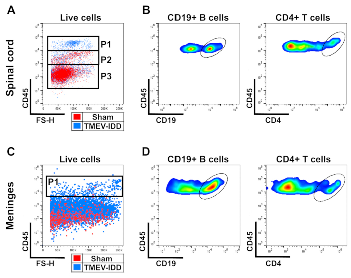
Flow cytometry diagrams of CD19+ B cells and CD4+ T cells in spinal cord/meninges, TMEV-IDD study.
Figure 1: Identification of CD45hi infiltrating immune cells in CNS tissues. (A) Gating strategy for the identification of CD45hi infiltrating immune cells (P1), CD45lo microglia (P2), and CD45- oligodendrocytes, astrocytes, and neurons among total liv...

Access restricted. Please log in or start a trial to view this content.

Disclosures

No conflicts of interest declared.

Materials

List of materials used in this article
NameCompanyCatalog NumberComments
Bovine Serum AlbuminThermoFisher Scientific37002D
CentrifugeBeckman CoulterAllegra X-12R centrifuge
Collagenase IWorthingtonLS004196
Conical tube, 15 mLVWR525-1069
Conical tube, 50 mLVWR89039-658
Cover glassHauser Scientific5000
Curved forcepsFine Science Tools11003-14
Disposable polystyrene tube, 14 mLFisher Scientific14-959-1B
DNase IWorthingtonLS002139
Dry iceAirgasN/A
Durmont #7ForcepsFine Science Tools11271-30
EDTA disodium salt dihydrateAmresco0105-500g
Ethanol, 100%anyN/A
Filter top tube, 5 mLVWR352235
Fixable viability stain 780Becton Dickinson565388
GlucoseFisher ChemicalD16-500
Hank's Balnced Salt Solution (HBSS)Corning21-020-CV
HemacytometerAndwin Scientific02-671-51B
HemostatFine Science Tools13004-14
HEPES (N-2-hydroxyethylpiperazine-N-2-ethane sulfonic acid)ThermoFisher Scientific15630080
KClFisher chemicalBP366-500
KH2PO4 (anhydrous)Sigma AldrichP5655-100G
Liquid NitrogenAirgasN/A
Mouse FC block (CD16/32)Becton Dickinson553141
Na2HPO4 (anhydrous)Fisher ChemicalS374-500
NaClFisher chemicalS671-500
Needle, 25 gaugeBecton Dickinson305122
Normal mouse serumThermoFisher Scientific31881
Nylon mesh strainerVWR352350
OCTSakura4583
Paraformaldehyde, 20%Electron Microscopy Sciences15713-SDiluted to 4% using 1 x PBS
Pasteur pipette, 9 inch, unpluggedFisher Scientific13-678-20C
PBS (1x)Corning21-040-CV
PE Rat Anti-Mouse CD4Becton Dickinson553730
PE-CF594 Rat Anti-Mouse CD19Becton Dickinson562329
Percoll density gradient mediaGE healthcare17-0891-01
PerCP-Cy5.5 Rat Anti-Mouse CD45Becton Dickinson550994
Petri dish, 100 mmVWR353003
pH meterFisher Scientific13-636-AB150
Pipet-AidDrummond Scientific Corporation4-000-101
Pipette 200 µlGilsonFA10005M
Pipette tips, 1 mLUSA Scientific1111-2831
Pipette tips, 200 µlUSA Scientific1111-1816
Pipette, 1 mLGilsonFA10006M
Prolong Diamond mountant with DAPIThermoFisher ScientificP36962
Purified Rat Anti-Mouse CD16/CD32Becton Dickinson553141
Rabbit anti-mouse CD3 (SP7 clone)Abcamab16669
Rabbit anti-mouse lamininAbcamab11575
Rat anti-mouse ERT-R7Abcamab51824
RPMI 1640Corning10-040-CV
Serological pipet, 1 mLVWR357521
Serological pipet, 10 mLVWR357551
Serological pipet, 5 mLVWR357543
Sodium hydroxideFisher ScientificS318-100
SucroseFisher chemicalS5-500
Surgical scissorsFine Science Tools14001-16
Surgical scissors, extra fineRobozRS-5882
Syringe, 10 mLBecton Dickinson302995
Syringe, 5 mLBecton Dickinson309646
Trypan blueGibco15250-061

Reprints and Permissions

Request permission to reuse the text or figures of this JoVE article

Request Permission

Tags

Enzymatic DigestionDensity Gradient CentrifugationImmune Cell IsolationCollagenase TreatmentDNase I DegradationEDTA InactivationRPMI Culture MediumLow Speed Centrifugation

Related Articles