Slice cultures facilitate the manipulation of embryo development by gene and pharmacological perturbations. However, culture conditions must ensure that normal development can proceed within the reduced environment of the slice. We illustrate a protocol that facilitates normal spinal cord development to proceed for at least 24 hr.
Method Article
Chicken Embryo Spinal Cord Slice Culture Protocol
* These authors contributed equally
In This Article
Summary
Abstract
Slice cultures can facilitate the manipulation of embryo development both pharmacologically and through gene manipulations. In this reduced system, potential lethal side effects due to systemic drug applications can be overcome. However, culture conditions must ensure that normal development proceeds within the reduced environment of the slice. We have focused on the development of the spinal cord, particularly that of spinal motor neurons. We systematically varied culture conditions of chicken embryo slices from the point at which most spinal motor neurons had been born. We assayed the number and type of motor neurons that survived during the culture period and the position of those motor neurons compared to that in vivo. We found that serum type and neurotrophic factors were required during the culture period and were able to keep motor neurons alive for at least 24 hr and allow those motor neurons to migrate to appropriate positions in the spinal cord. We present these culture conditions and the methodology of preparing the embryo slice cultures using eviscerated chicken embryos embedded in agarose and sliced using a vibratome.
Introduction
During normal embryo development, many different cell types are generated which must migrate from their point of origin to where they will eventually function in the mature organism. Within the developing spinal cord, progenitor cells are located in the ventricular zone at the midline and generate many subtypes of postmitotic neurons and glial cells which must migrate to integrate into functional neuronal circuits required for sensory processing and motor outputs1. Spinal motor neurons that control the contraction of limb muscles have been extensively studied and much is known of the molecules and mechanisms that drive the formation of their different subclasses. However, much less is known of the mechanisms by which motor neurons migrate, segregate and receive synaptic inputs.
Motor neuron subtype identity is acquired during development in a hierarchical fashion. Motor neuron subtypes can be broadly classed into distinct columns, divisions and pools which are defined by their axon projections. Motor columns project axons to either axial, visceral or limb muscles. Motor divisions subdivide the limb projecting motor neurons of the lateral motor column (LMC) into those projecting to ventral or dorsal muscles2. Within the divisions, clusters of motor neurons termed motor pools project to individual muscles in the limb2, 3. Limb-projecting motor neurons are defined by their expression of the transcription factor Foxp14, 5, whereas divisional identity is characterized by the expression of the homeodomain transcription factors Islet-1 (ventrally projecting) or Lhx-1 (dorsally projecting)6. Indeed, Lhx-1 expression is required for the appropriate dorsal projections of the motor neurons7. Each of these subtypes occupy distinct positions within the spinal cord; ventral projecting motor neurons come to reside medial to dorsally projecting motor neurons. This results in the LMC being segregated into so called LMCmedial (LMCm) and LMClateral (LMCl) divisions. Divisional and motor pool organization is acquired in two phases8. In the first phase, divisional segregation is achieved via a migration of motor neurons in a characteristic medial to lateral fashion. The LMCl cells are generated after the LMCm cells and so LMCl migrates through the LMCm to achieve its final settling position. The second phase takes motor neurons within a division and clusters them into motor neuron pools, presumably via a dorsal-ventral sorting of the motor neurons. Members of the catenin-dependent classical cadherin family have been shown to be crucial to both phases of motor neuron organization8-10. However, how cadherin function controls cell movement is poorly understood.
The acquisition of columnar, divisional and pool identities requires extrinsic signals that pattern intrinsic expression of transcription factors11. Additionally, around 50% of all motor neurons die via apoptosis during normal development in a process that requires extrinsic signals from the limb, largely through neurotrophic support of motor neuron survival. Thus, motor neuron development requires the appropriate environment to be maintained over a relatively long timecourse. For this reason, the practicalities of generating spinal cord slice cultures to maintain motor neuron survival require the elucidation of appropriate culture conditions. Previous embryo slice cultures have been used to follow the behavior of extrinsically added purified neuronal populations12-14. However, to our knowledge culture conditions that facilitate in situ motor neuron survival and migration within the slice itself have not been published. We modified a standard culture condition that facilitates the survival of purified, dissociated cranial motor neurons to facilitate motor neuron development within an embryo slice culture system. We also monitored the positioning of the surviving motor neurons and demonstrate that largely normal positions of the motor neuron divisions is acquired during the culture period.
Protocol
1. Make Required Solutions
- 1 M Phosphate buffer: Weigh out 109.4 g Na2HPO4 and 32 g NaH2PO4.H20, mix and dissolve in water to total volume of 1 liter.
- Phosphate Buffered Saline (PBS): Prepare 0.1 M Phosphate buffer, 0.15 M NaCl. 100 ml of PBS should suffice for the processing of slices from 10 embryos.
- Agarose: Heat PBS solution to around 70 °C and add solid low melting temperature agarose slowly whilst stirring continuously. Make the solution to a final concentration of 4% (w/v) agarose. Make a stock of 50 ml of this solution. Leave this solution in a waterbath kept at 48 °C until required. Any agarose solution unused after the experiment can be stored at 4 °C for several weeks.
- Antibody block solution: Add foetal calf serum and triton X-100 to a final concentration of 1% (v/v) foetal calf serum, 0.1% (v/v) Triton X-100 in PBS. 10 ml of block solution will suffice for the immunostaining of 5 slides of cryostat sections.
- Fixative: Prepare 0.75 mm NaOH, 4% (w/v) paraformaldehyde (CAUTION). To prepare fixative heat required amount of water to 70 °C, add concentrated NaOH to required final concentration, add solid paraformaldehyde and mix until dissolved. Cool on ice to 4 °C. 10 ml of this solution will suffice for 20 wells of embryo slices.
- Sucrose Solution: Prepare 10 ml of a 30% (w/v) sucrose solution containing 0.1 M Phosphate buffer. 10 ml of this solution will suffice for 10 wells of embryo slices.
- 70% (v/v) Ethanol. Dilute 70 ml of absolute ethanol with 30 ml of water.
- Culture Medium: Prepare a solution of 1% chicken embryo extract, 1% Penicillin/ Streptomycin, 0.35% L-Glutamine, 0.1% 2-mercaptoethanol, 4% chicken serum, 2% B27 supplement and 100 ng/ml ciliaryneurotrophic growth factor (CNTF) all diluted in neurobasal medium; prepared on ice and used the same day. 10 ml of culture medium is required for 20 wells of embryo slices.
2. Chick Embryo Preparation
- Place fertilized hens eggs with the longest side horizontally in a forced draft incubator at 38 °C until they develop to stage15 24. This usually requires approximately 4 days of incubation.
- On the second day of incubation, sterilize the egg shell by wiping with a tissue paper soaked in 70% ethanol. Carefully pierce the flat end of the egg shell using a 21 gauge needle attached to a 5 ml syringe and remove 5 ml of albumin. This lowers the embryo away from the shell so the embryo is not damaged when the shell is opened. Return the eggs to the incubator and incubate them for a further 2 days.
- Using blunted dumont #5 forceps carefully pierce the top middle of the shell and remove around 9 cm2 of shell. Cut around the embryo and carefully lift out the embryo and place in HBSS during dissection. The embryos should be staged according to Hamburger and Hamilton (1992)15. All embryos used should be at close to stage 24. Any embryos that show any signs of abnormality should be discarded.
- Remove the amnion membrane, chorio-allantoic membrane, the head, and allantois using two dumont #5 forceps. Eviscerate the embryo to remove all internal organs. To do this, cut open the ventral midline of the embryo with micro dissecting scissors, hold the embryo around the rostral trunk region with one pair of forceps, grab the rostral part of the gut and the heart and pull caudally. It is important that this is done cleanly. You should be able to see the embryo somites clearly. Cut the embryo in half, transversely between the upper and lower limb buds.Now trim the thoracic body wall using microdissection scissors.
3. Embryo Slice Preparation
- Remove the agarose solution from the water bath. Cut 5 cm from the bottom of a disposable 3 ml plastic Pasteur pipette.
- Gently remove the lumbar half of the dissected embryo using two forceps to cradle the embryo out of the dissection solution and place in a dry Petri dish. Using the Pasteur pipette, remove around 2 ml of agarose, pipette approximately 0.5 ml on top of the embryo and then suck up the embryo slice into the pipette. Transfer the embryo and agarose to a peel away plastic mould take care not to introduce any bubbles in the agarose. Gently lower the embryo to the bottom of the agarose in the plastic mould with the thoracic section facing downwards. The embryo should be positioned to be straight using a 21 gauge needle. The slices will also contain part of the developing limb and it is important that the orientation of the embryo in the agarose will allow this. Place the boat on ice to set the agarose. Take care that the embryo does not change orientation during this period (around 2 min).
- The Leica VT1000S vibratome (Speed 3, Frequency 4, 400 μm thick slices) should be set up with the slicing chamber filled with HBSS and surrounded by ice-cold water. The agarose block is then stuck to the vibratome platform with super glue. The agarose block is trimmed with a razor blade to leave the embryo piece with 1-3 mm of agarose surrounding it. The slices that include limb bud sections with a clear apical ectodermal rigde are selected and these slices are then placed in HBSS. Generally, there are only one to two suitable slices per embryo. Gently remove the agarose around the tissue slices using two needles. It is essential that this is done thoroughly and carefully.
4. Culturing Embryo Slices
- Add 500 ml of culture solution (outlined above) to the required number of wells of a 24-well ultra low attachment treated tissue culture plate. Transfer the slices carefully from the HBSS solution into the well. In general, we try to have two to three slices in each well. Place the 24-well plate in the tissue culture incubator at 37 °C with 5% CO2 for 24 hr.
5. Sectioning the Slices for Immunostaining
- Following the culture period, add 500 μl of unbuffered fix to each well of embryo slices and leave on ice for 20 min. The total solution is then removed carefully and three PBS washes are carried out, with 5-min intervals. The slices are then left in sucrose solution at 4 °C, until the slices sink, around 6 hr but can be left for longer (e.g. overnight).
- Remove the slices and dab off extra sucrose solution and equilibrate in around 1 ml O.C.T for around 5 min at 20 °C. Mount each slice flat in an O.C.T.-filled peel away plastic mold. After mounting, place the molds vertically on dry ice to solidify.
- Mount the O.C.T. blocks into the cryostat and equilibrate the blocks at -24 °C for around 20 min. Cut each slice with 15 mm sections mounted on Superfrost-Plus slides. Slides can be stored at -80 °C until they are needed.
6. Immunofluorescence
- Pipette 1-2 ml of PBS on the horizontal slides held in a humidified chamber with the slides raised from the bottom of the chamber (we use 1 or 2 ml plastic pipettes fixed with autoclave tape). Incubate the sections in PBS for 5 min at 20 °C to remove excess O.C.T. Remove the PBS solution from the slide and add 500 ml of block solution over the sections, incubate for 30 min at 20 °C. Remove the block solution and immediately add 500 ml of diluted primary antibody added to the slides, incubate at 4 °C for 18-22 hr.
- Remove primary antibody solution and immediately wash the slides with 1 ml of PBS for 5 min, three times at 20 °C to remove excess primary antibody solution. Add 500 ml of diluted secondary antibody over the sections. The slides are then left at 20 °C for 30 min. The secondary antibody solution is then removed and the slide washed 3 times 5 min in PBS at 20 °C.
- Following these washes, remove the final PBS wash, dab the edge of the slide to remove excess PBS, add two drops of vectashield on the slides and gently lay a glass coverslip over the sections, avoiding bubble formation. Remove excess Vectashield by blotting from the edge of the slides before imaging.
7. Imaging
- Image the immunostained sections. We use a Nikon Eclipse E80i fluorescence microscope equipped with a Hamammatsu ORCA ER digital camera and 10 X, 20X and 40 X objective lenses.
Results
The use of cultured slices to follow the development of interneuron and projection neurons has been influential in progressing our understanding of the generation of organization within the cortex16. In the spinal cord, motor neuron development has been followed using cultures of whole zebrafish embryos17. However, spinal motor neuron organization in zebrafish is relatively simple (owing to the lack of large limb muscles in fish). The molecular mechanisms that drive a hierarchy of spinal motor neuron organization during the development of higher vertebrates are currently poorly understood. Culture conditions that facilitate neuron survival and cell body migration in embryo slice cultures of higher vertebrates are thus required. Currently, spinal cord slice cultures of higher vertebrates have been used to seed dissociated neurons on top of the slices12-14, investigate fluorescently labelled axons in the periphery of the slice18, or of progenitor cell behavior in early spinal cord development19. Slice culture conditions for later spinal cords, particularly those that maintain motor neuron development are currently lacking.
To attempt to follow spinal neuronal development, particularly that of spinal motor neurons, we investigated various culture conditions that might promote motor neuron survival and the acquisition of initial order in motor organization through lateral migration of the neurons. Initial trials using stage 18 to stage 20 chick embryo spinal cord slices proved fruitless; we were not able to keep motor neurons alive for longer than a few hours and were unable to demonstrate the generation of substantial numbers of LMCl cells over the culture period (data not shown). We therefore investigated slice cultures of stage 24 embryos, a timepoint when the majority of LMCl and LMCm neurons have been generated, but when LMCl neuron migration is still in its infancy (Figure 1a-c). This lateral migration of LMCl neurons is largely complete by stage 27, around 24 to 36 hr later (Figure 1 d-f). Our assay could thus be simplified to being able to keep the neurons alive and to facilitate their migration laterally into the ventral horn. We started our experiments with relatively simple culture conditions containing just Hank's balanced salt solution with or without chicken serum or chicken embryo extract. In all cases, although we were able to maintain LMC neurons in the slice, we found little evidence of LMCl neurons being maintained (at least by expression of Lhx-1). Further, we found inappropriate expression of genes such as HB9 in the dorsal spinal cord, a situation that never occurs in vivo (data not shown). Thus, these simple culture conditions are not suitable for the investigation of the acquisition of motor neuron organization.
We thus sought out more complex conditions and as a base used a formulation that has been published to facilitate the survival of dissociated embryonic chicken cranial motor neurons. This condition20 (1% chick embryo extract, 1% Pen Strep, 0.35% L-Glutamine, 0.1% 2-mercaptoethanol, 2% horse serum, 2% B27 supplement and 50 ng/ml ciliaryneurotrophic growth factor (CNTF) in neurobasal medium (here called Guthrie medium) did keep more motor neurons alive than the more simple media. Additionally, it did not result in spurious expression of transcription factors in the dorsal spinal cord. However, the survival of LMCl neurons was poor (Figure 1 g-i, p). We therefore varied what we considered to be key factors in the Guthrie medium, namely the animal of origin of the serum, the concentration of the serum and the concentration of CNTF in the medium. We found that changing the serum from Horse serum to Chick serum and an increase in concentration of CNTF from 50 ng/ml to 100 ng/ml substantially increased the survival of the LMCl and LMCm cells and also allowed their migration into the ventral horn over the 24 hr of the culture conditions (Figure 1 j-l, p). The total number of motor neurons, the ratio of LMCm to LMCl and, indeed, the migration of the LMCl cells into the ventral horn appeared very similar to sections of embryos that had been allowed to develop in ovo rather than in the slice culture (Figure 1 k-o, p). Thus, we believe that based on transcription factor expression, cell number and position of motor neurons, our slice culture conditions recapitulate normal spinal motor neuron development at least over a 24 hr period.
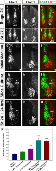
Figure 1. a-c. Status of generation of LMC (Foxp1 staining in b) and LMCl (Lhx-1 staining in a) at stage 24. c is a merge of the two channels. The midline is shown as a dotted line in a and D, V shows the orientation of the dorsoventral (D, V) axis of the spinal cord. d-f. Status of LMC (Foxp1 staining in d) and LMCl (Lhx-1 staining in e) organization at stage 27. f is a merge of the two channels. g-i. Lhx-1 (g) and Foxp1 (h) expression in sections of a slice cultured in a medium that supports cranial motor neuron cell survival (Guthrie medium, our "initial medium" see reference 20). Lhx-1 is no longer expressed in motor neurons, although there is some survival of LMC cells evidenced by Foxp1 expression. D, V in (g) shows the orientation of the dorsoventral (D, V) axis of the spinal cord. M-L shows the orientation of the mediolateral (M, L) axis of the spinal cord j-l. LMCl cells (Lhx-1 in the ventral horn in j) and the LMC in general (Foxp1 in k) can be observed in slices cultured in medium containing 4% chick serum and 100 ng/ml CNTF. Note that some LMCl cells are found in a lateral position in the ventral horn. The arrow in (j) shows the lateral position of some of the the LMCl cells. (l) is a merge of the two channels. m-o. Status of expression of Lhx-1 (m) and Foxp1 (n) in sections of an embryo kept in ovo during the slice culture of the embryo shown in j-l. (o) is a merge of the two channels. p. Quantitation of the percentage of LMCl (Foxp1+ve/ Lhx1+ve) cells versus LMC (Foxp1+ve/Lhx1-ve) cells found in slice cultures in different conditions compared to control embryos kept in ovo (which represent 100%). Student's t-test *** represents p<0.01. Click here to view larger figure.
Discussion
The protocol described here allows a chicken embryo slice to be incubated for more than one day whilst keeping motor neurons alive and continuing to migrate during the culture period. In elucidating the conditions to keep motor neurons alive, we have identified several key features required. It seems that up to 4% chick serum is critical to the survival of motor neurons and its replacement with serum from a different species is not tolerated. Interestingly, we found that the presence of higher concentrations of serum were detrimental to the slices as they promoted overgrowth of the tissue leading to gross distortions in the slice shape. More importantly, the presence of cilliary neurotrophic factor also proved critical. It remains a distinct possibility that the slices may be able to be cultured for considerably longer periods than just 24 hr, perhaps even allowing the visualization of sensory afferent input to the spinal cord. Additionally, the culture conditions here may also prove useful to slice cultures of the developing brainstem and midbrain/ cerebellum.
Perhaps the biggest potential benefit in the use of these slice culture conditions is that they facilitate the possibility for pharmacological manipulation of protein function whilst minimizing the potential for adverse effects on the rest of the embryo, such as the heart. A large number of inhibitors and agonists of different signalling pathways can be used to assess the migration of different spinal neuron subtypes and, potentially, their effect of the formation of local spinal circuits during development. Additionally, some genetic perturbations of protein expression, such as those that make use of siRNAs and morpholino oligonucleotides, suffer from the fact that they can only be introduced early in development (owing to limitations in in ovo electroporation procedures) and the effects only last for a short period of time (typically one day). Our slice culture is started at later stages and affords the possibility to use this technology later in development at the slicing stage to view their effects during the period of culture of the slice.
The slice culture protocol could also allow the visualization of both spinal motor neuron as well as dorsal interneuron migration in real time via time-lapse video microscopy. This could be achieved using phase contrast microscopy under white light or through the use of fluorescence imaging. If the latter technique were to be used then the introduction of fluorescent labels is needed. This could be achieved either by introduction of fluorescent dyes such as DiI or DiO via either retrograde labelling from the limb or anterograde labelling from the ventricle or in ovo electroporation of constructs to drive fluorescent proteins in a subset of neurons.
Disclosures
None of the authors have competing interests or conflicts of interest.
Acknowledgements
We would like to thank Sharon Boast for many insightful discussions and B. Novitch for the kind gift of the antibodies to Foxp1. This work as funded by a studentship from the Wellcome Trust to KCT and a project grant from the Wellcome Trust to SRP (Grant reference number 094399/B/10/Z).
Materials
| Name | Company | Catalog Number | Comments |
|---|---|---|---|
| Eggs | Henry Stewart Farms, UK | Fertilised Brown Bovan Gold Hen's eggs (Henry Stewart Farms, UK), stored at 14 °C until incubation | |
| 21 Gauge needle | BD-Microlance-3 | 304432 | |
| 5 ml syringe | BD Plastipak | 302187 | |
| #5 Dumont forceps | Roboz | RS 5045 | |
| Micro Dissecting scissors | Roboz | RS 5910SC | (3.5 in straight) |
| Embryo embedding moulds: Peel away moulds | Agar Scientific | G3764 | Pyramidal shaped 22 mm to 12 mm |
| Super glue | Power Flex, Loctite | ||
| Ethanol | SIGMA | 32221 | |
| Sodium di-hydrogen Phosphate, mono-hydrate | BDH | 102454R | |
| Di Sodium mono-hydrogen Phosphate | VWR/ BDH | 102494C | |
| Sodium Chloride | VWR/BDH | 27810.295 | |
| Low melting temperature agarose | Invitrogen | 16520 | |
| Fetal Bovine serum | Gibco | 10500-064 | |
| Triton X-100 | SIGMA | T8787 | |
| Sodium Hydroxide | FLUKA | 71689 | |
| Paraformaldehyde | VWR/BDH | 28794.295 | |
| Plastic disposable pasteur pipettes | ElKay Precision laboratory consumables | 127-P503-000 | Liquipette 3 ml volume |
| Hanks Balanced Salt Solution (HBSS) | Gibco | 14170 | |
| Sucrose | SIGMA | S0389 | |
| Sodium Hydrogen Carbonate | SIGMA | S5761 | |
| Chicken Embryo Extract | Seralabs | CE650-J | |
| Penicillin/ Streptomycin solution | Gibco | 15070 | |
| L-Glutamine solution | Gibco | 25030 | |
| 2-mercapt–thanol | Gibco | 21985 | |
| Horse serum | Southern Group Labs | 9028B | |
| B27 supplement | Invitrogen | 17504-044 | |
| Neurobasal medium | Gibco | Cat ~ 2103 | |
| CilliaryNeurotrophic factor (CNTF) | R and D Systems | 557-NT-010 | |
| Chick Serum | SIGMA | C5405 | |
| Primary antibodies | were purchased from Developmental Studies Hybridoma Bank, with the exception of anti-foxP1 antibodies, which were a kind gift from Bennett Novitch of UCLA. Primary antibodies dilutions were Rabbit anti-FoxP1 (1/32000), Mouse anti-Lhx1/2 (4F2) (1/50), Guinea Pig anti-FoxP1 (1/1000). All antibodies were stored at 4 °C. | ||
| Secondary antibodies | Donkey anti-Guinea Pig (cy3) (1:1000) purchased from Jackson Immunoreseach Laboratories Inc. Donkey anti-Mouse (Alexa Fluor 488) (1:1000), Donkey anti-Rabbit (Alexa Fluor 594) (1:1000) purchased from Invitrogen All antibodies were stored at 4 °C. | ||
| 24-well plate | Corning COSTAR | 3473 | ultralow attachment 24-well plate |
| O.C.T. | Tissue Tek, Sakura | 4583 | Optimum Cutting Temperature |
| Vectashield | Vector Labs | H1000 | Vectashield fluorescent mounting medium |
| Slides | Thermo Scientific | Superfrost-Plus positively-charged slides, 25 x 75 x 1.0 mm | |
| Cover Glass | VWR International | 631-0167 | 25x60 mm |
| Forced draft incubator: | Lyon Electric Company | kept humid by ensuring water levels always sufficient, maintained at 38 °C | |
| Cryostat | Brite, Huntingdon, UK | -36 °C | |
| Tissue Culture Incubator | DH Autoflow | Nuaire 38 °C, 5% CO2 | |
| Vibratome | Leica | Leica VT1000-S | |
| Microscope | Nikon | Nikon Eclipse E80i fluorescence microscope | |
| Digital Camera | Nikon | Nikon DS5M and Hamammatsu ORCA ER |
References
- Jessell, T. M. Neuronal specification in the spinal cord: Inductive signals and transcriptional codes. Nature Reviews Genetics. 1 (1), 20-29 (2000).
- Landmesser, L. Distribution Of Motoneurons Supplying Chick Hind Limb Muscles. Journal of Physiology-London. 284, 371-389 (1978).
- Romanes, G. J. The motor pools of the spinal cord. Progress in Brain Research. 11, 93-119 (1964).
- Dasen, J. S., De Camilli, A., Wang, B., Tucker, P. W., Jessell, T. M. Hox repertoires for motor neuron diversity and connectivity gated by a single accessory factor, FoxP1. Cell. 134 (2), 304-316 (2008).
- Rousso, D. L., Gaber, Z. B., Wellik, D., Morrisey, E. E., Novitch, B. G. Coordinated actions of the forkhead protein Foxp1 and Hox proteins in the columnar organization of spinal motor neurons. Neuron. 59 (2), 226-240 (2008).
- Tsuchida, T., Ensini, M., et al. Topographic organization of embryonic motor-neurons defined by expression of limhomeobox genes. Cell. 79 (6), 957-970 (1994).
- Kania, A., Johnson, R. L., Jessell, T. M. Coordinate roles for LIM homeobox genes in directing the dorsoventral trajectory of motor axons in the vertebrate limb. Cell. 102 (2), 161-173 (2000).
- Price, S. R., Garcia, N. V. D., Ranscht, B., Jessell, T. M. Regulation of motor neuron pool sorting by differential expression of type II cadherins. Cell. 109 (2), 205-216 (2002).
- Demireva, E. Y., Shapiro, L. S., Jessell, T. M., Zampieri, N. Motor Neuron Position and Topographic Order Imposed by β- and γ-Catenin Activities. Cell. 147 (3), 641-652 (2011).
- Bello, S. M., Millo, H., Rajebhosale, M., Price, S. R. Catenin-Dependent Cadherin Function Drives Divisional Segregation of Spinal Motor Neurons. Journal of Neuroscience. 32 (2), 490-505 (2012).
- Haase, G., Dessaud, E., et al. GDNF acts through PEA3 to regulate cell body positioning and muscle innervation of specific motor neuron pools. Neuron. 35 (5), 893-905 (2002).
- Hotary, K. B., Landmesser, L. T., Tosney, K. W. Embryo slices. Methods in Cell Biology. 51 (51), 109-124 (1996).
- Hotary, K. B., Tosney, K. W. Cellular interactions that guide sensory and motor neurites identified in an embryo slice preparation. Developmental Biology. 176 (1), 22-35 (1996).
- Krull, C. E., Tosney, K. Embryo slices and strips: Guidance and adhesion assays in the avian embryo. Methods in Cell Biology: New Methods in Avian Embryology. Bronner-Fraser, M. 87, 2nd Edition, Elsevier Academic Press Inc. San Diego. 97-113 (2008).
- Hamburger, V., Hamilton, H. L. A series of normal stages in the development of the chick embryo. (Reprinted From Journal Of Morphology, Vol. 88, 1951). Developmental Dynamics. 195 (4), 231-272 (1992).
- Rakic, P. A century of progress in corticoneurogenesis: From silver impregnation to genetic engineering. Cerebral Cortex. 16, I3-I17 (2006).
- Bingham, S. M., Sittaramane, V., Mapp, O., Patil, S., Prince, V. E., Chandrasekhar, A. Multiple mechanisms mediate motor neuron migration in the zebrafish hindbrain. Dev. Neurobiol. 70 (2), 87-99 (2010).
- Brachmann, I., Jakubick, V. C., Shaked, M., Unsicker, K., Tucker, K. L. A simple slice culture system for the imaging of nerve development in embryonic mouse. Developmental Dynamics. 236 (12), 3514-3523 (2007).
- Das, R. M., Wilcock, A. C., Swedlow, J. R., Storey, K. G. High-resolution Live Imaging of Cell Behavior in the Developing Neuroepithelium. J. Vis. Exp. (62), e3920(2012).
- Barnes, S. H., Price, S. R., Wentzel, C., Guthrie, S. C. Cadherin-7 and cadherin-6B differentially regulate the growth, branching and guidance of cranial motor axons. Development. 137 (5), 805-814 (2010).
Reprints and Permissions
