We describe here a method to isolate and purify dendritic cells from different anatomical compartments in the human female reproductive tract for the evaluation of their phenotypical and functional characteristics. This method can be adapted to isolate other immune cells or dendritic cells from other mucosal tissues.
Method Article
Isolation of Dendritic Cells from the Human Female Reproductive Tract for Phenotypical and Functional Studies
In This Article
Summary
Abstract
The characterization of the human dendritic cells (DCs) resident in mucosal tissues is challenging due to the difficulty in obtaining samples, and the low numbers of DCs present per tissue. Yet, as the phenotype and function of DCs is modified by the tissue environment, it is necessary to analyze tissue resident DC populations, since blood derived DCs incompletely reflect the complexities of DCs in tissues. Here we present a protocol to isolate DCs from the human female reproductive tract (FRT) using hysterectomy specimens that allows both phenotypical and functional analyses. The protocol consists of tissue digestion to generate a single cell mixed cell suspension, followed by positive magnetic bead selection. Our tissue digestion protocol does not cleave surface markers, which allows phenotypical and functional analysis of DCs in the steady state, without overnight incubation or cell activation. This protocol can be adapted for the isolation of other immune cell types or isolation of DCs from other tissues.
Introduction
The FRT has the dual function of protecting against pathogens while allowing implantation and pregnancy1. To accomplish this, the FRT is compartmentalized, with each anatomical region displaying unique histological, immunological, and functional characteristics1.
DCs present at mucosal surfaces make contact with microbes in the lung, the gut, and the genital tract, and provide immune surveillance for potential pathogens2. DCs have the unique ability to prime naïve T-cells and trigger adaptive immune responses3. DCs in the FRT are also specialized to tolerate foreign antigens, such as those found in sperm and the developing fetus, to allow successful pregnancy4. Therefore, depending on the location, the DC phenotype and function will be distinct. It is known that DCs are strongly influenced by the tissue environment, such that their number, phenotype, and functions are modified by the tissue environment in which they reside3. Therefore, to understand the role that FRT DCs play in infectious diseases, pregnancy, and cancer in the FRT, resident DCs need to be studied, since blood derived DC models are inadequate to address the regulatory complexities found in FRT tissues.
The characterization of human tissue resident DCs is challenging due to the low numbers of cells present in mucosal tissues and the difficulty obtaining human tissue samples. DCs have been studied in the FRT using immunohistochemistry5,6, which informs about the cell location within the tissue, but precludes functional studies, and is limited in the number of identifying cell markers that can be analyzed at once. Moreover, single cell isolation protocols for flow cytometric analysis have been developed7,8,9. Some of these protocols take advantage of the migratory capacity of DCs to isolate those cells that migrate. These methods usually require overnight incubations and selection of activated DCs, but do not allow for the study of DCs in the steady state.
Here, using hysterectomy specimens, we optimized a protocol to isolate DCs from different anatomical sites in the FRT, the endometrium (EM), the endocervix (CX), and the ectocervix (ECX), that enables both phenotypical and functional analyses. Using a non-proteolytic enzymatic digestion protocol, we can proceed immediately after tissue digestion to cell isolation and flow cytometric characterization without cell activation. Using multicolor flow cytometry and adapting functional studies for low numbers of cells, we can identify and characterize rare subsets of DCs in the different sites of the FRT.
Protocol
Studies involving human subjects were conducted according to the principles expressed in the Declaration of Helsinki. Studies were approved by Dartmouth College Institutional Review Board and the Committee for the Protection of Human Subjects (CPHS). Written informed consent was obtained before surgery from HIV-negative women undergoing hysterectomies at Dartmouth-Hitchcock Medical Center (Lebanon, NH). Trained pathologists selected tissue samples from the EM, CX, and ECX, free of pathological lesions and distant from the sites of pathology. Blood samples were obtained from volunteer healthy donors recruited at Dartmouth Hitchcock Medical Center. Blood donors were anonymous. Freshly isolated EM, CX, and ECX tissues from the operating room were transferred to Pathology for isolation and classification prior to transfer to our laboratory in separate, sterile polypropylene tubes.
1. Enzymatic Digestion of Tissues
NOTE: Use sterile materials and work in a biological safety cabinet.
- Prepare a modified Hank's Balanced Salt Solution (HBSS) using 1x HBSS with phenol red, 100 U/mL penicillin-streptomycin, 0.35 g/L NaHCO3, and 20 mM HEPES.
- Prepare the enzymatic cocktail (modified HBSS, 450 U/mL collagenase IV, 0.01% [wt/vol] deoxyribonuclease I, and 2 mg/mL D-glucose). Pass the solution through a sterile 0.22 µm filter. Ideally, prepare the digestion enzyme on the day of use, but it can be frozen at -20 °C for storage. Avoid repeated freezing and thawing cycles.
- Rinse the tissue with modified HBSS and place in a 100 x 15 mm2 Petri dish. Adjust the Petri dish size based on the amount of tissue available and the volume of enzyme cocktail used (see step 1.4.2 below).
NOTE: Tissues vary in size depending on the surgical sample obtained from the pathology, ranging from 1-10 g. - Physical processing with disposable scalpel and forceps:
- Add approximately 3 mL of digestion enzyme cocktail to the tissue to prevent drying while cutting.
- Use forceps to stabilize the tissue and mince with a scalpel to increase the surface area of the tissue exposed to the digestion enzyme cocktail. Cut the tissue into small pieces (< 2 mm on a side). Add sufficient digestion enzyme cocktail to cover all tissue pieces (5 mL/g of tissue). Place the lid on the Petri dish.
- Incubate at 37 °C with 5% CO2 on a rotator at approximately 80 rpm for 45 min. Ensure that the rotator speed thoroughly mixes the digestion enzyme cocktail without spilling or producing bubbles.
- Visualize the tissue preparation with an inverted light microscope with the 4X and 10X objectives.
NOTE: After digestion, small tissue fragments and a mixed cell suspension including visible epithelial glands should be present (see Figure 1A).
2. Physical Separation of Single Cells
- In a sterile environment, place a taut 250 µm mesh with holder into a 150 x 15 mm2 Petri dish. Ensure that the mesh is about 0.5 cm above the surface of the Petri dish.
- Wet the mesh with 2 mL of modified HBSS and transfer the digested tissue onto the mesh.
- Using the flat surface of a 10-mL syringe plunger, gently but firmly, grind the digested minced tissue through the mesh.
Caution: Grinding too aggressively will damage the mesh and increase cell mortality. This step will separate the epithelial cells and stromal cells (including immune cells) from the connective tissue and mucus. - Once the tissue fragments have been thoroughly dispersed, elevate the mesh and the holder 2-3 cm above the Petri dish and rinse with 3 mL of modified HBSS to recover additional cells.
- Collect the cell suspension. Place the 20 µm mesh with holder in a Petri dish and wet with 2 mL of modified HBSS. Pour the cell suspension through the mesh holding the mesh 2-3 cm above the Petri dish, and rinse with 6 mL of modified HBSS.
NOTE: This step will separate the epithelial sheets, which are retained on the top, from single cells that pass through the mesh. The flow-through is a mixed cell suspension, which includes immune cells and stromal fibroblasts. - Collect the mixed cell suspension and centrifuge at 500 x g for 10 min. Aspirate the liquid and resuspend the pellet in 4 mL of modified HBSS for density gradient centrifugation.
- Layer the mixed cell suspension over 3 mL of polysucrose solution and centrifuge at room temperature at 500 x g for 30 min with no brake. Collect the white band from the top of the polysucrose solution and wash with PBS.
NOTE: This fraction is the enriched mixed cell suspension, which is rich in immune cells and contains some stromal fibroblasts.
3. Dead Cell Removal
NOTE: If a large percentage of dead cells (> 20%) are present in the enriched mixed cell suspension after density gradient centrifugation, dead cell removal with magnetic beads is recommended. Dead cells need to be removed since they may interfere with the positive magnetic bead selection process.
- Count the cells using a hemocytometer on a light microscope with the 10X objective. Spin down all the cells in the enriched mixed cell suspension (500 x g, 10 min, 4 °C). Completely aspirate and discard the supernatant. Add 100 µL of dead cell removal beads per 1 x 10⁷ total cells (live and dead), according to the manufacturer's instructions. Incubate at room temperature for 15 min.
- Place a 30 µm filter on the magnetic column, apply the cell suspension and allow the bead-labeled cells to fall through the magnetic column.
- Rinse the column with dead cell removal buffer as recommended (3 mL). Collect the flow-through containing the live cells. The range of cell recovery after dead cell removal is presented in Figure 1B.
NOTE: These cells can be used to perform flow cytometry staining for phenotypical studies (section 5) or continue with DC isolation (section 4).
4. DC Purification by Positive Magnetic Bead Selection
- Following dead cell removal (step 3.2), spin the cells down, remove the supernatant, and resuspend the cell pellet in 80 µL magnetic selection buffer per 1 x 10⁷ cells (PBS, 0.5% heat-inactivated human AB serum (HS), 2 mM EDTA).
- For positive selection of DC populations, add CD1a or CD14 magnetic beads according to the manufacturer's instructions (20 µL magnetic beads per 1 x 10⁷ cells). Incubate for 15 min at 4°C.
- Wash the cells with the magnetic selection buffer, spin down, and remove buffer completely. Resuspend the cells in a minimum of 500 µL of magnetic selection buffer.
- Attach the column to the magnet and add a 30 µm filter on the top of the column to retain any remaining cell clumps. Rinse the filter and column with magnetic selection buffer as recommended.
- Apply the cell suspension to the column. Rinse 3 times with magnetic selection buffer.
- Transfer the column to a 15-mL tube, add 5 mL of magnetic selection buffer, and apply the plunger to release the selected cells from the column.
- To increase the purity, repeat steps 4.4-4.6 with the recovered cells using a new column. The range of cell recovery after magnetic bead selection is presented in Figure 1C.
5. Assessment of Cell Purity: Flow Cytometry and Microscopy
- Antibody staining for flow cytometry:
- Resuspend up to 1 x 10⁶ cells in 100 µL of PBS containing 20% heat-inactivated HS to block Fc receptors, and incubate for 10 min on ice.
- Add the desired fluorescently labeled antibodies and the reagent for identification of dead cells (for an example, see section 6.5). Set fluorescence minus one (FMO) controls to establish the gates, and use compensation beads to prepare single stain controls for compensation. Examples of antibodies used are given in Table 1. Incubate for 20 min at 4 °C, protected from light.
- Wash the cells with 2 mL of magnetic selection buffer.
NOTE: Presence of EDTA is important to avoid the formation of cell clumping for flow cytometric analysis. Spin down and discard the supernatant. - Fix the cells by adding 100 µL of 2% paraformaldehyde solution per tube.
- Analyze the samples in by a flow cytometer10,11.
- Spin the column (e.g., cytospin) and use Giemsa staining to analyze morphology:
- Resuspend the cells in 200 µL of magnetic selection buffer.
- Load in a spin column and spin for 5 min. Let the slide dry completely.
- Fix the slide by immersion in 100% methanol for 2 min. Rinse the slide with deionized water and allow it to air dry.
- Perform a standard Wrights-Giemsa staining. Cover the cells with Eosin and incubate for 10 min, rinse with deionized water, and let it dry. Cover the cells with Wrights-Geimsa stain, incubate for 1 min, rinse with deionized water, and air dry.
- Examine the preparations with an inverted microscope (Figure 2A, 40X objective).
6. Allogeneic Stimulation Assay to Assess DC Cell Function
- Thaw the frozen peripheral blood mononuclear cells (PBMCs)
- Warm the cell culture media with 10% HS to 37 °C.
- Thaw the PBMCs by placing the cryotube in a 37 °C water bath, until a small amount of frozen cell media remains in the cryotube. In a sterile environment, transfer the contents of the cryotube to 10 mL of pre-warmed cell culture media with 10% HS in a 15-mL tube.
- Centrifuge at ~ 500 x g for 7 min. Remove the media, disperse the pellet, resuspend the PBMCs in 10 mL of cell culture media with 10% HS, and centrifuge the cells.
- Repeat the wash step a third time. Count the cells.
- Naïve T-cell selection:
- Use a negative selection magnetic antibody cell selection kit to isolate naïve T-cells according to the manufacturer's protocol.
- After isolation, check the purity using CD45RA and CCR7 antibodies.
NOTE: Typical purity is > 99% naïve T-cells.
- Naïve T-cell proliferation dye staining:
- Prepare 600 µL of 2x solution of cell proliferation dye, protecting the dye from exposure to light.
- Wash the naïve T-cells 2x with PBS. Resuspend cells in 500 µL PBS.
- While gently vortexing the cells in the biosafety cabinet, add 500 µL of proliferation dye to the naïve T-cells.
- Incubate the cells for 10 min at 37°C.
- Stop the cell labeling by adding 5 mL of cell culture media with 10% HS to the cells. Incubate on ice for 5 min, protected from light.
- Centrifuge the cells at ~ 500 x g for 7 min, aspirate the media, and resuspend the cells in 4 °C of cell culture media with 10% HS. Repeat two more times for a total of 3 washes. Take an aliquot of cells to count prior to the last centrifugation.
NOTE: Typically, final naïve T-cell numbers will range between 5% and 15% of the initial PBMC cell count. - Resuspend the cells with the cell culture media with 10% HS at the desired cell concentration.
- DC-allogeneic naïve T-cell co-culture:
- Plate the isolated DCs and naïve T-cells in a 96-well, round-bottom plate, at a 1:15 ratio of DCs to T-cells. Ensure that the optimal number for DCs ranges between 3,000 and 5,000 cells/well.
- Centrifuge the plate at ~ 500 x g for 3 min to ensure the cells are located at the center of the well. Cell-to-cell contact is important for proper signaling to occur.
- Incubate at 37 °C for 6 days. Monitor the cluster of cells, which appear in the wells as proliferation occurs, with microscopy (Figure 3A).
- Flow cytometry staining to assess proliferation:
- After 6 days, cells can be examined by flow cytometry. Stain the co-culture for flow cytometry.
- Prepare a 2x solution of yellow viability dye (maximum emission 572 nm) in PBS.
NOTE: The use of PBS without serum is important, as serum proteins interfere with the dye). - Centrifuge the plate with cells at ~ 500 x g for 5 min.
- Remove 100 µL of supernatant.
NOTE: At this point collect and store the supernatant for separate analysis of soluble factors. - Wash the cells two times with PBS: add 150 µL/well of PBS, centrifuge at ~ 500 x g for 5 min, remove 150 µL of supernatant with a pipette. Ensure that there is 50 µL of supernatant remaining in each well.
- Add 50 µL of 2x viability dye to each well. Incubate at room temperature for 10 min, protected from light.
- During the incubation, prepare a master mixture of desired antibody markers. For example: CD3 APC/Cy7, CD4 PE, CD8 FITC, etc.
- Add the antibody mixture to each well. Incubate at room temperature for 10 min, protected from light.
- Wash the cells 2x with PBS + 2% HS: add 150 µL/well of PBS + 2% HS. Centrifuge at ~ 500 x g for 5 min. Remove 150 µL of supernatant with a pipette.
- Fix the cells by adding 100 µL/well of 2% para-formaldehyde. If needed: transfer the cells and media to polypropylene flow cytometry tubes. Tubes can be kept at 4 °C and protected from the light, until the flow cytometric analysis.
- Read tubes in a flow cytometer.
Results
Following tissue digestion, the release of epithelial sheets and glands can be observed, as shown in Figure 1A; this is a positive control that indicates the enzymatic digestion was successful. The number of total viable cells and DCs recovered per gram of tissue are also shown in Figure 1B and Figure 1C, respectively. Immune cells represent between 5-30% of the stromal cells prior to density centrifugation12,13. Figure 2 shows the morphology (Figure 2A) and purity (Figure 2B, C) of the isolated DCs. When two rounds of selection are performed as indicated in the protocol, the expected purity ranges between 85-92% (Figure 2C). The number of recovered cells is variable and depends on the initial tissue size and donor variability. Characterization of the isolated cells have been described13. Figure 3 shows a representative allogeneic stimulation assay to assess DC functionality. Figure 3A shows the characteristic T-cell clumps that appear when proliferation occur, and Figure 3B shows the proliferation quantification by flow cytometry. Other functional assays, such as viral capture or cytokine and chemokine production can also be performed13. Figure 4 shows the gating strategy to identify different DCs populations in the distinct FRT anatomical compartments after flow-cytometric analysis of enriched mixed cell suspensions (Figure 4A). The titration of antibodies and negative controls are important, as DCs and macrophages display high autofluorescence. FMO controls are shown in Figure 4B. Figure 4C shows examples of how to identify specific DC subsets, such as CD1c+, CD207+, or CD103+ DCs.
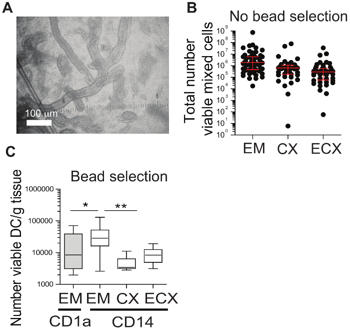
Figure 1: Tissue processing and cell availability. (A) Representative image of epithelial sheets and glands released after tissue digestion. (B) Range of total number of viable cells recovered after tissue processing and dead cell removal in each FRT compartment: endometrium (EM), endocervix (CX), and ectocervix (ECX). Every dot represents cells from a different subject. (C) Number of DCs recovered per gram of tissue after magnetic bead isolation. B and C are adapted from13. Please click here to view a larger version of this figure.
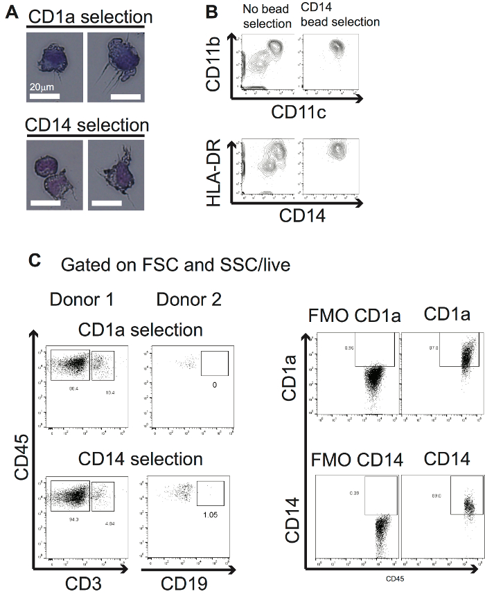
Figure 2: Morphology and purity of the isolated cells. (A) Dendritic morphology of the isolated cells determined by microscopy following Giemsa staining. (B) Expression of phenotypic markers before and after bead isolation, as determined by flow cytometry. (C) Representative purity obtained after CD1a+ and CD14+ bead isolation. Adapted from13. Please click here to view a larger version of this figure.
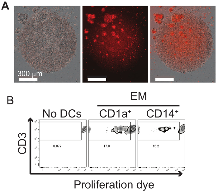
Figure 3: Proliferation assay. (A) Representative microscopy images of T-cell cluster formation during proliferation: phase-contrast (left), proliferation dye staining (middle), and overlay (right). (B) Representative example of induction of proliferation after co-culture of isolated endometrial CD1a+ or CD14+ DCs and allogeneic naïve T-cells obtained from frozen PBMCs. B is adapted from13. Please click here to view a larger version of this figure.
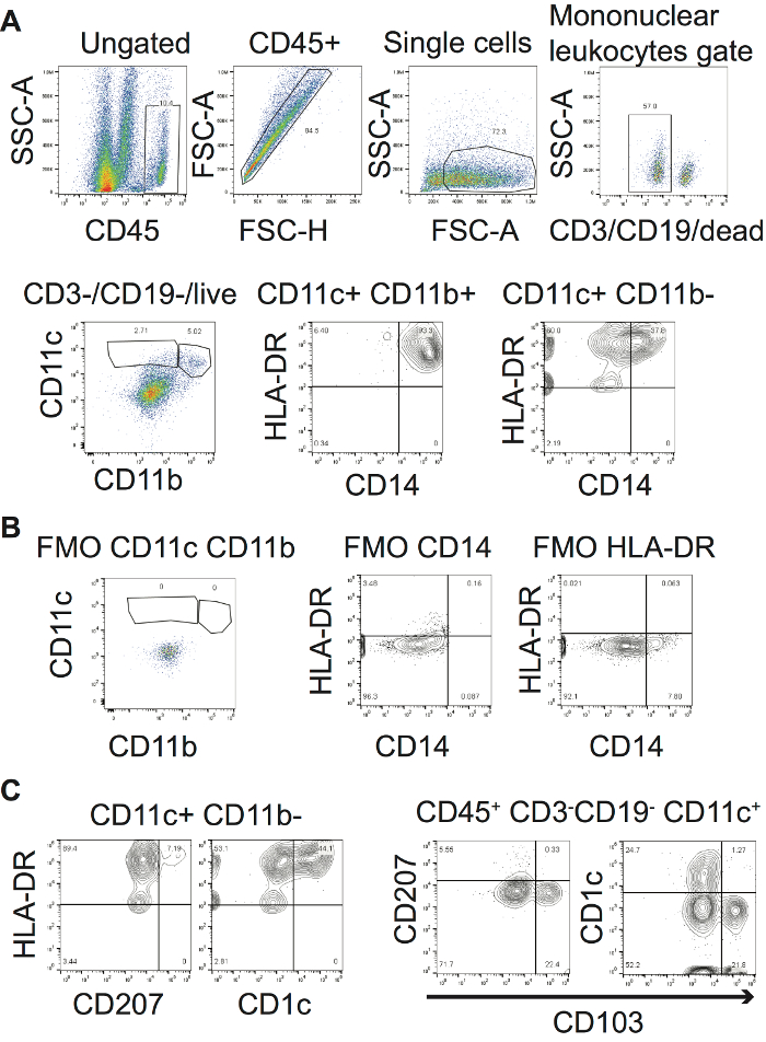
Figure 4: Phenotypical characterization of DCs in the enriched mixed cell suspensions from the FRT. (A) Gating strategy for the identification of DCs in the mixed cell suspension. Each gated population in multicolor plots is shown in the next panel. Contour plots show the CD11c+ CD11b+ and CD11c+ CD11b- gates, respectively. Markers that identify unwanted cells can be stained using the same channel to exclude them from the analysis; for example, dead cells, CD3+, and CD19+ cells. (B) FMO controls to establish appropriate gates. (C) Discrimination of DC subsets following the gating strategy. Contour plots were chosen instead of multicolor plots to more accurately represent the population distribution when the cell numbers were low10. Low cell numbers are expected at the end of the gating strategy, as tissue DCs are rare populations. Adapted from13. Please click here to view a larger version of this figure.
| Cell marker | Fluorochrome | Clone | Concentration | Amount of antibody per sample |
| (in 100 µl of buffer) | ||||
| CD45 | vioblue450 | HI30 | 1.0 µg/5µl | 0.4 µg |
| CD11b | PE | ICRF44 | 1.0 µg/5µl | 0.4 µg |
| CD11c | PerCp/Cy5.5 | Bu15 | 200 µg/ml | 0.4 µg |
| CCR5 | PE-Cy7 | 2D7 | 0.25 µg/5µl | 0.1 µg |
| CCR7 | PE-Cy7 | REA108 | 99 µg/ml | 0.2 µg |
| HLA-DR | BV-570 | L243 | 100 µg/ml | 0.2 µg |
| HLA-DR | FITC | AC122 | 82.5 µg/ml | 0.16 µg |
| CD3 | APC | UCHT1 | 16 µg/ml | 0.03 µg |
| CD3 | viogreen | REA614 | 137 µg/ml | 0.27 µg |
| CD163 | APC | GHI/61 | 80 µg/ml | 0.16 µg |
| CD207 | APC | 1OE2 | 200 µg/ml | 0.4 µg |
| CD1a | FITC | HI149 | 0.5 µg/5µl | 0.2 µg |
| CD1a | AF-700 | HI149 | 0.5 mg/ml | 1.0 µg |
| CD103 | PE-Cy7 | B-Ly7 | 0.25 µg/5µl | 0.1 µg |
| CD83 | PE | HB15e | 0.25 µg/5µl | 0.1 µg |
| CD14 | APC-Fire | 63D3 | 400 µg/ml | 0.8 µg |
| CD209 | FITC, PE, APC | 120507 | 1.25 µg/0.25ml | 0.01 µg |
| Zombie yellow viability dye | ||||
| 7AAD | 0.1mg/ml | 0.2ug |
Table 1: Example of antibodies for the characterization of DCs in the FRT by flow cytometry.
Discussion
Mucosal DCs are a rare cell population strongly influenced by the tissue environment, which changes their phenotype and function once they enter the tissues3. While blood derived DCs are a very useful model, they do not fully represent the diversity of DC populations found in tissues. Therefore, to understand the unique characteristics of mucosal DCs, isolation of primary cells from tissues is necessary. Isolation of DCs from different anatomical sites in the FRT offers the opportunity to study DC function in vitro and compare between DC subsets and anatomical locations.
Here we provide a protocol that allows the purification of DCs from human FRT tissues for in vitro assessment of functional characteristics. Since DCs are found at mucosal sites in low numbers, we developed a protocol to enrich the tissue cell preparation by density gradient centrifugation and dead cell removal prior to cell isolation with magnetic beads. Proper removal of dead cells is key, as they will interfere with bead isolation. This technique provides high purity of positively isolated cells (≈ 90%), in a relatively short amount of time (4-6 h), and without the need for overnight incubation, in vitro cell culture, or migration prior to isolation, each of which can activate DCs and alter their phenotypic characteristics. Our protocol does not trigger cell activation, as measured by the lack of up-regulation of maturation markers (CD86, HLA-DR, CD83)13. Another advantage is the use of a non-proteolytic enzymatic digestion, which does not cleave surface markers and allows for immediate cell isolation or phenotypical analysis following tissue digestion14.
Critical to this protocol is the generation of a single cell suspension to avoid the blockage of the magnetic columns. To ensure this, dead cells must be removed prior to the magnetic cell selection, filters need to be used on top of the magnetic columns, and tissues larger than 3 g should be divided between two columns. The use of a second column after the first round of purification is key to high cell purity.
One limitation of the isolation method is the inherent low number of DCs in FRT tissues, which usually does not allow for good cell recovery of tissues smaller than 1 g. Under these circumstances, however, phenotypical analyses can still be performed using the enriched mixed cell suspension. This is possible due to the non-proteolytic digestion method used, which does not cleave surface markers and allows for immediate analysis of cell phenotype after tissue digestion14. Additionally, the patient age can be a factor that influences the number of cells recovered.
Using this protocol, we have previously demonstrated that CD1a+ isolation provides a phenotypically more homogeneous population than CD14+ selection13; however, CD1a+ DCs are difficult to recover from CX and ECX. While CD14 can also be expressed on macrophages, we found that the FRT CD14+ isolated population is rich in DCs, based on co-expression of DC markers and their ability to stimulate allogeneic naïve T-cells, which is a hallmark function of DCs13.
Since the number of DCs recovered is generally low (< 100,000 total cells), functional studies need to be optimized for low cell numbers. Here we show the optimization of an allogeneic stimulation assay, which can be performed with only 3,000 DCs/well. We have carried out other function studies with these cells, including hormonal responsiveness, cytokine, and antimicrobial peptide production upon HIV-stimulation and HIV-capture by FRT DCs13. Once the DCs are isolated and plated, assays should be performed within 24 h, as cell viability decreases after that time.
This protocol can be adapted to isolate different DC subsets, or other cell types by selecting the proper markers coupled to the magnetic beads and combining sequential positive and negative bead selection to remove unwanted cell types. One caution, though, is that with every round of selection some percentage of cells will be lost, so for the isolation of FRT DCs, which are already rare, and the tissue size is generally small, multiple rounds of selection with different markers (and the necessary washes associated) are not desirable. Moreover, this protocol can be adapted to isolate other immune cells or DCs from other tissues14,15.
Disclosures
The authors have nothing to disclose
Acknowledgements
Study supported by NIH grants AI102838 and AI117739 (CRW). We thank Richard Rossoll for technical assistance. Flow cytometric analysis was carried out in DartLab, the Shared Resource at Dartmouthsupported by (P30CA023108-37) and (P30GM103415-15).
Materials
| Name | Company | Catalog Number | Comments |
|---|---|---|---|
| Hank's Balanced Salt Solution (HBSS) | Hyclone | SH30015.03 | |
| Penicillin-streptomycin | Hyclone | SV30010 | |
| HEPES (1M) | Hyclone | 15630-080 | |
| Collagenase IV | Sigma | C5138 | |
| Deoxyribonuclease I | Worthington Biochemical | LS002140 | |
| D-glucose | Sigma Aldritch | 50-99-7 | |
| 0.22 um Stericup 500 mL filter | Millipore | SCGPU05RE | |
| 100mm x 15mm polystyrene petri dish | Fisherbrand | FB0875712 | |
| 150mm x 15mm polystyrene petri dish | Fisherbrand | FB0875714 | |
| 150mm x 25mm polystyrene dish | Corning | 430599 | Treated cell culture dish |
| Isotemp Incubator | Fisher Scientific | FICO3500TABB | 5.0% CO2 |
| American Rotator V | American DADE | R4140 | |
| 250 um nylon mesh | Sefar | 03-250/50 | |
| 20 um nylon mesh | Sefar | 03-20/14 | |
| Beckman GS-6R Centrifuge | Beckman | 358702 | |
| X-VIVO 15 with Gentamicin L-Gln, Phenol Red, 1 L | Lonza | 04-418Q | |
| Human AB Serum | Valley Biomedical | HP1022 | |
| Histopaque-1077 | Sigma Aldritch | 10771 | |
| Phosphate Buffer Solution (PBS) | National Diagnostics | CL-253 | pH 7.4 |
| Dead Cell Removal Kit | Miltenyi Biotec | 130-090-101 | |
| Pre-separation filter (30um) | Miltenyi Biotec | 130-041-407 | |
| LS column | Miltenyi Biotec | 130-042-410 | |
| Quadro MACS Separator | Miltenyi Biotec | 130-090-976 | |
| MACS multi-stand | Miltenyi Biotec | 130-042-303 | |
| EDTA | USB | 15694 | |
| CD1a Microbeads, human | Miltenyi Biotec | 130-051-001 | |
| CD14 Microbeads, human | Miltenyi Biotec | 130-050-201 | |
| eFluor 670 cell proliferation dye | eBiosciences | 65-0840-85 | |
| 96 well round bottom plate | Falcon | 9/8/2866 | |
| Zombie yellow viability dye | Biolegend | 423104 | |
| CD3 APC/Cy7, anti-human | Tonbo Biosciences | 25-0038-T100 | |
| CD4 PE, anti-human | eBiosciences | 12-0048-42 | |
| CD8a FITC, anti-human | Tonbo Biosciences | 35-0086-T100 | |
| Gallios Flow Cytometer | Beckman Coulter Life Sciences | B43618 | 10 color, VBR |
| MACSquant Analyzer 10 | Miltenyi Biotec | 130-096-343 | 8 color, VBR |
References
- Wira, C. R., Rodriguez-Garcia, M., Patel, M. V. The role of sex hormones in immune protection of the female reproductive tract. Nat Rev Immunol. 15 (4), 217-230 (2015).
- Banchereau, J., Steinman, R. M. Dendritic cells and the control of immunity. Nature. 392 (6673), 245-252 (1998).
- Schlitzer, A., McGovern, N., Ginhoux, F. Dendritic cells and monocyte-derived cells: Two complementary and integrated functional systems. Semin Cell Dev Biol. 41, 9-22 (2015).
- Tagliani, E., Erlebacher, A. Dendritic cell function at the maternal-fetal interface. Expert Rev Clin Immunol. 7 (5), 593-602 (2011).
- Kaldensjo, T., et al. Detection of intraepithelial and stromal Langerin and CCR5 positive cells in the human endometrium: potential targets for HIV infection. PLoS One. 6 (6), e21344(2011).
- Schulke, L., Manconi, F., Markham, R., Fraser, I. S. Endometrial dendritic cell populations during the normal menstrual cycle. Hum Reprod. 23 (7), 1574-1580 (2008).
- Ballweber, L., et al. Vaginal langerhans cells nonproductively transporting HIV-1 mediate infection of T cells. J Virol. 85 (24), 13443-13447 (2011).
- Hladik, F., et al. Initial events in establishing vaginal entry and infection by human immunodeficiency virus type-1. Immunity. 26 (2), 257-270 (2007).
- Duluc, D., et al. Functional diversity of human vaginal APC subsets in directing T-cell responses. Mucosal Immunol. 6 (3), 626-638 (2013).
- Herzenberg, L. A., Tung, J., Moore, W. A., Parks, D. R. Interpreting flow cytometry data: a guide for the perplexed. Nat Immunol. 7 (7), 681-685 (2006).
- Juno, J. A., Boily-Larouche, G., Lajoie, J., Fowke, K. R. Collection, isolation, and flow cytometric analysis of human endocervical samples. J Vis Exp. (89), (2014).
- Givan, A. L., et al. Flow cytometric analysis of leukocytes in the human female reproductive tract: comparison of fallopian tube, uterus, cervix, and vagina. Am J Reprod Immunol. 38 (5), 350-359 (1997).
- Rodriguez-Garcia, M., et al. Dendritic cells from the human female reproductive tract rapidly capture and respond to HIV. Mucosal Immunol. 10 (2), 531-544 (2017).
- Rodriguez-Garcia, M., Barr, F. D., Crist, S. G., Fahey, J. V., Wira, C. R. Phenotype and susceptibility to HIV infection of CD4+ Th17 cells in the human female reproductive tract. Mucosal Immunol. 7 (6), 1375-1385 (2014).
- Shen, Z., Rodriguez-Garcia, M., Ochsenbauer, C., Wira, C. R. Characterization of immune cells and infection by HIV in human ovarian tissues. Am J Reprod Immunol. , (2017).
Reprints and Permissions
