Acute myocardial infarction in mice induces acute but incompletely characterized changes in left ventricular (LV) function. LV catheterization in mice undergoing coronary artery occlusion serves as a novel method for a real-time evaluation of LV function.
Method Article
Real-time Pressure-volume Analysis of Acute Myocardial Infarction in Mice
In This Article
Summary
Abstract
Acute myocardial infarction can lead to acute heart failure and cardiogenic shock. The evaluation of hemodynamics is critical for the evaluation of any potential therapeutic approach directed against acute left ventricular (LV) dysfunction. Current imaging modalities (e.g., echocardiography and magnetic resonance imaging) have several limitations since data on LV pressure cannot directly be measured. LV catheterization in mice undergoing coronary artery occlusion could serve as a novel method for a real-time evaluation of LV function.
At the beginning of the procedure, mice were anesthetized followed by endotracheal intubation. For LV catheterization, the right carotid artery was exposed via middle-neck incision. The catheter was introduced and placed into the LV cavity. Left thoracotomy was conducted and the left main coronary artery (LCA) was ligated. To induce reperfusion, the suture was released after 45 min. Pressure-volume data was recorded at all times.
Ligation of the LCA caused a decrease in LV systolic function as evidenced by a 30% reduction in stroke volume, LV ejection fraction (EF), and cardiac output. Maximum dP/dt as a parameter for LV contractility was also significantly reduced and diastolic function was severely impaired (minimum dP/dt -40%). Reperfusion over a period of 20 min did not lead to a complete recovery of LV function.
Real-time pressure-volume analysis served as a valid procedure for monitoring cardiac function during acute myocardial infarction in mice. Maintaining stable anesthesia and a standardized surgical approach was crucial to ensure valid results. As the early phase of acute myocardial infarction is critical for morbidity and mortality, the delineated method could be beneficial for preclinical evaluation of new strategies for cardioprotection.
Introduction
Cardiovascular disease is the most common cause of death in western civilization1. Acute myocardial infarction is a critical event, which is associated with high acute and chronic mortality2. Even if revascularization is achieved via emergency percutaneous coronary intervention (PCI), mortality remains high, particularly within the first 48 h after onset of symptoms in patients with acute myocardial infarction3. Cardiogenic shock caused by acute reduction in left ventricular (LV) function is a major cause for in-hospital mortality in these patients3. This early reduction in LV function is caused by myocardial damage following ischemia and reperfusion. This so-called ischemia/reperfusion (I/R) injury is mediated by changes in cellular metabolome such as exaggerated generation of reactive oxygen species4,5.
To explore possible protective mechanisms leading to a decrease in myocardial damage in a preclinical setting, reliable mouse models are essential including methods for evaluation of post-I/R LV function6. In this setting, transthoracic echocardiography7 and magnetic resonance imaging (MRI)6 are widely used for functional phenotyping8,9. However, these methods are not suitable for the assessment of severe LV dysfunction and cardiogenic shock in an ongoing acute myocardial infarction and cannot directly show data on LV pressure. The Langendorff apparatus using isolated heart in an ex vivo assay provides information about the underlying pathomechanisms of early-phase I/R injury10. This method is limited due to its inability to reproduce in vivo adaptive mechanisms such as regulation of the autonomous nervous system or hormonal regulation and acid-base homeostasis. There is currently no method available for a complete functional phenotyping of cardiogenic shock and left ventricular dysfunction during an ongoing myocardial I/R injury.
A synchronized approach with combination of pressure-volume (PV) catheterization and transient surgical left main coronary artery (LCA) occlusion could be beneficial but technically challenging. Stable extracardiac hemodynamics during I/R injury are essential for valid results since unstable anesthesia or blood loss could heavily influence the results. A novel approach for hemodynamic phenotyping of I/R injury via LV PV catheterization and transient LCA occlusion could bring new insights on cardiogenic shock and LV dysfunction in acute myocardial infarction and serve as a method for future analysis on cardioprotection.
Protocol
All experiments were completed in accordance and compliance with all relevant regulations (‘European Convention for the Protection of Vertebrate Animals used for Experimental and other Scientific Purposes’ (Directive 2010/63/EU) and animal care was in accordance with institutional guidelines. All experiments have been performed with male C57BL/6JRj mice at the age of 6 months.
1. Preparation
- Prepare a surgical microscope and a heating pad as well as a rectal probe to monitor the body temperature. Clean and sterilize all surgical instruments.
- Prepare 2 pieces of 10 cm 5-0 silk thread for vessel ligation, 5 cm of 6-0 polypropylene thread, and 2 mm silicon tube for ligation of the LCA.
- Prepare the PV catheter calibration cuvette by pre-heating it to 37 °C and prepare a 100 µL Hamilton syringe filled with 15% sodium chloride (NaCl) in H2O for saline calibration.
- Place the PV catheter (3 cm, 1.4 F) in 37 °C pre-heated 0.9% NaCl in H2O (saline) at least 30 min before measurement. Connect the catheter to the data acquisition device and connect the device to an analog/digital converter. Connect both devices to a computer.
- Set up the software. Perform software-guided pressure calibration and conductance calibration as demanded by the software-guided workflow8.
2. Anesthesia and Analgesia
- Anesthetize the mouse using ketamine 100 mg/kg body weight and xylazine hydrochloride 10 mg/kg body weight by intraperitoneal injection. At the beginning of I/R surgery, administer 0.05 mg/kg body weight buprenorphine intraperitoneally to maintain analgesia.
- After 10 min, perform an endotracheal intubation using a 20 G intravenous (iv) catheter and ventilate the mouse with 40% oxygen (O2) and 2% (v) isoflurane. Set appropriate ventilation parameters (e.g., 220 µL stroke volume, 150/min for a 25 - 30 g C57BL/6JRj mouse).
- Continuously monitor body temperature via rectal probe. Fasten mouse on the heated plate with the head pointing towards the investigator. Mouse normal body temperature is 36.5 - 38 °C. Maintain body temperature within 1 degree by adjustment of heated plate temperature.
3. Left Ventricular Catheterization
- Disinfect chest and neck with three alternating scrubs of betadine and 70% alcohol. Wait for skin disinfectant to dry. Remove the chest hair by using a small animal shaving system.
- Perform a 10 mm longitudinal median incision 5 mm beneath the bottom lip towards the sternum using small surgical scissors.
- Dissect the left and right part of the submandibular gland via blunt preparation using a forceps. Separate muscle and fat tissue in the right paratracheal region to expose the right commune carotid artery. Mobilize and separate the vessel for a total length of 5 - 10 mm from connective tissue via careful blunt preparation alongside the vessel with a bent forceps.
NOTE: Avoid mechanical manipulation of the vagus nerve or the carotid body at all time as this can cause severe hypotension and bradycardia. - Pass the two prepared silk threads under the vessel. Ligate the distal vessel with a tight knot and place a loose knot on the proximal exposed region that still allows passage of the catheter.
- Fix the threads of the cranial (tight) knot next to the head of the mice to apply a light tension on the vessel as this will facilitate the introduction of the catheter. Place a hemostat vascular clamp on the proximal vessel proximal of the loose knot to reversibly block blood flow.
- Perform a wedge-shaped incision 1 mm proximal to the cranial knot to open the vessel with micro scissors.
NOTE: A small drop of blood will indicate proper execution of this step. - Insert the catheter carefully for 10 mm. Start recording of catheter data.
NOTE: Stretching the incision with a forceps can make this process easier. - Extract the vascular clamp. Add 1 - 2 drops of saline to the incision to facilitate catheter movement. Continue introducing the catheter for approximately another 10 mm. After passing the proximal knot with the sensor tip, fasten the knot carefully just enough to prevent blood reflux alongside the thinner parts of the catheter without impairing catheter movement.
NOTE: The size of the sensor at the tip of the catheter prevents reflux of blood when extracting the vascular clamp. - Gently continue inserting the catheter until pressure analysis shows arterial blood pressure profile indicating that the catheter is placed in the aorta (Figure 3A).
NOTE: The catheter reaching the aortic valve will be indicated by light resistance and pulse-synchronized motion of the catheter. - When experiencing resistance trying to advance through the aortic valve, pull the catheter back 5 mm and advance again until LV catheterization will be indicated in a change in PV analysis as diastolic pressure will reach 0 - 20 mmHg (Figure 3B). Note changes in volume monitoring to further confirm left ventricular placement of the sensor tip (Figure 3C). Fasten the proximal knot more tightly to prevent catheter movement.
4. Ischemia/Reperfusion Surgery
- Perform skin incision from caudal sternum towards the left axilla for a total length of 15 mm. Proceed with blunt preparation of the two muscle layers until the ribs can be visualized.
- Open the thorax via incision between the third and fourth left rib. Use surgical hooks to gain access to the pericardium. Resect the pericardium above the heart. Before continuing with LCA ligation, wait 30 s without touching the animal to record PV data for valid analysis.
- Localize the LCA emerging under the left auricle and descending at the left side of the heart towards the apex. Use a 6-0 polypropylene suture to encircle the artery with a loop 2 mm underneath the left auricle. Place a small silicon tube under the loop and place a tight knot above.
NOTE: Distal myocardium turning grey serves as positive control for LCA occlusion6. I/R surgery should be performed within 5 min independently from the operating investigator. - Cut the suture at 1 mm length. Release the surgical hooks and manually close the muscle layers above the incision. Wait 45 min while continuously recording PV data.
- After 45 min, re-open the incision and remove the silicon tube to induce reperfusion. Record data for another 20 min.
NOTE: A change to a red color as seen before ischemia indicates successful reperfusion.
5. Calibration
NOTE: The calibration of the PV catheter system consists of 4 mandatory steps, two of which have to be performed after the measurement. Calibration should be repeated after every experiment to ensure valid results.
- Perform pressure calibration and conductance calibration before the experiment as described in Step 1.5.
- Perform saline calibration when the catheter is still placed in the left ventricle after the experiment itself is finished. Localize the right jugular vein lateral of the carotid artery in the prepared area. Inject 10 µL 25% NaCl in H2O via a Hamilton syringe while recording data.
- Calculate the calibration using the acquisition software by highlighting the ascending phase in volume curve (Figure 5C). Repeat this process for a total of 3 times. Avoid loss of blood after syringe extraction by using a vascular clamp to rapidly compress the puncture.
- Perform volume calibration to calibrate volume data acquisition by analysis of standardized volumes. Obtain approximately 500 µL mouse blood from cardiac puncture with a slightly heparinized 1 mL syringe (e.g., 5 µL per 200 IE heparin). Pull back the PV catheter 10 - 15 mm to avoid damaging the catheter.
- Fill the obtained blood into the 37 °C pre-heated calibration cuvette (Figure 5A). Avoid bubbles as it may interfere with the results. Add the catheter tip into each well and record data. Obtain a standard curve by software-guided analysis (Figure 5B). Repeat the process for a total of 3 times.
- Euthanize mice by exsanguination or cervical dislocation while sufficient isoflurane anesthesia is maintained at all time.
6. Data Analysis
- After completing the calibration steps, perform software-guided data analysis. Therefore, highlight the appropriate section (at least ten cycles) within the Analyze section of the PV Workflow and perform baseline analysis. Exclude cycles with deviations due to ventilation or manipulation if necessary (Figure 3D).
- Perform PV baseline analysis before passage of the aortic valve (arterial pressure only), immediately before and after LCA occlusion. Proceed by conducting PV baseline analysis and in intervals of 5 min. while ischemia and after reperfusion. At the end of the experiment, perform analysis of pressure data after retraction of the catheter from the left ventricle (arterial pressure).
- Analyze at least 10 consecutive cycles to avoid sampling error. When experiencing strong interference of obtained values with the ventilation, transient interruption of the ventilation for a maximum of 5 s can be considered.
- Use the following parameters that are calculated in Baseline Analysis (Figure 3D) to characterize LV function:
- Stroke volume (µL)
- Ejection fraction: Stroke volume / end-diastolic volume (%)
- Cardiac output: Stroke volume * heart rate (µL/min)
- Cardiac index: Cardiac output / body surface area (µL/(min*cm²)
- Stroke work: Inner area of PV curve (mmHg*µL)
- Maximum pressure (Pmax); Mean pressure (Pmean)
- max dP/dt (mmHg/s) as a parameter of LV systolic function
- min dP/dt (mmHg/s) as a parameter for LV compliance
- Time constant of isovolumetric relaxation: Tau (ms)
Results
After LV catheterization, reversible LCA ligation was performed for 45 min followed by 10 min of reperfusion. PV data was recorded at all times (Figure 1).
Correct placement of the PV catheter was confirmed by obtaining the characteristic LV PV graph (Figure 2A). LV catheter placement showed the typical ventricular pressure range with a minimum of 0 - 20 mmHg whereas false placement of the PV-catheter in the aorta would have shown a typical arterial pressure curve with a minimum pressure of 30 - 60 mmHg (diastolic blood pressure) and a small excursion at the end of systole indicating the aortic valve closing (Figure 3B and 3C). Successful occlusion of the LCA was visually confirmed by blanching of the distal LV myocardium (Figure 2B).
After LCA occlusion, PV data was acquired in 5 min intervals. Pressure data analysis demonstrated no changes in maximum LV systolic pressure indicating preserved peripheral perfusion and stable anesthesia (Figure 4A). Analysis of LV volume revealed a significant decrease in both EF (52% vs. 40%, p = 0.008) and absolute stroke volume (Figure 4B and 4C). These changes happened within the early phase of ischemia and LV functional data remained unchanged in the later phase of ischemia. Maximum dP/dt as a parameter of LV contractility showed a 30% reduction in mice undergoing myocardial ischemia. Stroke work was 30% reduced (Figure 4D and 4E). As a parameter for diastolic function, minimum dP/dt was significantly decreased indicating impaired LV compliance (Figure 4F). Reperfusion by extraction of the silicon tube was visually validated. Reperfusion did not show significant changes in PV data analysis within a period of 20 min (Figure 4A-4D). Sham-operated animals did not show a significant reduction in LV systolic or diastolic parameters (Figure 4I-4J).
At the end of data acquisition, cuvette calibration and saline calibration were performed (Figure 5).
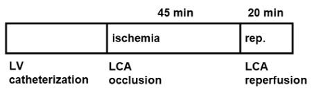
Figure 1: Scheme of the method. Sequence of left ventricular (LV) catheterization, left main coronary artery (LCA) occlusion and reperfusion.
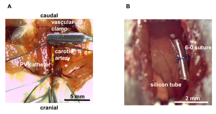
Figure 2: Surgical procedures. (A) Left ventricular catheterization placed via the right common carotid artery. (B) Left main coronary artery occlusion with polypropylene suture and silicon tube. Please click here to view a larger version of this figure.
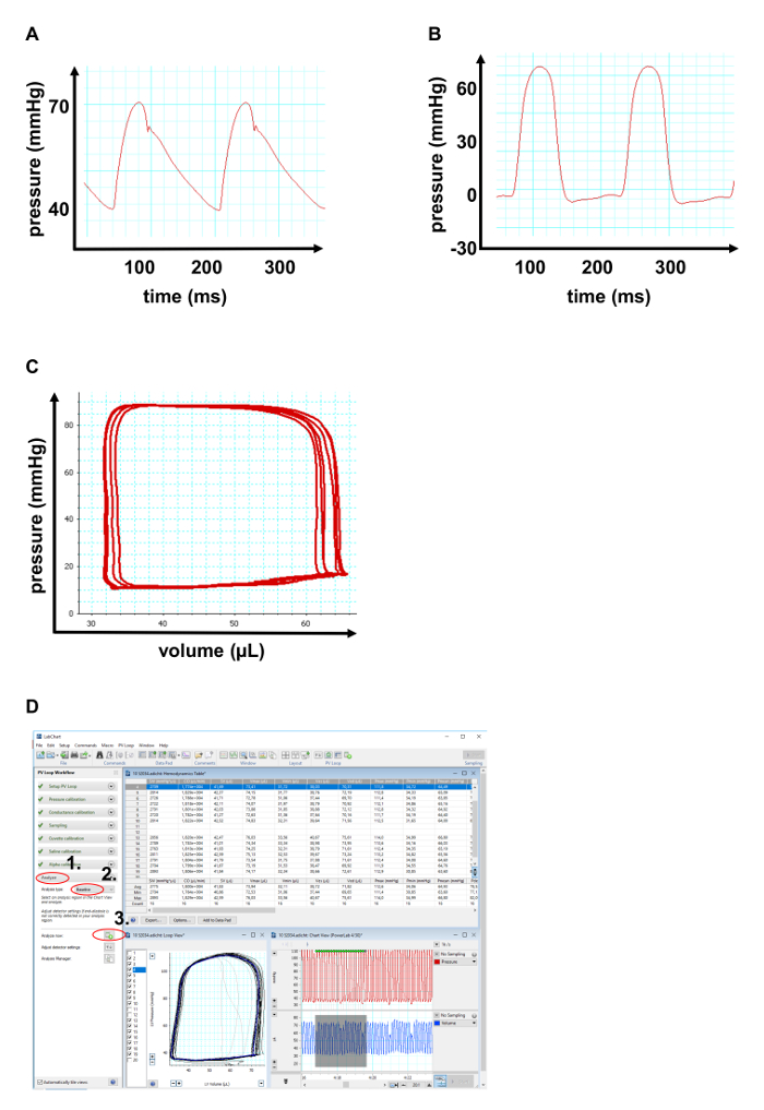
Figure 3: Representative pressure-volume data. (A) Representative arterial pressure indicated by a minimal pressure of >30 mmHg and a typical excursion at the end of systole indicating closing of the aortic valve. (B) Representative left ventricular pressure data showing diastolic values <20 mmHg. (C) Representative left ventricular pressure-volume diagram. (D) Screenshot of software-based PV baseline analysis. Please click here to view a larger version of this figure.
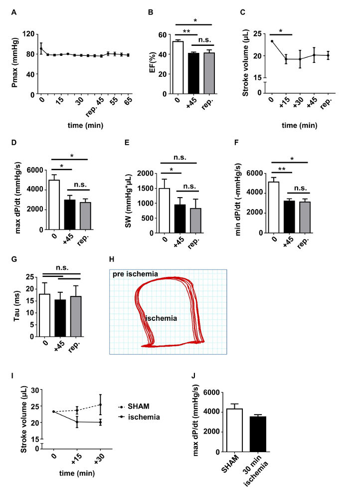
Figure 4: Pressure/volume data in mice undergoing ischemia/reperfusion. (A) Systolic left ventricular blood pressure (Pmax). (B) Left ventricular (LV) ejection fraction (EF) (%). (C) LV stroke volume (µL). (D) Maximum dP/dT (max dP/dt) (mmHg/s). (E) LV Stroke work (SW). (F) Minimum dP/dt (min dP/dt) (mmHg/s). (G) Time constant of isovolumetric relaxation Tau (ms). (H) Pressure/volume diagram before and 45 min after induction of myocardial ischemia. (I-J) Stroke volume (SV) (µL) and maximum dP/dt (max dP/dt) (mmHg/s) in sham-operated animals compared to animals after 15/30 min ischemia. Data (A-G) are presented as mean ± SEM. * p <0.05 via Student's t-test or ratio-paired t-test, n = 4 mice/group (A+D-G)) or n = 3 mice/group (B, C). +45: 45 min ischemia; rep.: reperfusion. Please click here to view a larger version of this figure.
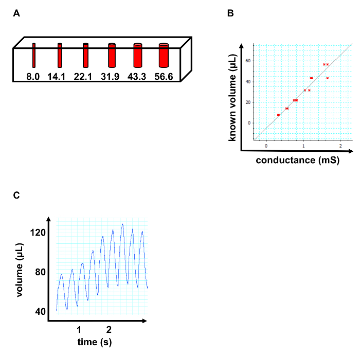
Figure 5: Post-hoc calibration. (A) Schematic of calibration cuvette. Volumes in µL. (B) Representative linear regression analysis of obtained volume data to perform cuvette calibration. (C) Representative volume data after injection of 10 µL of 25% sodium chloride in H2O into the right jugular vein to perform saline calibration. Please click here to view a larger version of this figure.
Discussion
PV monitoring of LV hemodynamics in acute myocardial infarction serves as a novel method for real-time in vivo assessment of cardiogenic shock and impaired LV function in I/R injury. PV catheterization can provide a broad spectrum of parameters with regard to LV systolic and diastolic function. In addition to the LV volumetric parameters typically obtained by echocardiography or MRI (chamber volumes, EF, stroke volume, and cardiac output), PV analysis yields a more complete profile of LV function by simultaneously providing measures of LV systolic performance (contractility dP/dt, stroke work) and LV compliance (-dP/dt, Tau) as a parameter for diastolic function.
As acute heart failure in patients with acute myocardial infarction is a critical predictor for early in-hospital morbidity and mortality2, monitoring of acute hemodynamic impairment and cardiogenic shock in acute myocardial infarction could serve as a valuable tool for identifying possible protective mechanisms in an experimental setting.
Several factors turned out to be critical for successful data acquisition. Stable anesthesia was crucial for valid PV data since isoflurane showed a strong cardiodepressive effect with drops in pressure, LV EF, and stroke volume. Atraumatic preparation of the carotid artery was important to avoid hypovolemia due to blood loss. Furthermore, compression or injury of the vagus nerve and the carotid body could result in severe impairment of hemodynamics.
Saline calibration and cuvette calibration appeared to be another critical step to maintain valid data. For saline calibration, injection of 15% NaCl solution led to increased conductance indicated by a temporary increase in volume level (Figure 5C). Maintaining the same speed when injecting was crucial for stable data. When conducting cuvette calibration, it was important to avoid bubbles within the cuvettes to ensure valid results.
The obtained PV data furthermore indicate the importance of a simultaneous acquisition of pressure and volume data for a valid hemodynamic characterization since pressure data alone did not show significant changes throughout the experiment (Figure 4A). The combined PV analysis offered both baseline parameters for LV systolic function (e.g., ejection fraction) as well as parameters for LV contractility (dP/dt) and LV relaxation (-dP/dt, Tau).
Interestingly, acute occlusion of the LCA in patients usually causes a severe deficit of LV function with immediate necessity for mechanical hemodynamic support and is associated with a high mortality rate11,12. LCA occlusion in mice showed less hemodynamic impairment and LCA occlusion-associated death during the procedure was not observed. As a sign of persisting hemodynamic stability during ischemia, systolic LV blood pressure was stable at all time (Figure 4A). However, this effect could be caused by more distal ligations in mice compared to LCA occlusions in humans.
Taken together, real-time hemodynamic monitoring of acute myocardial infarction in mice could serve as a new method for studying cardioprotective mechanisms in severe LV dysfunction aiming to improve early-phase treatment of patients undergoing acute myocardial infarction.
Disclosures
The authors have nothing to disclose.
Acknowledgements
The authors acknowledge the following funding sources: Else Kröner-Fresenius-Stiftung (Tienush Rassaf); Hans und Gertie Fischer Stiftung (Tienush Rassaf), grant from the medical faculty, University Duisburg-Essen, Germany (Tienush Rassaf, Lars Michel); Ernst- und Berta Grimmke-Stiftung (Christos Rammos).
Materials
| Name | Company | Catalog Number | Comments |
|---|---|---|---|
| Betaisodona Loesung | Mundipharma | 4162-1606/89x30mm | Povidon-Iod |
| Calibration cuvette | Millar instruments | 910-1049 | Calibration cuvette |
| Contura professional hair trimmer | Wella | HS-60 | Small animal shaving system |
| Eclipse Needle 27G | BD | REF 305770 | 27G needle |
| Forceps | FST | 11203-25, 11069-08, 11616-15, 11506-12, 11051-10 | Surgical forceps |
| Forceps | Aesculap Braun | BN731R, BD 311R | Surgical forceps |
| Foris FS2434 | Eizo | 0FTD2033 | Monitor |
| Hamilton Syringe 100 µl needle | Hamilton | 80621 | 100µl syringe with needle |
| Heated Small animal OP table | Harvard Apparatus | 15001 | Heated OP table |
| Heparin-Natrium 25000 | Ratiopharm | N68542.04 | Heparin |
| Ketamin 10% 100 mg/ml | bela-pharm | FS1670041 | Ketamin |
| Labchart Pro 8 + Pro modules | AD Instruments | MLS260/8 | PV data analysis software |
| LAS EZ | Leica | LAS EZ | Microscope camera software |
| Leica IC80 HD | Leica | IC80 HD | Microscope camera |
| Leica M80 | Leica | M80 | Microscope |
| Micro-tip catheter transducer | Millar instruments | SPR-839 | PV catheter |
| MiniVent | Harvard Apparatus | 845 | ventilation |
| MPVS Ultra | Millar instruments | PL3508B48/M | PV catheter data acquisition device |
| Octenisept | Schülke | 20000832-A | disinfectant |
| Plastipak 1ml | PD | REF 303172 | 1ml syringe |
| PowerLab 8/35 | AD Instruments | PL3508 | analog/digital converter |
| Prolene 6-0 | Ethicon | XNEH7814.P31 | Polypropylene suture |
| Retraction Kit | FST | 18200-20 | retraction of surgical situs |
| Seraflex 5-0 | Naila | IC108000 | silk suture |
| Small and micro-scissors | FST Essen | 14059-11, 15007-08, 14064-11 | Surgical scissors |
| Small silicon tube | Reichelt Chemietechnik | tube for LCA occlusion | |
| Sodium Chloride | Sigma-Aldrich | S7653 | Sodium Chloride |
| testo 108 | testo | 5631080 | rectal thermometer |
| Thinkcentre desktop computer | Lenovo | PC0EJS2V | Computer |
| Vasofix Safety 20G | Braun | 4269110S-01 | intubation catheter |
| Windows 10 | Microsoft | KW9-00240 | Operating system |
| Xylazin 2% | Ceva | 6324464.00.00 | Xylazine hydrochloride |
References
- Sanchis-Gomar, F., Perez-Quilis, C., Leischik, R., Lucia, A. Epidemiology of coronary heart disease and acute coronary syndrome. Annals of Translational Medicine. 4 (13), 256(2016).
- Anderson, J. L., Morrow, D. A. Acute Myocardial Infarction. New England Journal of Medicine. 376 (21), 2053-2064 (2017).
- McNamara, R. L., et al. Predicting in-hospital mortality in patients with acute myocardial infarction. Journal of the American College of Cardiology. 68 (6), 626-635 (2016).
- Kurian, G. A., Rajagopal, R., Vedantham, S., Rajesh, M. The Role of Oxidative Stress in Myocardial Ischemia and Reperfusion Injury and Remodeling: Revisited. Oxidative Medicine and Cellular Longevity. , (2016).
- Turer, A. T., Hill, J. A. Pathogenesis of myocardial ischemia-reperfusion injury and rationale for therapy. American Journal of Cardiology. 106 (3), 360-368 (2010).
- Totzeck, M., Hendgen-Cotta, U. B., French, B. A., Rassaf, T. A practical approach to remote ischemic preconditioning and ischemic preconditioning against myocardial ischemia/reperfusion injury. Journal of Biological Methods. 3 (4), (2016).
- Respress, J. L., Wehrens, X. H. Transthoracic echocardiography in mice. Journal of Visualized Experiments. (39), (2010).
- Pacher, P., Nagayama, T., Mukhopadhyay, P., Batkai, S., Kass, D. A. Measurement of cardiac function using pressure-volume conductance catheter technique in mice and rats. Nature Protocols. 3 (9), 1422-1434 (2008).
- Zhang, B., Davis, J. P., Ziolo, M. T. Cardiac catheterization in mice to measure the pressure volume relationship: Investigating the Bowditch effect. Journal of Visualized Experiments. (100), e52618(2015).
- Rossello, X., Hall, A. R., Bell, R. M., Yellon, D. M. Characterization of the Langendorff perfused isolated mouse heart model of global ischemia-reperfusion injury: Impact of ischemia and reperfusion length on infarct size and LDH release. Journal of Cardiovascular Pharmacology and Therapeutics. 21 (3), 286-295 (2016).
- Task Force on the management of ST-segment elevation acute myocardial infarction of the European Society of Cardiology (ESC). ESC Guidelines for the management of acute myocardial infarction in patients presenting with ST-segment elevation. European Heart Journal. 33 (20), 2569-2619 (2012).
- Shigemitsu, O., et al. Acute myocardial infarction due to left main coronary artery occlusion. Therapeutic strategy. Japanese Journal of Thoracic and Cardiovascular Surgery. 50 (4), 146-151 (2002).
Reprints and Permissions
