Retinal explants obtained from wild-type macaques were cultured in vitro. Retinal degeneration and the cGMP-PKG signaling pathway was induced using the PDE6 inhibitor zaprinast. cGMP accumulation in the explants at different zaprinast concentrations was verified using immunofluorescence.
Method Article
Organotypic Retinal Explant Cultures from Macaque Monkey
In This Article
Summary
Abstract
Hereditary retinal degeneration (RD) is characterized by progressive photoreceptor cell death. Overactivation of the cyclic guanosine monophosphate (cGMP)-dependent protein kinase (PKG) pathway in photoreceptor cells causes photoreceptor cell death, especially in models harboring phosphodiesterase 6b (PDE6b) mutations. Previous studies on RD have used mainly murine models such as rd1 or rd10 mice. Given the genetic and physiological differences between mice and humans, it is important to understand to which extent the retinas of primates and rodents are comparable. Macaques share a high level of genetic similarity with humans. Therefore, wild-type macaques (aged 1-3 years) were selected for the in vitro culture of retinal explants that included the retina-retinal pigment epithelium (RPE)-choroid complex. These explants were treated with different concentrations of the PDE6 inhibitor zaprinast to induce the cGMP-PKG signaling pathway and simulate RD pathogenesis. cGMP accumulation and cell death in primate retinal explants were subsequently verified using immunofluorescence and the TUNEL assay. The primate retinal model established in this study may serve for relevant and effective studies into the mechanisms of cGMP-PKG-dependent RD, as well as for the development of future treatment approaches.
Introduction
Hereditary retinal degeneration (RD) is characterized by progressive photoreceptor cell death and is caused by mutations in a wide variety of pathogenic genes1. The end result of RD is vision loss and in the vast majority of cases the disease remains untreatable to this day. Therefore, it is important to study the cellular mechanisms leading to photoreceptor death using models that faithfully represent the human disease condition. Here, primate-based models are of particular interest due to their closeness to humans. Notably, such models may advance the development of appropriate therapeutic interventions that can halt or delay photoreceptor cell death.
Previous research on the mechanisms of cell death in RD has demonstrated that the decrease or loss of phosphodiesterase 6 (PDE6) activity caused by RD-triggering gene mutations leads to reduced hydrolysis of cyclic guanosine monophosphate (cGMP)2,3. cGMP is a specific agonist of the cyclic nucleotide-gated ion channels (CNGCs) in the rod outer segments (ROSs) and is also a key molecule responsible for the conversion of light signals into electrical signals in vertebrate photoreceptor cells4. Reduced cGMP hydrolysis causes the accumulation of cGMP in ROSs, leading to the opening of CNGCs 5. Consequently, the phototransduction pathways are activated, resulting in an increase in cation concentrations in photoreceptor cells. This process imposes a metabolic burden on photoreceptors, which when overactivated, for instance, by mutations in PDE6, may cause cell death.
Many studies have shown that a significant overaccumulation of cGMP in photoreceptors of mouse models with different RD gene mutations may cause the activation of cGMP-dependent protein kinase (PKG)3,6. This leads to a substantial increase in dying, TUNEL-positive cells and a gradual thinning of the photoreceptor cell layer. Previous studies suggest that PKG overactivation caused by elevated cGMP levels is a necessary and sufficient condition for the induction of photoreceptor cell death2,5. Studies on different mouse models of RD have also shown that PKG activation induced by elevated cGMP levels in photoreceptors, leads to overactivation of downstream effectors such as poly-ADP-ribose polymerase 1 (PARP1), histone deacetylase (HDAC), and calpain2,7,8,9. This implies causal associations between these different target proteins and photoreceptor cell death.
However, previous research on the pathology, toxicopharmacology, and therapy of RD was mainly based on mouse models for RD10,11,12. Nevertheless, immense difficulties remain in the clinical translation of these results. This is owing to the considerable genetic and physiological differences between mice and humans, especially with respect to the retinal structure. In contrast, non-human primates (NHPs) also share a high degree of similarity with humans with respect to genetic characteristics, physiological patterns, and environmental factor regulation. For example, optogenetic therapy was investigated as a means to restore retinal activity in an NHP model13. Lingam and colleagues demonstrated that good manufacturing practice-grade human-induced pluripotent stem cell-derived retinal photoreceptor precursor cells may rescue cone photoreceptor damage in NHP14. Therefore, NHP models are important for the exploration of RD pathogenesis and the development of effective treatment methods. In particular, NHP models of RD, exhibiting pathogenic mechanisms similar to those in humans, could play a critical role in studies on the development and in vivo toxicopharmacology analysis of new drugs.
In view of the long life-cycle, high level of technical difficulties, and high cost involved in establishing in vivo primate models, we established an in vitro non-human primate (NHP) model using cultures of explanted macaque retina. First, wild-type macaques aged 1-3 years were selected for in vitro culture of retinal explants, which included the retina-RPE-choroid complex. Explants were then treated with different concentrations of the PDE6 inhibitor zaprinast (100 µM, 200 µM, and 400 µM) to induce the cGMP-PKG signaling pathway. Photoreceptor cell death was quantified and analyzed using the TUNEL assay, and cGMP accumulation in explants was verified via immunofluorescence. Given the high degree of similarity with respect to cell distribution and morphology, retinal layer thickness, and other physiological characteristics of the retina between monkeys and humans, the establishment of the cGMP-PKG signaling pathway in the in vitro retinal model may facilitate future research on the pathogenesis of RD as well as studies into the development and toxicopharmacology of new drugs for RD treatment.
Protocol
The animal study was reviewed and approved by the Ethics Review Committee of Institute of Zoology, Chinese Academy of Sciences (IACUC-PE-2022-06-002), and animal ethics review and animal protocol of Yunnan University (YNU20220149).
1. Preparation of retinal explants
- Obtain primate eyeballs from wild-type macaques, aged 1 to 3 years old, store in tissue storage solution, and transport on ice within 3 h of enucleation after the monkeys were sacrificed or after natural death.
- For the preparation of proteinase K solution, dissolve 25 mg of proteinase K in 250 µL of distilled water, and then add 225 µL of the solution to 18.5 mL of basal medium (R16).
- Wash the eyeballs in 10 mL of 5% povidone-iodine (povidone-iodine: water = 1:19) for 30 s and dip them into 5% penicillin/streptomycin (PEN/STREP):phosphate-buffered saline = 1:19 for 1 min to avoid bacterial contamination. Then, incubate the eyeballs in 10 mL of R16 medium for 5 min.
- Pre-heat the proteinase K solution in a 37 °C incubator. Then, immerse the eyeballs in 10 mL of proteinase K solution and incubate for 2 min. Immerse the eyeballs in 10 mL of R16 + fetal bovine serum (1:1) solution, incubate for 5 min, and then transfer the eyeballs to 10 mL of fresh R16 for a final wash.
- Separate out the sclera, cornea, iris, lens, and vitreous body, and cut the retina from 4 sides with tweezers and scissors (Figure 1A-L).
- Cut retinal explants with trephine blades into five sections-a 4 mm trephine blade for the nasal/temporal/superior/inferior side and a 6 mm trephine blade for the central side (containing optic disk and macula; Figure 1M-O). Cut at the following distance from the optic nerve head: 1.5 mm for nasal, 4 mm for temporal, and 2 mm for both superior and inferior side.
- Transfer the retina explants (containing retina and choroid) with eyelid depressor and place them in the middle of the insert, photoreceptor side up (Figure 1P). Place approximately 1 mL of complete medium (R16 supplemented with BSA, transferrin, progesterone, insulin, T3, corticosterone, vitamin B1, vitamin B12, retinol, retinyl acetate, DL-tocopherol, tocopheryl acetate, linoleic acid, L-cysteine HCl, glutathione, glutamine, vitamin C) below the membrane on the plate to fully cover the retina.
2. Primate retinal explant culture
- Culture retinal explants in complete medium and place in a 37 °C incubator aerated with humidified 5% CO2.
- Apply drug treatment at appropriate concentration to the retinal culture medium (e.g., zaprinast at concentrations of 100 µM, 200 µM, or 400 µM). Use at least three explants per treatment condition and use explants without drug as control. Treat with drug for 4 days.
3. Fixing and cryo-sectioning
- Incubate retinal explants with 1 mL of paraformaldehyde (PFA) for 45 min, rinse them briefly with 1 mL of phosphate-buffered saline (PBS), and then incubate with 10% sucrose for 10 min, 20% sucrose for 20 min, and 30% sucrose for 30 min (1 mL for each retina explant).
- Cut retinal explants using trephine blades to ensure consistency in the size of the explants obtained from different sites with the following characteristics: four quadrants in the periphery, 6 mm at the center. Then, transfer the retinal explants into a tinbox (about 1.5 cm x 1.5 cm x 1.5 cm) with O.C.T. embedding medium to cover the tissue. Immediately, freeze the tissue sections in liquid nitrogen and place in a -20 °C refrigerator for storage.
- Use a sharp blade to trim the agar into a small block containing the tissue sample, cut the sample blocks into 10 µm sections, and place them on adhesion microscope slides using a soft brush. Then, dry for 45 min at 40 °C and store at -20 °C until use.
4. Immunohistochemistry (Figure 2)
- cGMP staining
- Dry the slides and draw a hydrophobic ring around the retinal sections with a liquid blocker pen to cover the samples.
- Immerse slides in 200 µL of 0.3% phosphate buffer saline and Triton X-100 per slide (PBST) for 10 min at room temperature, and then in blocking solution (5% normal donkey serum, 0.3% PBST, 1% BSA, 200 µL per slide) for 1 h at room temperature.
- Incubate the samples overnight with the primary antibody (1:250, sheep anti-cGMP, 200 µL per slide) prepared in the blocking solution at 4 °C, and then rinse with PBS for 10 min (200 µL per slide) 3x.
- Incubate the samples with secondary antibody (1:300, Donkey anti sheep Alxea Fluor 488, 200 µL per slide) for 1 h at room temperature, and then rinse with PBS for 10 min, 3x. Cover the samples with an antifade mounting medium containing 4',6-diamidino-2-phenylindole (DAPI), and store at 4 °C for at least 30 min before imaging.
- TUNEL staining
- Dry the slides at room temperature for 15 min and draw a water-repellent barrier ring around retinal tissue specimens with the liquid blocker pen, and then incubate them in 40 mL of PBS for 15 min.
- Incubate slides in 42 mL (per five slides) of TBS (0.05 M Tris-buffer) at 37 °C. Aspirate out the TBS, and incubate the samples with 6 µL of proteinase K for 5 min. Then, wash slides 3x with TBS for 5 min each time.
- Expose the slides to 40 mL of ethanol-acetic acid solution in the choplin, cover with a transparent sheet, and incubate at -20 °C for 5 min. Wash the slides 3x with TBS for 5 min each time.
- Expose the slides to blocking solution (1% BSA; 200-300 µL per slide as per requirement) and incubate in a humidity chamber for 1 h.
- Prepare TUNEL kit solution, TMR red, in the following proportion: 62.50 µL of blocking solution (1% BSA), 56.25 µL of labeling solution (TMR-dUTP), and 6.25 µL of the enzyme (proportion for each slide). Add approximately 130 µL of this solution per slide and incubate at 37 °C for 1 h. Then, wash the slides with PBS for 5 min (200 µL per slide), 2x.
- Place cover slides containing 1-2 drops of antifade mounting medium with DAPI over the samples, and then incubate the slides at 4 °C for at least 30 min before imaging.
- cGMP staining combined with TUNEL staining
- cGMP staining was followed by TUNEL staining. Follow the steps of TUNEL staining from step 4.2.1 to step 4.2.5, and then continue the steps of cGMP staining from step 4.1.2 to step 4.1.4.
- Perform light and fluorescence microscopy with camera parameters as follows: exposure time = 125 ms, ROI size = 2752 x 2208, Color = B/M. Capture images using the software. Picture four different fields with a digital camera at 20x magnification from each section and obtain representative pictures from the central areas of the retina using DAPI (465 nm), EGFP (509 nm), TMP (578 nm), with Z-Stack scanning having an interval = 1 µm and optional = 1.251 µm.
Results
In this study, Macaque monkey retinal explant culture was performed using explants containing the retina-RPE-choroid complex (Figure 1, Supplementary Figure S1). Compared with the in vitro culture of retinal cells using the retina without the attached RPE and choroid, our explant culture facilitates better cell survival and accordingly, prolongs the survival of photoreceptor cells.
We used different concentrations of the PDE6 inhibitor zaprinast (100 µM, 200 µM, and 400 µM; Supplementary Figure S2) to induce an accumulation of cGMP in photoreceptors and subsequent activation of PKG in vitro. Each concentration treatment was given for 4 days, respectively. The treatment of the in vitro retinal model with 400 µM zaprinast exhibited the largest number of TUNEL-positive cells, a significant cGMP signal, and low toxicity for the RPE and neuronal cells in the inner retina. The high proportion of TUNEL-positive cells in Figure 2 suggested increasing cGMP activity may enhance zaprinast-induced retinal cell degeneration and cGMP accumulation plays a role in photoreceptor degeneration. The macaque retinal degeneration model established in this study will serve for comprehensive investigations on the mechanisms of cGMP-PKG-dependent RP.

Figure 1: Production process of retinal explant culturing in 1-year-old macaques. (A-L) Separate out the sclera, cornea, iris, lens, and vitreous body, and cut the retina from four sides with tweezers and scissors. (M-O) Cut the retinal explants with trephine blades into five sections - a 4 mm trephine blade for the nasal/temporal/superior/inferior side, and a 6 mm trephine blade for the central side (containing optic disk and macula). (P) Transfer the retinal explants (containing retina and choroid) butterflied open to the membrane and place the membrane on a 6-well culture plate. Please click here to view a larger version of this figure.
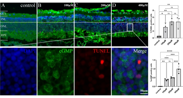
Figure 2: Evaluation of cGMP activities within the ONL of retinal explants of 1-year-old monkey. Explants were treated with different concentrations of the PDE6 inhibitor zaprinast (100 µM, 200 µM, and 400 µM). The number of TUNEL (red) and cGMP (green) positive cells in the three different concentrations was strongly increased in ONL when compared with control (A-D). Compared with the control group, the TUNEL/cGMP-positive cells have significant differences in the 100 µM, 200 µM, and 400 µM groups. Nuclei were stained with DAPI (blue). An area of the retina treated with 400 µM zaprinast was magnified to observe cGMP (green) and TUNEL (red) positive cells alone, as well as a merged image. Error bars represent standard deviation (SD); * = p ≤ 0.05; ** = p ≤ 0.01; *** = p ≤ 0.001; **** = p ≤ 0.0001. Abbreviations: GCs = ganglion cells, INL = inner nuclear layer, ONL = outer nuclear layer, RPE = retinal pigment epithelium, cGMP = cyclic guanosine monophosphate, PDE6 = phosphodiesterase 6. Scale bar = 50 µm. Please click here to view a larger version of this figure.
Supplementary Figure S1: Location of retinal punches used for explant cultures. Explants derived from nasal punches were located 1.5 mm from the optic nerve head, while this distance was 4 mm for temporal and 2 mm for superior and inferior punches, respectively. Retinal explants were obtained by cutting with trephine blades at five locations - a 4 mm trephine blade for the nasal/temporal/superior/inferior side and a 6 mm trephine blade for the central side (containing optic disk and macula). Please click here to download this File.
Supplementary Figure S2: TUNEL staining for dying cells. Control retina (A) and retinas treated with Zaprinast at concentrations of 100 µM, 200 µM, or 400 µM (B-D). Please click here to download this File.
Discussion
Visual phototransduction refers to the biological process by which light signals are converted to electrical signals by photoreceptor cells within the retina of the eye. Photoreceptor cells are polarized neurons capable of phototransduction, and there are two different types of photoreceptors termed rods and cones after the shapes of their outer segments. Rods are responsible for the scotopic vision and cones are responsible for photopic and high acuity vision. Hereditary RD relates to neurodegenerative diseases characterized by progressive retinal photoreceptor cell death15.
Current research on RD has mainly been based on mouse models of RD, including in vitro retinal explant cultures derived from murine RD models16. However, there exist considerable genetic and physiological differences, especially in retinal structure, between mice and humans. In contrast, NHPs share a high degree of similarity with humans, making NHP models most appropriate for the investigation of RD pathogenesis and therapy development.
In this study, we established an in vitro NHP model by culturing retinal explants of macaques. Compared with routine single-cell primary culture and an in vitro culture of immortalized cell lines, an in vitro retinal explant culture can preserve retinal tissue conformation and intercellular interactions, which enables better simulation of in vivo conditions. In addition, it largely reduces the number of animals used and shortens the experimental duration, and provides a relatively controllable experimental approach for investigating the effects of various factors involved in retinal development or degeneration.
Successful preparation of retinal explants is crucial to the successful establishment of in vitro retinal models. Based on our previous research experience of preparing retinal explants for mice, we summarize here several key points for monkey retinal explant preparation:
1) Explant preparation must be performed as soon as possible after animal sacrifice and enucleation. The use of a tissue storage solution for eyeball storage before use in explant preparation is recommended as it enables better maintenance of cell viability compared with phosphate-buffered saline.
2) Considering the large size of monkey eyeballs and the long periods of external environment exposure, washing the eyeballs with diluted povidone iodophor and a dual antibiotic solution prior to retinal explant preparation may reduce contaminations. However, washing should not be performed for an excessively long duration. The addition of antibiotics during explant culture is not recommended as it may affect cell viability.
3) Enucleated eyeballs were subjected to a sequential separation of the sclera, cornea, iris, lens, and vitreous body followed by the slicing of the retina. Unlike mouse eyeballs, which are small and in which the sclera and uvea are tightly attached to each other, monkey eyeballs are relatively large and possess a tough sclera and suprachoroidal space. Therefore, scissors are required for the separation of the sclera. Although this is not a difficult step in the handling of enucleated monkey eyeballs, care must be exercised to avoid damaging the retina and uvea. In addition, the tough suspensory ligament of the monkey lens has to be cut with scissors before the careful removal of the lens. Separation of the vitreous body is one of the most time-consuming steps of the explant preparation process due to the large vitreous chamber of the monkey eye, the high viscosity of the vitreous body, and the firm attachment of the vitreous body with the retina in monkeys aged 1-3 years. The vitreous body was removed to the greatest extent possible using scissors and a pipette was used to facilitate the subsequent transfer and culture of the retinal explants. We are currently in the process of exploring faster and more effective methods for vitreous body removal. Retinal explants (5 mm) were cut using trephine blades to ensure consistency in the size of the explants obtained from different sites (site characteristics: four quadrants in the periphery, 6 mm at the center). During the transfer of explants to culture inserts, the retina and choroid may become separated. Therefore, gentle and nimble handling is required to ensure that the explants are transferred during the first attempt itself, as repeated attempts may cause retinal damage. Lastly, mechanical damage should be minimized to the greatest extent possible during the entire retinal explant preparation process to avoid damaging the retinal structure. The time taken for explant preparation should also be minimized to avoid inadvertent cell death.
4) Retinal explant culture was performed using explants containing the retina-RPE-choroid complex. Compared with the in vitro culture of retinal cells using the retina without the attached RPE and choroid, our explant culture allows for better cell survival and accordingly prolongs the survival of photoreceptor cells. During the culture process, the retina faces upward and the choroid faces downward. Compared with mouse retinal explants, monkey retinal explants are larger in size, which places greater demands on culture nutrition. Therefore, we performed daily culture medium changes for the monkey retinal explant culture instead of adopting a culture medium change frequency of once every 2 days, which was used previously for the mouse retinal explant culture.
Taken together, we have established an in vitro NHP model through the culture of the retinal explants of macaques and their treatment with zaprinast to induce the cGMP-PKG signaling pathway similar to that observed in RD pathogenesis. Zaprinast is a PDE6 inhibitor, and PDE6 maintains the balance of the intracellular concentration of cGMP that activates PKG17,18. The cGMP-PKG pathway may negatively regulate oxidative phosphorylation and mitochondrial pathways, thereby affecting retinal degeneration19. The induction of the cGMP-PKG signaling pathway in this in vitro NHP retinal model establishes it as a favorable model for future research on the pathogenesis of RD, as well as for the development and toxicopharmacology testing of new drugs for RD treatment. Nevertheless, the use of this model has some limitations, notably, its relatively short culturing periods, and the relatively high costs for primates. During our future research, this in vitro model will be used for the post-intervention functional evaluation of the retina using MEA and µERG. Information and cellular locations of PKG targets will be combined with the measurement of activities of downstream effectors (e.g., calpain, PARP, and HDAC) and investigations into the pertinent neurodegenerative mechanisms.
Disclosures
All authors declare no conflicts of interest.
Acknowledgements
This study was supported by grants from the National Natural Science Foundation of China (No. 81960180), the Zinke heritage foundation, and the Charlotte and Tistou Kerstan Foundation, Yunnan Eye Disease Clinical Medical Center (ZX2019-02-01). We thank Prof. Longbao Lv (Institute of Zoology, Chinese Academy of Sciences, Kunming, China) for sharing the monkey eyeballs used in this study.
Materials
| Name | Company | Catalog Number | Comments |
|---|---|---|---|
| Bovine Serum Albumin (BSA) | Sigma | B2064 | Blocking solution |
| Corticosterone | Sigma | C2505 | Supplements of Complete Medium |
| DL-tocopherol | Sigma | T1539 | Supplements of Complete Medium |
| Donkey anti sheep, Alxea Fluor 488 | Life technologies corporation | A11015 | Secondary antibody of cGMP |
| Ethanol-acetic acid solution | Shyuanye | R20492 | Fixing liquid |
| Fetal Bovine Serum | Gemini | 900-108 | Blocking solution |
| Fluorescence microscope | Carl Zeiss | Axio Imager.M2 | Immunofluorescence imaging |
| Glutamine | Sigma | G8540 | Supplements of Complete Medium |
| Glutathione | Sigma | G6013 | Supplements of Complete Medium |
| In Situ Cell Death Detection Kit, TMR red | Roche | 12156792910 | TUNEL assay |
| Insulin | Sigma | 16634 | Supplements of Complete Medium |
| L-cysteine HCl | Sigma | C7477 | Supplements of Complete Medium |
| Linoleic acid | Sigma | L1012 | Supplements of Complete Medium |
| MACS Tissue Storage Solution | Miltenyi | 130-100-008 | Optimized storage of fresh organ and tissue samples |
| Normal Donkey Serum | Solarbio | SL050 | Blocking solution |
| Paraformaldehyde(PFA) | Biosharp | BL539A | Fixing agent |
| PEN. / STREP. 100× | Millipore | TMS-AB2-C | Penicillin / Streptomycin antibiotics |
| Phosphate buffer saline(PBS) | Solarbio | P1010 | Buffer solution |
| Povidone-iodine | Shanghailikang | 310411 | Disinfector agent |
| Progesterone | Sigma | P8783 | Supplements of Complete Medium |
| Proteinase K | Millpore | 539480 | Break down protein |
| R16 medium | Life technologies corporation | 074-90743A | Basic medium |
| Retinol | Sigma | R7632 | Supplements of Complete Medium |
| Retinyl acetate | Sigma | R7882 | Supplements of Complete Medium |
| Sheep anti-cGMP | Jan de Vente, Maastricht University, the Netherlands | Primary antibody of cGMP | |
| Sucrose | GHTECH | 57-50-1 | Dehydrating agent |
| T3 | Sigma | T6397 | Supplements of Complete Medium |
| Tissue-Tek medium (O.C.T. Compound) | SAKURA | 4583 | Embedding medium |
| Tocopheryl acetate | Sigma | T1157 | Supplements of Complete Medium |
| Transferrin | Sigma | T1283 | Supplements of Complete Medium |
| Transwell | Corning Incorporated | 3412 | Cell / tissue culture |
| Tris-buffer (TBS) | Solarbio | T1080 | Blocking buffer |
| Triton X-100 | Solarbio | 9002-93-1 | Surface active agent |
| VECTASHIELD Medium with DAPI | Vector | H-1200 | Mounting medium |
| Vitamin B1 | Sigma | T1270 | Supplements of Complete Medium |
| Vitamin B12 | Sigma | V6629 | Supplements of Complete Medium |
| Vitamin C | Sigma | A4034 | Supplements of Complete Medium |
| Zaprinast | Sigma | Z0878 | PDE6 inhibitor |
| Zeiss Imager M2 Microscope | Zeiss, Oberkochen,Germany | upright microscope | |
| LSM 900 Airyscan | high resolution laser scanning microscope | ||
| Zeiss Axiocam | Zeiss, Oberkochen,Germany | digital camera | |
| Zeiss Axiovision4.7 | |||
| Adobe | |||
| Illustrator CC 2021 (Adobe Systems Incorporated, San Jose, CA) | |||
| Primate eyeballs from wildtype macaque | KUNMING INSTITUTE OF ZOOLOGY | SYXK ( ) K2017 -0008 ) K2017 -0008 | |
| Super Pap Pen Pen (Liquid Blocker, Diado, 0010, Japan | |||
| TUNEL kit solution (REF12156792910, Roche,Germany), |
References
- O'Neal, T. B., Luther, E. E. StatPearls. , StatPearls Publishing. Copyright 2022, StatPearls Publishing LLC (2022).
- Power, M., et al. Cellular mechanisms of hereditary photoreceptor degeneration - Focus on cGMP. Progress in Retinal and Eye Research. 74, 100772(2020).
- Paquet-Durand, F., Hauck, S. M., van Veen, T., Ueffing, M., Ekström, P. PKG activity causes photoreceptor cell death in two retinitis pigmentosa models. Journal of Neurochemistry. 108 (3), 796-810 (2009).
- Tolone, A., Belhadj, S., Rentsch, A., Schwede, F., Paquet-Durand, F. The cGMP pathway and inherited photoreceptor degeneration: Targets, compounds, and biomarkers. Genes (Basel). 10 (6), 453(2019).
- Arango-Gonzalez, B., et al. Identification of a common non-apoptotic cell death mechanism in hereditary retinal degeneration. PLoS One. 9 (11), 112142(2014).
- Mencl, S., Trifunović, D., Zrenner, E., Paquet-Durand, F. PKG-dependent cell death in 661W cone photoreceptor-like cell cultures (experimental study). Advances in Experimental Medicine and Biology. 1074, 511-517 (2018).
- Power, M. J., et al. Systematic spatiotemporal mapping reveals divergent cell death pathways in three mouse models of hereditary retinal degeneration. Journal of Comparative Neurology. 528 (7), 1113-1139 (2020).
- Sancho-Pelluz, J., et al. Excessive HDAC activation is critical for neurodegeneration in the rd1 mouse. Cell Death & Disease. 1 (2), 24(2010).
- Kulkarni, M., Trifunović, D., Schubert, T., Euler, T., Paquet-Durand, F. Calcium dynamics change in degenerating cone photoreceptors. Human Molecular Genetics. 25 (17), 3729-3740 (2016).
- Trifunović, D., et al. cGMP-dependent cone photoreceptor degeneration in the cpfl1 mouse retina. Journal of Comparative Neurology. 518 (17), 3604-3617 (2010).
- Samardzija, M., et al. HDAC inhibition ameliorates cone survival in retinitis pigmentosa mice. Cell Death & Differentiation. 28 (4), 1317-1332 (2021).
- Schön, C., et al. Gene therapy successfully delays degeneration in a mouse model of PDE6A-linked Retinitis Pigmentosa (RP43). Human Gene Therapy. 28 (12), 1180-1188 (2017).
- McGregor, J. E., et al. Optogenetic therapy restores retinal activity in primate for at least a year following photoreceptor ablation. Molecular Therapy. 30 (3), 1315-1328 (2022).
- Lingam, S., et al. cGMP-grade human iPSC-derived retinal photoreceptor precursor cells rescue cone photoreceptor damage in non-human primates. Stem Cell Research & Therapy. 12 (1), 464(2021).
- Das, S., et al. The role of cGMP-signalling and calcium-signalling in photoreceptor cell death: perspectives for therapy development. Pflugers Archiv. 473 (9), 1411-1421 (2021).
- Hoon, M., Okawa, H., Della Santina, L., Wong, R. O. Functional architecture of the retina: Development and disease. Progress in Retinal and Eye Research. 42, 44-84 (2014).
- Schnichels, S., et al. Retina in a dish: Cell cultures, retinal explants and animal models for common diseases of the retina. Progress in Retinal and Eye Research. 81, 100880(2021).
- Maryam, A., et al. The molecular organization of human cGMP specific Phosphodiesterase 6 (PDE6): Structural implications of somatic mutations in cancer and retinitis pigmentosa. Computational and Structural Biotechnology Journal. 17, 378-389 (2019).
- Huang, L., Kutluer, M., Adani, E., Comitato, A., Marigo, V. New in vitro cellular model for molecular studies of retinitis pigmentosa. International Journal of Molecular Sciences. 22 (12), 6440(2021).
- Zhou, J., Rasmussen, M., Ekström, P. cGMP-PKG dependent transcriptome in normal and degenerating retinas: Novel insights into the retinitis pigmentosa pathology. Experimental Eye Research. 212, 108752(2021).
Reprints and Permissions
