The present protocol provides a step-by-step procedure for rapid and simultaneous optical clearing, muti-round labeling, and 3D volumetric reconstruction of tens of postmortem human brain sections by combining the (SWITCH - H2O2 - Antigen Retrieval - 2,2'-thiodiethanol [TDE]) SHORT tissue transformation technique with light-sheet fluorescence microscopy imaging in a routinely high-throughput protocol.
Method Article
Optical Clearing and Labeling for Light-sheet Fluorescence Microscopy in Large-scale Human Brain Imaging
* These authors contributed equally
In This Article
Summary
Abstract
Despite the numerous clearing techniques that emerged in the last decade, processing postmortem human brains remains a challenging task due to its dimensions and complexity, which make imaging with micrometer resolution particularly difficult. This paper presents a protocol to perform the reconstruction of volumetric portions of the human brain by simultaneously processing tens of sections with the SHORT (SWITCH - H2O2 - Antigen Retrieval - 2,2'-thiodiethanol [TDE]) tissue transformation protocol, which enables clearing, labeling, and sequential imaging of the samples with light-sheet fluorescence microscopy (LSFM). SHORT provides rapid tissue clearing and homogeneous multi-labeling of thick slices with several neuronal markers, enabling the identification of different neuronal subpopulations in both white and grey matter. After clearing, the slices are imaged via LSFM with micrometer resolution and in multiple channels simultaneously for a rapid 3D reconstruction. By combining SHORT with LSFM analysis within a routinely high-throughput protocol, it is possible to obtain the 3D cytoarchitecture reconstruction of large volumetric areas at high resolution in a short time, thus enabling comprehensive structural characterization of the human brain.
Introduction
Analyzing the 3D molecular organization and cytoarchitecture of large volumes of the human brain requires optical transparency of specimens, achieved through protocols with extensive processing time. Optical clearing techniques were developed to minimize heterogeneity in refractive index (RI) within the tissues, thereby reducing light scattering and increasing the light penetration depth for high-resolution imaging1,2,3,4,5. Current advances in clearing and deep tissue-labeling methods allow volumetric imaging of intact rodent organs and embryos by exploiting cutting-edge microscopy techniques6,7,8,9,10,11,12.
However, volumetric 3D reconstruction of large areas of the postmortem human brain still represents a challenging task compared with model organisms. The complex biological composition and the variable postmortem fixation and storage conditions can compromise the tissue clearing efficiency, the antibody penetration depth, and the epitope recognition13,14,15,16,17,18,19. Moreover, mechanical tissue sectioning and subsequent clearing and labeling of each slice is still required to achieve an efficient clearing and uniform labeling of large human brain areas, resulting in long processing times and the need for sophisticated custom equipment, compared with model organisms15,20,21,22.
The SWITCH - H2O2 - antigen Retrieval -TDE (SHORT) tissue transformation technique has been developed specifically to analyze large volumes of the human brain18,23. This method employs the tissue structural preservation of the SWITCH protocol11 and high concentrations of peroxide hydrogen to decrease tissue autofluorescence, in combination with epitope restoration. SHORT allows uniform staining of human brain slices with markers for different neuronal subtypes, glial cells, vasculature, and myelinated fibers18,24. Its results are compatible with the analysis of both low- and high-density proteins. The resulting high transparency levels and uniform labeling enable volumetric reconstruction of thick slices with fluorescence microscopy, in particular, for fast acquisition light-sheet apparatus can be used18,24,25,26,27.
In this work, we describe how the SHORT tissue transformation technique can be used for simultaneous clearing and multi-round labeling of tens of formalin-fixed human brain sections. Four different fluorescent markers can be used together, leading to the identification of different cellular sub-populations. After clearing, high-resolution volumetric imaging can be performed with fluorescence microscopy. Here, we used a custom-made inverted LSFM18,24,25,26,27, which enables fast optical sectioning of the sample and rapid acquisition of multiple channels in parallel. With this routinely high-throughput protocol, it is possible to obtain a comprehensive cellular and structural characterization with a sub-cellular resolution of large areas of the human brain as already demonstrated in the mapping of an entire Broca's area23.
Protocol
Formalin-fixed human tissue samples were provided by the Department of Neuropathology at the Massachusetts General Hospital (MGH) Autopsy Service (Boston, USA). Written consent was obtained from healthy participants prior to death, following IRB-approved tissue collection protocols from the Partners Institutional Biosafety Committee (PIBC, protocol 2003P001937). The authorization documents are kept with the MGH Autopsy Services in Boston, MA, United States, and are available upon request.
1. Agarose embedding and sample cutting
- Prepare a 4% w/v agarose solution in 1x phosphate-buffered saline (PBS, pH 7.4) in a beaker and embed the tissue block inside. Wait until it reaches room temperature (RT) and then store at 4 °C for 24 h.
- To obtain high-precision sections, glue the sample to the vibratome's specimen disc and fill the tray with cold 1x PBS (pH 7.4). Adjust the vibratome parameters such as frequency, amplitude, and speed according to the type of tissue to be sliced (values used: frequency = 5 (50 Hz); amplitude = 0.4; speed = 3 (0.15 mm/s).
NOTE: Different vibratomes should be used depending on the sample volume. For samples with a maximum volume of 70 x 40 x 15 mm, a vibratome (e.g., Leica VT1000 S) can be used; for larger samples, it is possible to use a compresstome (e.g., VF-900-0Z Microtome18) or a custom-made apparatus21. Here, we present slices cut with a thickness of either 400 µm or 500 µm. - After cutting, put all the slices in a preserver solution composed of 1x PBS (pH 7.4) with 0.01% w/v Sodium Azide (NaN3) and store at 4 °C.
NOTE: Before embedding, wait until the agarose solution reaches 37 °C. A higher temperature might damage the specimen. It is recommended not to leave the samples in PFA for more than 48 h as prolonged fixation might interfere with the labeling procedure by damaging antigens and increasing tissue autofluorescence. For long-term storage of human tissues, PBS with NaN3 is used to prevent microbial contamination. Due to the toxicity of NaN3, prepare the preserver solution under a chemical hood.
2. Tissue fixation
NOTE: All the solutions used in the following protocol are prepared in large volumes, sufficient to process all the slices of the same tissue block and minimize the technical variability and reduce the time for individual steps.
- Prepare the Switch-off solution with 50% v/v 1x PBS pH 3, 25% v/v 0.1 M hydrochloric acid (HCl), 25% v/v 0.1 M potassium hydrogen phthalate (KHP), and fresh 4% v/v glutaraldehyde. Place all the samples in dishes with screw lids (70 x 23 mm) and incubate them with the Switch-off solution with gentle shaking at 4 °C for 1 day, protecting them from light with aluminum foil.
- Prepare the Switch-on solution with 1x PBS (pH 7.4) and fresh 1% v/v glutaraldehyde. Place all the samples in new dishes and incubate them with the Switch-on solution with gentle shaking at 4 °C for 1 day, protecting them from light with aluminum foil.
NOTE: Due to the high toxicity and light sensitivity of glutaraldehyde, both Switch-off and Switch-on solutions must always be prepared fresh and kept under the chemical hood on ice. Always fill the dish with 20 mL of each solution and seal it with parafilm.
3. Inactivation and clearing
NOTE: Reactive glutaraldehyde in the samples must be inactivated by incubation with an inactivation solution consisting of 1x PBS pH 7.4, 4% w/v acetamide, 4% w/v glycine, pH 9.0. The solution can be stored at 4 °C for up to 3 months. To remove lipids and make the tissue transparent, we use the clearing solution consisting of 200 mM sodium dodecyl sulfate (SDS), 20 mM sodium sulfite (Na2SO3), and 20 mM boric acid (H3BO3), pH 9.0. The solution must be stored at RT as SDS precipitates at 4 °C. All the following steps will be done in tubes filled with the solution.
- Move the samples from the dishes to tubes and wash for 3 x 2 h with 1x PBS pH 7.4 at RT in gentle shaking.
NOTE: For step 3.1, it is necessary to work under the chemical hood because of the glutaraldehyde toxicity. The acetamide, SDS, and boric acid are also toxic reagents, and they must be handled under a chemical hood. Always fill the tube with 50 mL of each solution. - Incubate the samples in inactivation solution at 37 °C in a water bath overnight (o/n).
- Wash for 3 x 2 h in 1x PBS pH 7.4 at RT in gentle shaking.
- Incubate the samples in clearing solution at 55 °C in a water bath for 3-7 days. Change the solution every 2 days (Table 1).
4. Immunolabeling
NOTE: Before the labeling step, it is necessary to remove the residual SDS, reduce the autofluorescence, and unmask the epitopes with an antigen retrieval solution consisting of 10 mM Tris base, 1 mM EDTA, and 0.05% v/v Tween 20, pH 9. The solution's pH of 9 is optimized for an efficient retrieval process; Tris base acts as a buffering agent to maintain a stable pH environment; EDTA, which is a chelating agent, enhances the antigen's accessibility. The nonionic detergent Tween 20 aids in improving the permeability of the tissue. This solution can be stored at 4 °C. It is important to note that hydrogen peroxide combined with the antigen retrieval step have a synergistic effect in reducing the autofluorescence signal, by breaking down and/or modifying the endogenous fluorophores responsible for non-specific background signal (such as lipofuscin).
- Prepare 1x PBS with 0.1% v/v Triton X-100 (PBST), prewarm it to 37 °C, and wash the samples 3 x 3 h during the day in gentle shaking at 37 °C in the incubator. Leave the last wash o/n.
NOTE: Store the PBST stock solution at 4 °C. It is crucial to remove SDS from the tissue as it can form insoluble precipitates at low temperatures that may interfere with subsequent acquisition processes. - The day after, add 10 mL of 30% v/v hydrogen peroxide (H2O2) to each sample and leave it with gentle shaking at RT for 45 min.
- Wash 3 x 10 min with 1x PBS (pH 7.4) with gentle shaking at RT.
- Preheat the antigen retrieval solution at 95 °C in a water bath; transfer the samples to the heated solution and leave them at 95 °C for 10 min.
- Allow the specimens to cool down to RT for 40 min in gentle shaking.
- Wash 3 x 5 min with deionized water in gentle shaking.
- Equilibrate the samples in PBS at 4 °C for 15 min.
- Prepare fresh antibody solution made of PBST with 0.01% w/v NaN3 (see the NOTE for step 1.3) and add primary antibodies. Incubate the samples with the solution in dishes with screw lids at 37 °C for n days (1-7) in an incubator with gentle shaking and protection from light (Table 2).
NOTE: The incubation time is size- and thick-dependent (Table 1 and Table 2). - After n days, move the samples into tubes and wash 3 x 2 h with prewarmed PBST at 37 °C with gentle shaking in the incubator.
- Add secondary antibodies in PBST with 0.01% w/v NaN3 and incubate the samples in new dishes with screw lids at 37 °C for n days (1-6) in an incubator with gentle shaking and protected from light (Table 1 and Table 2).
- After n days, transfer the samples to tubes and wash 3 x 3 h during the day with prewarmed PBST at 37 °C in a gentle shaking incubator. Leave the last wash o/n.
NOTE: As Triton X-100 and Tween 20 are viscous, pipette slowly to allow the tip to fill. Always fill the tube with 50 mL of each solution. Use 10 mL of antibody solutions for each sample and seal the dish with parafilm to prevent evaporation of the solution. Since the antibody solutions contain NaN3, they can be stored at 4 °C and reused to label new specimens within 2 weeks.
5. Refractive index matching
NOTE: To achieve a high level of transparency, it is necessary to homogenize the tissue refractive index. Here we use 2,2'-thiodiethanol (TDE) diluted in 1x PBS. TDE solutions must be stored at RT.
- Transfer the samples to new dishes, add 30% v/v TDE, and leave them for 4 h with gentle shaking at RT.
- Remove the 30% v/v TDE solution, add 68% v/v TDE to the samples, and leave them with gentle shaking at RT.
NOTE: As TDE is viscous, pipette slowly to allow the tip to fill. Inhalation of TDE vapor or mist can irritate the respiratory system. Therefore, it is advisable to work in a well-ventilated area or use fume hoods to minimize exposure. TDE solutions must be stored at RT. Use 10 mL of TDE solutions for each sample.
6. Sample mounting
NOTE: To facilitate sample mounting and LSFM image acquisition, we use a custom-made, sealed sample holder (termed "sandwich"), which consists of three parts: a microscope slide, a spacer, and a coverslip.
- Put the steel spacer (56 x 56 x 0.5 mm3) over the microscope slide (60 x 60 x 1 mm3) and lay the sample on the microscopy slide.
- Carefully put the quartz coverslip (60 x 60 x 0.5 mm3), clip everything together, and prepare a two-component silicon glue in a 1:1 ratio. Use a quartz coverslip to match the refractive index and reduce the optical aberration during the acquisition process.
- Using a 1 mL syringe, fill the space between the spacer and the coverslip with a two-component silicon glue and let it dry for 10 min.
- Remove the clips and use a small needle to slowly fill the entire sandwich with 68% v/v TDE.
- Make fresh glue and seal the sandwich.
NOTE: If the TDE accidentally reaches the spacer in step 6.1, the glue will not cure. Make sure that no air bubbles form during step 6.2.
7. Stripping
NOTE: The structural preservation and the enhanced epitope accessibility of SHORT allow multi-round labeling of slices by removing the antibodies and restaining the samples with other markers.
- Open the sandwich with a blade, put the specimens in new dishes with screw lids filled with 30% v/v TDE, and leave them for 3 h.
- Transfer the samples to tubes, wash for 3 x 2 h in 1x PBS (pH 7.4) at RT with gentle shaking, and leave the last wash o/n.
- Place the samples in clearing solution in a water bath at 80 °C for 4 h.
- Wash for 3 x 2 h in 1x PBST (pH 7.4) at 37 °C in gentle shaking and leave the last wash o/n. Now the sample is ready to be labeled again starting from step 4.8.
NOTE: Always fill the tube with 50 mL of each solution. Use 10 mL of TDE solution for each sample.
Results
The protocol described here enables the simultaneous treatment of multiple slices, ranging in thickness from 100 µm to 500 µm, using the SHORT method. This approach significantly reduces the overall processing time for the entire procedure. In this work, we provide a comprehensive description of the entire pipeline (Figure 1) for processing multiple postmortem human brain thick sections simultaneously and we demonstrate the protocol on 24 slices at once (Figure 2A). The duration for clearing and co-labeling ranges from 11 to 30 days, considering the size and thickness of the samples (Table 1), excluding imaging and postimaging processing. Following delipidation and refractive index matching in TDE, optimal and homogeneous transparency of all the tissue slices is achieved in both grey and white matter (Figure 2B).
The image acquisition can be performed with different advanced fluorescence microscopes, including confocal25 and two-photon microscopy14. Here, we used a custom-made inverted LSFM18,23 equipped with four excitation laser lines: 405 nm, 488 nm, 561 nm, and 638 nm, capable of acquiring four different biological features labeled with fluorophore-conjugated antibodies. Figure 3A shows representative images of a 400 µm-thick slice from a human Broca's area co-labeled for NeuN, a pan-neuronal marker, Somatostatin (SST), and Calretinin (CR), which label two different subpopulations of interneurons and the nuclear marker DAPI (Figure 3A,B). In the 400 µm thick slab, we observed that the global fluorescence along Z is not limited to the surface, but it is almost uniform throughout the tissue depth, although the signal intensity slightly decreases in the middle (Figure 3C).
To achieve a more comprehensive cellular characterization of human brain tissue, it is possible to perform multi-round labeling of the same specimens by stripping up the antibodies and relabeling the sample as demonstrated in the SHORT original paper. Figure 4 (adapted from Pesce et al.18) demonstrates the potential application of seven different antibodies on a 500 µm-thick tissue slice processed with SHORT in three consecutive rounds of immunostaining. The antibodies used include NeuN, SST, CR, Parvalbumin (PV), and vasoactive intestinal peptide (VIP) as inhibitory neuronal markers, glial fibrillary acidic protein (GFAP) for glial cells, and vimentin (VIM) for vasculature (Table 2). The results showcase the remarkable ability of SHORT to preserve tissue information throughout various stripping procedures, effectively reduce autofluorescence signals, and achieve efficient specimen clearing.
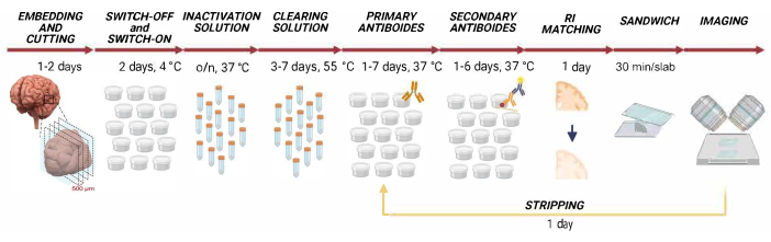
Figure 1: Timeline of SHORT. Scheme representing the steps required to process the samples with the SHORT protocol. In each step, tens of human brain tissue slabs are processed together. After embedding in agarose and slicing, samples are placed in Switch-on and Switch-off solutions for 1 day each; the inactivation steps are performed overnight while the delipidation through incubation in clearing solution requires 3-7 days. Afterward, samples are incubated with primary and secondary antibody solutions for a maximum of 7 and 6 days, respectively. After refractive index matching, samples are mounted in the sample holder (sandwich) for LSFM imaging and storage. At any time after sandwich assembly, samples can be stripped and relabeled for the detection of different markers. Abbreviations: TDE = 2,2'-thiodiethanol; SHORT = SWITCH - H2O2 - Antigen Retrieval -TDE; o/n = overnight; RI = refractive index; LSFM = light-sheet fluorescence microscopy. Please click here to view a larger version of this figure.
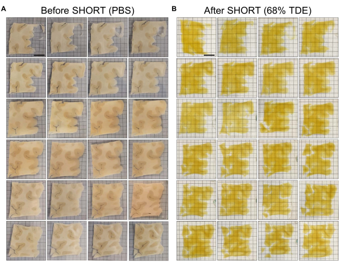
Figure 2: Human brain slices from a Broca area's block processed simultaneously. (A) Twenty-four consecutive slices from Broca's area of 4 x 4 x 0.04 cm3 in PBS before processing with the SHORT method. (B) The same specimens in A shown after the SHORT processing. Scale bar = 1 cm. Abbreviations: TDE = 2,2'-thiodiethanol; SHORT = SWITCH - H2O2 - Antigen Retrieval -TDE; PBS = phosphate-buffered saline. Please click here to view a larger version of this figure.

Figure 3. Representative LSFM images of a clarified slice labeled with four different markers. (A) Maximum intensity projection images (resolution of 3.3 x 3.3 x 3.3 µm3) showing a mesoscopic reconstruction of a SHORT-processed slice of Broca's area labeled for NeuN, Somatostatin, Calretinin, and the nuclear marker DAPI. On the right, the merged image (NeuN-SST-CR-DAPI) is shown. Scale bar = 0.5 cm. (B) Magnified insert of the merged image in A. Scale bar = 100 µm. (C) High resolution (0.55 x 0.55 x 3.3 µm) yz images from (B). Scale bar = 100 µm. Abbreviations: TDE = 2,2'-thiodiethanol; SHORT = SWITCH - H2O2 - Antigen Retrieval -TDE; LSFM = light-sheet fluorescence microscopy; NeuN = Neuronal Nuclear antigen; SST = somatostatin; CR = calretinin; DAPI = 4',6-diamidino-2-phenylindole. Please click here to view a larger version of this figure.
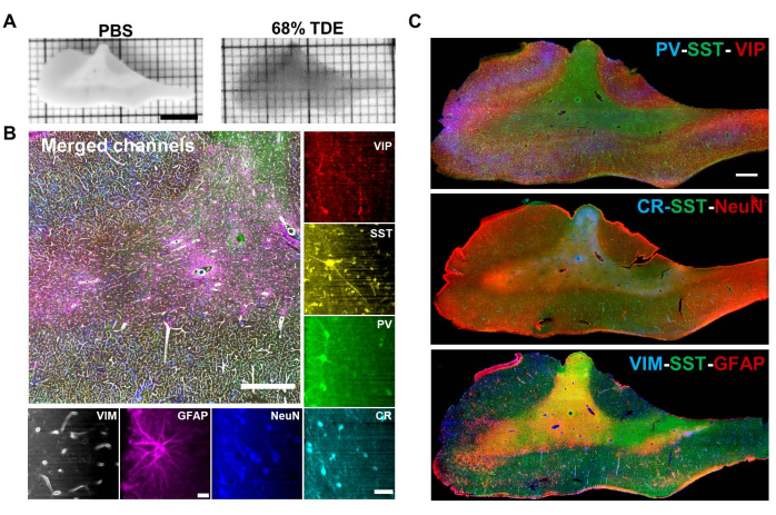
Figure 4: Representative LSFM images of multi-round staining in SHORT-processed slices. (A) Images of a human brain slice preclearing (in PBS) and after the multi-round labeling with refractive index matching (68% TDE). Scale bar = 1 cm. (B) Merging of the various channels acquired during three subsequent multi-rounds. The image shows both white and grey matter labeled with Vasoactive Intestinal Peptide, Somatostatin, Parvalbumin, Calretinin, Neuronal Nuclear antigen, Glial Fibrillary Acidic Protein, and Vimentin. High-resolution (resolution of 0.55 x 0.55 x 3.3 µm), single-channel images are shown on the right and below the image. Scale bar = 100 µm for all images, except for GFAP (scale bar 10 = µm). (C) Maximum intensity projection images of 500 µm-thick slice showing the signal from seven different antibodies used in three sequential rounds of immunostaining. Round 1: PV-SST-VIP; round 2: CR-SST-NeuN; round 3: VIM-SST-GFAP. Scale bar = 1 mm. This figure was modified from Pesce et al.18. Abbreviations: TDE = 2,2'-thiodiethanol; SHORT = SWITCH - H2O2 - Antigen Retrieval -TDE; LSFM = light-sheet fluorescence microscopy; NeuN = Neuronal Nuclear antigen; SST = somatostatin; CR = calretinin; PV = parvalbumin; GFAP = glial fibrillary acidic protein; VIM = vimentin; VIP = vasoactive intestinal peptide. Please click here to view a larger version of this figure.
Table 1: Representative time length of SHORT for samples with a range of areas and thickness. Specimens with different areas and thicknesses require a variable incubation time in clearing and antibody solutions, resulting in distinct time lengths for the whole SHORT pipeline. Please click here to download this Table.
Table 2: Antibodies compatible with SHORT. The table lists all the antibodies that were tested with the SHORT protocol and showed specific staining in 400-500 µm thick human brain specimens. The P/M column denotes polyclonal and monoclonal antibodies. Please click here to download this Table.
Discussion
High-resolution imaging and 3D reconstruction of large human brain areas require mechanical tissue sectioning followed by optical clearing and immunolabeling of single slices. The protocol presented here describes how the SHORT tissue transformation method can be used for rapid and simultaneous processing of multiple human brain thick sections for 3D brain reconstruction with a subcellular resolution with LSFM.
Unlike other approaches, with the SHORT method the clearing and multi-labeling steps do not require sophisticated customized equipment. A bulk of specimens can be processed altogether, reducing the processing time of large human brain tissue blocks and minimizing the technical variability.
Decolorization and epitope unmasking treatments are crucial when working with aged, formalin-fixed human tissues. The hydrogen peroxide and the alkaline antigen retrieval solutions can also be used in combination with other clearing methods such as the iDISCO6,28, CLARITY, SWITCH, and SHIELD24.
To ensure the preservation of epitopal information and achieve uniform clearing of both gray and white matter, it is necessary to transform the sample into a gel/tissue hybrid that is resistant to heat and chemical reagents. Glutaraldehyde was selected for its excellent crosslinking capability and its ability to penetrate the tissue slab at low pH (pH = 3). The concentration of glutaraldehyde is a critical factor in determining the strength and clarity of tissue. A higher concentration leads to better preservation of the tissue structure but also increases the time required for clearing. This choice enables the desired outcomes in terms of epitope preservation and homogeneous clearing. In the case of the switch-off solution (protocol step 2.1), the concentration of glutaraldehyde has been carefully chosen and optimized to strike a balance between effective crosslinking and rapid clearing18,25. SHORT is specifically tailored for working with formalin-fixed human brain slices from Broca's area, hippocampus, and cortical cortex18,23,25. However, when working with other biological models or different brain regions, some modifications may be necessary due to variations in the composition of gray and white matter.
One critical task of this protocol is also the optimization of the incubation time in clearing (protocol step 3.4) and antibody solutions (protocol steps 4.8 and 4.10), mainly related to the specimen size and the tissue biological composition. We estimated that 500 µm represent the tissue thickness limit to achieve optimal transparency in both white and gray matter and uniform labeling throughout the tissue depth for several antibodies staining (Table 2). A detailed description of these processes can be found in Pesce et al.18 and Scardigli et al.24. Different brain areas might have a different composition in lipids and proteins, and aged brains develop a high content of lipofuscin-type pigments and neuromelanin that produce a strong autofluorescence signal and alter the probe diffusion throughout the tissue. As an example, a 400 µm thick slice of 16 cm2 from human Broca's area needs 7 days of treatment with the clearing solution to reach a high level of delipidation. To ensure uniform staining throughout the tissue depth, it requires incubation with primary and secondary antibody solutions for 7 and 6 days, respectively (Figure 3). A fine optimization for the clearing and labeling steps is therefore required before the application of the high-throughput protocol described here.
SHORT enables storing samples in the custom-made imaging holder since it has been demonstrated that the fluorescent signal is preserved for at least one year. Researchers can therefore prepare all the sandwiches in advance and store them for the imaging step that can be done subsequently. Notably, the microscope slide, the spacer, and the cover glass used to prepare the sandwiches can be customized according to the size of the specimen.
The samples processed with the SHORT protocol described here were analyzed using a custom-made inverted LSFM18,23. This setup comprises two arms equipped with 12x custom-made objectives with a refractive index (RI) correction collar. Each objective acts as an illumination and detection arm at the same time, allowing the simultaneous acquisition of two channels at a volumetric speed of 0.16 cm3/h. As the specimen holder can accommodate two sandwiches at a time, we are able to consecutively image four channels in two slices in 10 h. Because of the mild toxicity of TDE vapor for the respiratory system, to avoid spherical aberration, the specimen holder is filled with 91% v/v glycerol/water solution that matches the RI (1.46) of both the quartz coverslip and the TDE solution. The 91% v/v glycerol/water solution must be changed every 3 days to avoid dust contamination. Therefore, our LSFM enables fast acquisition of specimens with low photobleaching at an isotropic resolution of 3.6 µm after postprocessing18,23,29. The large amount of data produced was processed using a defined pipeline for re-slicing and stitching of the image stacks using a self-developed software, ZetaStitcher (https://github.com/lens-biophotonics/ZetaStitcher)23,29.
Although we used SHORT in combination with light-sheet fluorescence microscopy, different kinds of fluorescence imaging techniques can be employed for the acquisition depending on the availability of the laboratory and the specific applications. For example, confocal microscopy25 or two-photon microscopy14 can be used to analyze cleared specimens. This type of analysis is recommended for samples with relatively small volumes since they are scanning-based systems and the acquisition time for large tissue areas is very long30. By achieving faster imaging and decoupling lateral and axial resolution, light-sheet fluorescence microscopy is the preferred method for imaging large volumes of cleared samples3,4,18,31,32,33.
In summary, we described a rapid high-throughput protocol for simultaneous clearing, homogeneous co-labeling, and 3D volumetric reconstruction of dozens of postmortem human brain sections. By combining SHORT with LSFM imaging and computational analysis, it is possible to obtain three-dimensional data for a comprehensive cell census and proteomic analysis of the human brain.
Disclosures
The authors declare that the research was conducted in the absence of any commercial or financial relationships that could be construed as a potential conflict of interest.
Acknowledgements
We thank Bruce Fischl, Massachusetts General Hospital, A.A. Martinos Center for Biomedical Imaging, Department of Radiology, for providing the human brain specimens analyzed in this study. This project received funding from the European Union's Horizon 2020 Research and Innovation Framework Programme under grant agreement No. 654148 (Laserlab-Europe), from the European Union's Horizon 2020 Framework Programme for Research and Innovation under the Specific Grant Agreement No. 785907 (Human Brain Project SGA2) and No. 945539 (Human Brain Project SGA3), from the General Hospital Corporation Center of the National Institutes of Health under award number U01 MH117023, and from the Italian Ministry for Education in the framework of Euro-Bioimaging Italian Node (ESFRI research infrastructure). Finally, this research was carried out with the contribution of "Fondazione CR Firenze." The content of this work is solely the responsibility of the authors and does not necessarily represent the official views of the National Institutes of Health. Figure 1 was created with BioRender.com.
Materials
| Name | Company | Catalog Number | Comments |
|---|---|---|---|
| 2,2'-thiodiethanol | Merck Life Science S.R.L. | 166782 | |
| Acetamide >= 99.0% (GC) | Merck Life Science S.R.L. | 160 | |
| Agarose High EEO | Merck Life Science S.R.L. | A9793 | |
| Boric Acid | Merck Life Science S.R.L. | B7901 | |
| Compressome VF-900-0Z Microtome | Precisionary | / | |
| Coverslips | LaserOptex | / | customized |
| Ethylenediaminetetraacetic acid disodium salt dihydrate | Merck Life Science S.R.L. | E5134 | |
| Glutaraldehyde | Merck Life Science S.R.L. | G7651 | |
| Glycine | Santa Cruz Biotechnology | SC_29096 | |
| Hydrogen Peroxide 30% | Merck Life Science S.R.L. | ||
| Incubator ISS-4075 | Lab companion | / | |
| Light-sheet fluorescence microscopy (LSFM) | / | / | custom-made |
| Loctite Attak | Henkel Italia srl | / | |
| Microscope slides | Laborchimica | / | customized |
| Phospate buffer saline tablet | Merck Life Science S.R.L. | P4417 | |
| Picodent Twinsil | Picodent | 13005002 | out of production |
| Potassium Hydrogen Phtalate | Merck Life Science S.R.L. | P1088 | |
| Sodium Azide | Merck Life Science S.R.L. | S2002 | |
| Sodium Dodecyl Sulfate | Merck Life Science S.R.L. | L3771 | |
| Sodium Sulfite | Merck Life Science S.R.L. | S0505 | |
| Spacers | Microlaser srl | customized | |
| Sputum Containers (dishes with screw lids) | Paul Boettger GmbH & Co. KG | 07.061.2000 | |
| Tris Base | PanReac AppliChem (ITW reagents) | A4577,0500 | |
| Triton X-100 | Merck Life Science S.R.L. | T8787 | |
| Tubes | Sarstedt | 62 547254 | |
| Tween 20 | Merck Life Science S.R.L. | P9416 | |
| Vibratome VT1000S | Leica Biosystem | / | |
| Water bath | Memmert | WNB 7-45 | |
| Antibodies and Dyes | |||
| Alexa Fluor 488 AffiniPure Alpaca Anti-Rabbit IgG (H+L) | Jackson Immuno Reasearch | 611-545-215 | Dilution used, 1:200 |
| Alexa Fluor 488 AffiniPure Bovine Anti-Goat IgG (H+L) | Jackson Immuno Reasearch | 805-545-180 | Dilution used, 1:200 |
| Alexa Fluor 647 AffiniPure Alpaca Anti-Rabbit IgG (H+L) | Jackson Immuno Reasearch | 611-605-215 | Dilution used, 1:200 |
| Anti-NeuN Antibody | Merck Life Science S.R.L. | ABN91 | Dilution used, 1:100 |
| Anti-Parvalbumin antibody (PV) | Abcam | ab32895 | Dilution used, 1:200 |
| Anti-Vimentin antibody [V9] - Cytoskeleton Marker (VIM) | Abcam | ab8069 | Dilution used, 1:200 |
| Calretinin Polyclonal antibody | ProteinTech | 12278_1_AP | Dilution used, 1:200 |
| DAPI | ThermoFisher | D3571 | Dilution used, 1:100 |
| Donkey Anti-Mouse IgG H&L (Alexa Fluor 568) | Abcam | ab175700 | Dilution used, 1:200 |
| Donkey Anti-Mouse IgG H&L (Alexa Fluor 647) | Abcam | ab150107 | Dilution used, 1:200 |
| Donkey Anti-Rabbit IgG H&L (Alexa Fluor 568) | Abcam | ab175470 | Dilution used, 1:200 |
| Donkey Anti-Rat IgG H&L (Alexa Fluor 568) preadsorbed | Abcam | ab175475 | Dilution used, 1:200 |
| Goat Anti-Chicken IgY H&L (Alexa Fluor 488) | Abcam | ab150169 | Dilution used, 1:500 |
| Goat Anti-Chicken IgY H&L (Alexa Fluor 568) | Abcam | ab175711 | Dilution used, 1:500 |
| Goat Anti-Chicken IgY H&L (Alexa Fluor 647) | Abcam | ab150171 | Dilution used, 1:500 |
| Goat Anti-Rabbit IgG H&L (Alexa Fluor 488) | Abcam | ab150077 | Dilution used, 1:200 |
| Recombinant Alexa Fluor 488 Anti-GFAP antibody | Abcam | ab194324 | Dilution used, 1:200 |
| Somatostatin Antibody YC7 | Santa Cruz Biotechnology | sc-47706 | Dilution used, 1:200 |
| Vasoactive intestinal peptide (VIP) | ProteinTech | 16233-1-AP | Dilution used, 1:200 |
References
- Costantini, I., Cicchi, R., Silvestri, L., Vanzi, F., Pavone, F. S. In-vivo and ex-vivo optical clearing methods for biological tissues: review. Biomedical Optics Express. 10 (10), 5251(2019).
- Richardson, D. S., et al. Tissue clearing. Nature Reviews Methods Primers. 1 (1), 84(2021).
- Ueda, H. R., et al. Tissue clearing and its applications in neuroscience. Nature Reviews Neuroscience. 21 (2), 61-79 (2020).
- Weiss, K. R., Voigt, F. F., Shepherd, D. P., Huisken, J. Tutorial: practical considerations for tissue clearing and imaging. Nature Protocols. 16 (6), 2732-2748 (2021).
- Tainaka, K., Kuno, A., Kubota, S. I., Murakami, T., Ueda, H. R. Chemical principles in tissue clearing and staining protocols for whole-body cell profiling. Annual Review of Cell and Developmental Biology. 32 (1), 713-741 (2016).
- Renier, N., et al. iDISCO: A simple, rapid method to immunolabel large tissue samples for volume imaging. Cell. 159 (4), 896-910 (2014).
- Pan, C., et al. Shrinkage-mediated imaging of entire organs and organisms using uDISCO. Nature Methods. 13 (10), 859-867 (2016).
- Lee, E., et al. ACT-PRESTO: Rapid and consistent tissue clearing and labeling method for 3-dimensional (3D) imaging. Scientific Reports. 6 (1), 18631(2016).
- Susaki, E. A., Ueda, H. R. Whole-body and whole-organ clearing and imaging techniques with single-cell resolution: toward organism-level systems biology in mammals. Cell Chemical Biology. 23 (1), 137-157 (2016).
- Lee, H., Park, J. -H., Seo, I., Park, S. -H., Kim, S. Improved application of the electrophoretic tissue clearing technology, CLARITY, to intact solid organs including brain, pancreas, liver, kidney, lung, and intestine. BMC Developmental Biology. 14 (1), 48(2014).
- Murray, E., et al. Simple, Scalable proteomic imaging for high-dimensional profiling of intact systems. Cell. 163 (6), 1500-1514 (2015).
- Cai, R., et al. Panoptic imaging of transparent mice reveals whole-body neuronal projections and skull-meninges connections. Nature Neuroscience. 22 (2), 317-327 (2019).
- Ueda, H. R., et al. Whole-brain profiling of cells and circuits in mammals by tissue clearing and light-sheet microscopy. Neuron. 106 (3), 369-387 (2020).
- Costantini, I., et al. Large-scale, cell-resolution volumetric mapping allows layer-specific investigation of human brain cytoarchitecture. Biomedical Optics Express. 12 (6), 3684(2021).
- Lai, H. M., et al. Next generation histology methods for three-dimensional imaging of fresh and archival human brain tissues. Nature Communications. 9 (1), 1066(2018).
- Zhao, S., et al. Cellular and molecular probing of intact human organs. Cell. 180 (4), 796-812.e19 (2020).
- Costantini, I., et al. Autofluorescence enhancement for label-free imaging of myelinated fibers in mammalian brains. Scientific Reports. 11 (1), 8038(2021).
- Pesce, L., et al. 3D molecular phenotyping of cleared human brain tissues with light-sheet fluorescence microscopy. Communications Biology. 5 (1), 447(2022).
- Schueth, A., et al. Efficient 3D light-sheet imaging of very large-scale optically cleared human brain and prostate tissue samples. Communications Biology. 6 (1), 170(2023).
- Morawski, M., et al. Developing 3D microscopy with CLARITY on human brain tissue: Towards a tool for informing and validating MRI-based histology. NeuroImage. 182, 417-428 (2018).
- Park, J., et al. Integrated platform for multi-scale molecular imaging and phenotyping of the human brain. bioRxiv. , (2022).
- Chung, K., et al. Structural and molecular interrogation of intact biological systems. Nature. 497 (7449), 332-337 (2013).
- Costantini, I., et al. A cellular resolution atlas of Broca's area. Science Advances. (9), eadg3844(2023).
- Scardigli, M., et al. Comparison of different tissue clearing methods for three-dimensional reconstruction of human brain cellular anatomy using advanced imaging techniques. Frontiers in Neuroanatomy. 15, 752234(2021).
- Pesce, L., et al. Exploring the human cerebral cortex using confocal microscopy. Progress in Biophysics and Molecular Biology. 168, 3-9 (2022).
- Keller, P. J., Dodt, H. -U. Light sheet microscopy of living or cleared specimens. Current Opinion in Neurobiology. 22 (1), 138-143 (2012).
- Power, R. M., Huisken, J. A guide to light-sheet fluorescence microscopy for multiscale imaging. Nature Methods. 14 (4), 360-373 (2017).
- Belle, M., et al. Tridimensional visualization and analysis of early human development. Cell. 169 (1), 161-173.e12 (2017).
- Sorelli, M., et al. Fiber enhancement and 3D orientation analysis in label-free two-photon fluorescence microscopy. Scientific Reports. 13 (1), 4160(2023).
- Helmchen, F., Denk, W. Deep tissue two-photon microscopy. Nature Methods. 2 (12), 932-940 (2005).
- Vieites-Prado, A., Renier, N. Tissue clearing and 3D imaging in developmental biology. Development. 148 (18), dev199369(2021).
- Costantini, I., et al. Editorial: The human brain multiscale imaging challenge. Frontiers in Neuroanatomy. (16), 1060405(2022).
- Costantini, I., et al. A versatile clearing agent for multi-modal brain imaging. Scientific Reports. 5 (1), 9808(2015).
Reprints and Permissions
