This protocol presents a reliable method to assess blood-brain barrier (BBB) integrity in adult zebrafish (Danio rerio) model using Evan's blue dye-based assessment, enabling both qualitative and quantitative analysis of neurovascular disruption.
A subscription to JoVE is required to view this content. Sign in or start your free trial.
Method Article
Visualizing the Gatekeeper: Evan's Blue Dye-Based Assessment of Blood-Brain Barrier Permeability in Adult Zebrafish
In This Article
Summary
Abstract
The Blood-Brain Barrier (BBB) is a semi-permeable interface that maintains central nervous system (CNS) homeostasis by regulating the movement of substances into and out of the brain, thereby protecting neural tissue from potentially harmful agents. Disruption of the BBB is a well-established feature in many neurodegenerative diseases, including Parkinson's disease (PD) and Alzheimer's disease (AD), where increased permeability contributes to and exacerbates disease progression. Although the adult zebrafish (Danio rerio) is increasingly recognized as a valuable model for studying neurodegenerative diseases, standardized methods for evaluating BBB integrity in this species remain limited. This protocol describes a simple, reproducible, and cost-effective approach to assess BBB permeability in adult zebrafish by analysing Evan's Blue (EB) dye extravasation from the neurovasculature into brain tissue. The method combines qualitative visualization with quantitative image analysis of EB distribution to detect BBB disruption. Key steps include proper intraperitoneal injection of EB dye, confirmation of systemic dye circulation, careful brain dissection, and consistent imaging and greyscale intensity measurement. Positive and negative controls are incorporated to validate dye penetration and support accurate interpretation. Representative results demonstrate greater EB extravasation in Parkinsonian zebrafish brains compared to saline-injected controls, indicating increased BBB permeability. This technique requires minimal specialized equipment and is suitable for laboratories with limited resources. Overall, this protocol offers a practical tool for investigating BBB integrity in adult zebrafish models and can be adapted for diverse applications, including studies of disease mechanisms and the evaluation of therapeutic interventions targeting neurovascular function.
Introduction
The Blood-Brain Barrier (BBB) is a selective, semi-permeable physiological interface formed primarily by specialized cerebral endothelial cells, supported by pericytes and astrocytes, that regulates molecular exchange between the bloodstream and the brain's interstitial space to preserve the Central Nervous System (CNS) microenvironment. Its integrity is essential for preserving CNS homeostasis and protecting neural tissue from harmful or toxic substances1. Disruption of the BBB is a well-recognized pathological feature in various neurodegenerative diseases, including Parkinson's disease (PD), Alzheimer's disease, and amyotrophic lateral sclerosis2,3. Compromised BBB function leads to increased permeability, allowing neurotoxic compounds and inflammatory mediators to infiltrate brain tissue, thereby exacerbating disease progression4. As such, evaluating BBB integrity is critical for gaining deeper insights into the neurovascular and inflammatory mechanisms underlying these disorders.
While in vivo rodent and in vitro models remain the gold standards for BBB research5, zebrafish (Danio rerio) have emerged as a valuable vertebrate model in neuroscience due to their genetic tractability, cost-effectiveness, ease of handling, and conserved neuroanatomical and BBB characteristics with mammals6. Despite their increasing use in neurodegenerative studies, methods to assess BBB integrity in adult zebrafish are still limited7,8. Techniques such as fluorescent tracer injections or transgenic reporters have been extensively used in larvae9,10, but pose significant challenges in adults due to pigmentation and decreased transparency. Hence, the objective of the present study is to establish a simple, reproducible, and visually demonstrable protocol to assess BBB permeability in adult zebrafish using Evan's Blue (EB) dye extravasation. This method incorporates both qualitative observation and quantitative measurement of dye leakage into brain tissue, providing a practical tool for evaluating neurovascular integrity under pathological conditions.
Evan's Blue dye, which binds to serum albumin and normally cannot cross the intact BBB11, offers a feasible alternative for assessing barrier disruption12. EB extravasation has been widely applied in rodent models to detect BBB leakage in conditions such as traumatic brain injury, stroke, and neuroinflammation13. However, its application in adult zebrafish has remained largely unexplored. This study presents a novel adaptation of the EB dye technique optimized for adult zebrafish, enabling reliable qualitative and quantitative evaluation of BBB permeability. The method is cost-effective, requires minimal specialized equipment, and can be easily scaled for multiple animals, offering a practical advantage over more complex imaging-based approaches typically used in larvae.
Using this protocol, we assessed BBB integrity in both healthy and Parkinsonian adult zebrafish. To induce Parkinsonism in zebrafish, the neurotoxin 1-methyl-4-phenyl-1,2,3,6-tetrahydropyridine (MPTP) was administered intraperitoneally, following a previously described method14. The results revealed significantly greater EB extravasation in the brains of Parkinsonian zebrafish, consistent with the well-documented BBB disruption observed in PD. These findings validate the sensitivity and reliability of the method, supporting its utility in linking vascular pathology to molecular and behavioural outcomes. This technique thus represents a valuable tool for researchers investigating neurovascular contributions to neurodegenerative diseases and may be applied in both genetic and toxin-induced zebrafish models.
Access restricted. Please log in or start a trial to view this content.
Protocol
This protocol was followed and was permitted by the Institutional Animal Care and Use Committee (IACUC) under the International Islamic University Malaysia (IIUM), with reference to the animal ethics approval (ref. no.: IIUM/504/14/2/IACUC).
1. Prepare Evans Blue dye stock solution
- Dissolve 0.1 g of EB dye in 10 mL of 1x phosphate-buffered saline (PBS) to prepare a 1% (w/v) stock solution. Mix thoroughly until the dye is completely dissolved.
CAUTION: EB is categorized as a Carcinogen Category 1B. Avoid direct contact with skin and eyes. Use protective clothing, gloves, safety glasses, and a dust respirator. Avoid inhaling dust and follow the manufacturer's handling instructions. Do not dispose of Evans Blue dye (solid or solution) in the sink. Retain waste in labelled original containers. Dispose of according to national and local hazardous waste regulations. - Filter the solution using a 0.45 µm syringe filter. Collect the filtrate into a sterile 15 mL centrifuge tube. Wrap the tube in aluminium foil and store at 4 °C.
NOTE: A smaller pore size filter (e.g., 0.2 µm) may also be used. The stock solution can be stored at 4 °C for up to two weeks, or at -20 °C for up to one month, with aliquoting recommended to minimize repeated freeze-thaw cycles when stored frozen.
2. Administer Evans Blue dye to adult zebrafish
- Anaesthetize the zebrafish via hypothermic shock by gradually lowering water temperature from 12 °C to 7 °C, until the fish no longer responds to tactile stimuli.
NOTE: Gradual temperature reduction can be achieved by adding small amounts of crushed ice to the water slowly over several minutes, allowing the temperature to decrease progressively. Tricaine immersion is also a suitable alternative anaesthetic method. - Inject 20 µL of 1% EB dye into the intraperitoneal (IP) cavity using a 31 G insulin syringe. Apply gentle pressure at the injection site for 10 s to prevent backflow.
NOTE: The zebrafish intraperitoneal cavity is located posterior to the pelvic girdle. To increase the success rate of EB administration, fast the fish for 24 h prior to the injection. This will empty the abdominal cavity to provide better access and accuracy to the intraperitoneal space. - Transfer the fish to a recovery tank and allow recovery for 30 min. Monitor for injury, mortality, or behavioural changes.
NOTE: To help zebrafish with recovery, use a slightly reduced water temperature (20-24 °C) for the recovery tank to provide a mild anaesthetic effect and help numb potential post-injection discomfort. Injected fish will exhibit a blue hue, indicating successful dye circulation (Figure 1). - After 30 min, euthanize the fish via hypothermic shock at 1 °C to 3 °C. Then, decapitate the fish using a sterile blade or surgical scissors.
NOTE: To aid in brain dissection later, leave extra flesh behind the head when decapitating. - Rinse the head twice in 1x PBS (1:20 fish to PBS ratio) to remove excess blood and EB dye.
NOTE: Insufficient washing may lead to false-positive results and compromise the accuracy of the analysis. To prevent this, wash the brain tissue thoroughly at least twice with 1x PBS, ensuring that all visible blood and free-flowing dye are removed. If any residual dye remains, a third wash is strongly recommended. - Fix the head in 4% paraformaldehyde (PFA) at 4 °C overnight (1:20 fish to fixative ratio). Agitation is not required during fixation, as the small size of the zebrafish head and sufficient fixative volume allow effective penetration under static conditions.
CAUTION: PFA is harmful if swallowed, inhaled, or in contact with skin. Prepare the PFA solution under a fume hood. Use appropriate personal protective equipment and avoid inhalation of dust. Follow all safety guidelines as per the manufacturer. Treat PFA as hazardous waste. Avoid disposing of it down the drain. Collect and label the waste in sealed containers and dispose of it in accordance with local and national regulations. - Dissect the zebrafish brain
- Place the fixed head in a clean petri dish and dissect the brain as previously described15, using pointed-end tweezers. Continuously add 1x PBS dropwise during dissection to keep the tissue hydrated and prevent drying.
- Prepare the agarose plate by dissolving 1 g of agarose in 100 mL of distilled water. Microwave it until fully melted, pour it into a petri dish, and allow it to solidify.
- Rinse the dissected brain twice in two separate 1x PBS baths. Then, place the tissue on a 1% agarose plate to retain moisture and prevent shrinkage.
NOTE: Insufficient washing may lead to false-positive results and compromise the accuracy of the analysis. To prevent this, wash the brain tissue thoroughly at least twice with 1× PBS, ensuring that all visible blood and free-flowing dye are removed. If any residual dye remains, a third wash is strongly recommended.
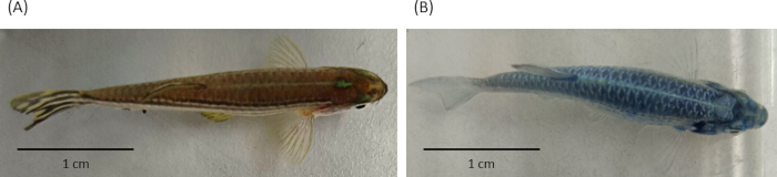
Figure 1: Comparison of skin coloration between a normal and an EB-injected zebrafish. (A) Normal zebrafish displaying a typical brownish skin tone. (B) EB-injected zebrafish exhibiting a noticeable bluish hue, indicating successful systemic circulation of the dye. Scale bar = 1 cm. Please click here to view a larger version of this figure.
3. Establish positive control via stab wound injury (SWI)
NOTE: Stab wound injury breaks the BBB and enables EB extravasation at the site of injury.
- Anaesthetize the fish using the hypothermic shock method (see step 2.1). Once anaesthetized, inject 20 µL of 1% EB dye and allow the fish to recover for 30 min as described in steps 2.2 and 2.3.
- After 30 min recovery period, prepare a sterile 31 G needle attached to an insulin syringe and perform a stab wound injury procedure as follows:
- Anaesthetize the fish again.
- Position the fish upright on a flat, cold surface (e.g., ice gel pack). Insert the needle into one side of the telencephalon at a 90° angle to a depth not exceeding 1.5 mm (Figure 2).
NOTE: To prevent direct skin contact with the ice gel pack and avoid potential skin damage, place a layer of gauze or paper towel between the fish and the cold surface (Figure 2). Alternatively, the fish can be positioned in a shallow petri dish placed on top of the cold pack to maintain cooling without direct contact. Mark 1.5 mm on the needle to prevent excessive penetration. - Transfer the fish to a recovery tank and allow to recover for 30 min.
- Euthanize, decapitate, fix, and dissect the brain as described in steps 2.4 to 2.7.
- For negative control, repeat steps 2.1 to 2.7 using 20 µL of 1x PBS instead of EB dye.

Figure 2: Stab wound injury for positive control. (A) Briefly insert a 31G needle into the telencephalon (red circle) to facilitate EB dye penetration into the brain tissue; Side-by-side comparison of an uninjured brain and a stabbed brain, (B) dorsal view of the stab site (red circle), (C) lateral view of the stab site. Scale bar = 1 mm. Please click here to view a larger version of this figure.
4. Qualitative analysis of EB extravasation
- Observe dissected brain tissues under a stereomicroscope. Optimize image settings (zoom, white balance, exposure, contrast, etc.) to ensure consistency.
- Capture images using a microscope-mounted camera and save in .TIFF format.
NOTE: Use identical imaging settings for all samples to ensure comparative validity.
5. Quantitative analysis of EB extravasation
- Download and install Fiji (a link to download the software is available in the Table of Materials). Open the image in Fiji.
- Perform colour deconvolution by clicking: Image > Colour > Colour Deconvolution > RGB, resulting in three separate images representing red, green, and blue hues.
NOTE: This step isolates the blue hue from the other colour components, ensuring that only the intensity of the blue dye is measured. - Activate the blue channel image (usually labelled "Color_3") by clicking the image. Convert it to 8-bit grayscale by clicking: Image > Type > 8-bit.
NOTE: Grayscale conversion minimizes colour artifacts and enhances intensity-based analysis. - Measure the grey intensity of the 8-bit image.
- Set measurements by selecting: Analyse > Set Measurements; select Area and Mean grey value.
- Using the rectangle selection tool, draw a uniform-sized region of interest (ROI) and measure the grey intensity by selecting Analyze > Measure, or pressing Ctrl + M.
NOTE: The same ROI size must be used across all samples. The absolute dimensions of the ROI may vary depending on the magnification and resolution of the image acquired, so researchers should standardize ROI size within their dataset to ensure comparability. Each pixel in the 8-bit grayscale image has an intensity value ranging from 0 to 255, where 0 represents pure black, 255 represents pure white, and values in between represent various shades of grey. - To reduce bias, draw three to five ROIs within anatomically consistent regions of the brain across all samples. Avoid regions with artefacts or inconsistent staining.
NOTE: Placement consistency can be achieved by referencing anatomical landmarks and ensuring ROI positioning is symmetrical across experimental groups. - Average the mean grey values from the ROIs for each image to obtain a single intensity value per brain. Use this average for statistical comparison between groups.
Access restricted. Please log in or start a trial to view this content.
Results
This protocol was tested on a Parkinsonian zebrafish model. Compromise of the BBB is a well-documented phenomenon in PD patients16,17,18 and in various Parkinsonian animal models19,20,21. To investigate BBB integrity in the adult zebrafish Parkinsonian model, EB dye extravasation into brain tissues was compared between saline-injected (...
Access restricted. Please log in or start a trial to view this content.
Discussion
Evaluating the integrity of the BBB is essential for understanding the pathophysiology of brain-related diseases, as a compromised BBB can both cause and worsen disease progression4. Although the adult zebrafish has emerged as an effective model for studying neurodegenerative diseases, methods for assessing BBB integrity in this species remain underdeveloped8. The protocol presented here provides a straightforward, cost-effective approach for evaluating BBB permeability in ...
Access restricted. Please log in or start a trial to view this content.
Disclosures
The authors declare no conflicts of interest.
Acknowledgements
This research was funded by the Malaysian Ministry of Higher Education (MoHE) under the Fundamental Research Grant Scheme (FRGS), grant number FRGS19-125-0734. The publication aid for this study was supported by the Faculty of Medicine, Universiti Kebangsaan Malaysia (UKM).
Access restricted. Please log in or start a trial to view this content.
Materials
| Name | Company | Catalog Number | Comments |
|---|---|---|---|
| Agarose powder | Vivantis | PC0701 | |
| Evan's blue dye | Sigma-Aldrich | E2129 | |
| Fiji ImageJ | National Institutes of Health, USA | Link to download: https://imagej.net/software/fiji/downloads | |
| Microscope camera | ShenZhen Hayear Electronics Co. Ltd. | HY-500M | |
| Paraformaldehyde (PFA) powder | Sigma-Aldrich | 158127 | |
| Phosphate-buffered saline (PBS) | Sigma-Aldrich | P4417 | |
| Sodium chloride (NaCl) | HmbG Chemicals | 7647-14-5 | |
| Stereomicroscope | Olympus Corporation | SZ40 | |
| Syringe filter unit, 0.45µm | Merck Millipore | SLHV004SL |
References
- Kadry, H., Noorani, B., Cucullo, L. A blood-brain barrier overview on structure, function, impairment, and biomarkers of integrity. Fluids Barriers CNS. 17, 1-24 (2020).
- Knox, E. G., Aburto, M. R., Clarke, G., Cryan, J. F., O'Driscoll, C. M. The blood-brain barrier in aging and neurodegeneration. Mol Psychiatry. 27 (6), 2659-2673 (2022).
- Sweeney, M. D., Sagare, A. P., Zlokovic, B. V. Blood-brain barrier breakdown in Alzheimer disease and other neurodegenerative disorders. Nat Rev Neurol. 14 (3), 133-150 (2018).
- Kim, S., Jung, U. J., Kim, S. R. The crucial role of the blood-brain barrier in neurodegenerative diseases: Mechanisms of disruption and therapeutic implications. J Clin Med. 14 (2), 386(2025).
- Guarino, V., et al. Advancements in modelling human blood-brain barrier on a chip. Biofabrication. 15 (2), 022003(2023).
- Kalueff, A. V., Stewart, A. M., Gerlai, R. Zebrafish as an emerging model for studying complex brain disorders. Trends Pharmacol Sci. 35 (2), 63-75 (2014).
- Eliceiri, B. P., Gonzalez, A. M., Baird, A. The blood-brain and other neural barriers: reviews and protocols. , Humana Press. Totowa, NJ. (2010).
- Li, Y., et al. Zebrafish: A promising in vivo model for assessing the delivery of natural products, fluorescence dyes and drugs across the blood-brain barrier. Pharmacol Res. 125, 246-257 (2017).
- Fleming, A., Diekmann, H., Goldsmith, P. Functional characterisation of the maturation of the blood-brain barrier in larval zebrafish. PLoS One. 8 (10), e77548(2013).
- Kislyuk, S., van den Bosch, W., Adams, E., De Witte, P., Cabooter, D. Development of a sensitive and quantitative capillary LC-UV method to study the uptake of pharmaceuticals in zebrafish brain. Anal Bioanal Chem. 410, 2751-2764 (2018).
- Jaffer, H., Adjei, I. M., Labhasetwar, V. Optical imaging to map blood-brain barrier leakage. Sci Rep. 3 (1), 3117(2013).
- Yao, L., Xue, X., Yu, P., Ni, Y., Chen, F. Evans blue dye: A revisit of its applications in biomedicine. Contrast Media Mol Imaging. 2018 (1), 7628037(2018).
- Goldim, M. P. S., Della Giustina, A., Petronilho, F. Using Evans blue dye to determine blood-brain barrier integrity in rodents. Curr Protoc Immunol. 126 (1), e83(2019).
- Razali, K., Kumar, J., Mohamed, W. M. Characterizing the adult zebrafish model of Parkinson's disease: a systematic review of dynamic changes in behavior and physiology post-MPTP administration. Front Neurosci. 18, 1432102(2024).
- Lopez-Ramirez, M. A., Calvo, C. F., Ristori, E., Thomas, J. L., Nicoli, S. Isolation and culture of adult zebrafish brain-derived neurospheres. J Vis Exp. (108), e53617(2016).
- Al-Bachari, S., Naish, J. H., Parker, G. J., Emsley, H. C., Parkes, L. M. Blood-brain barrier leakage is increased in Parkinson's disease. Front Physiol. 11, 593026(2020).
- Gray, M. T., Woulfe, J. M. Striatal blood-brain barrier permeability in Parkinson's disease. J Cereb Blood Flow Metab. 35 (5), 747-750 (2015).
- Hourfar, H., et al. The impact of α-synuclein aggregates on blood-brain barrier integrity in the presence of neurovascular unit cells. Int J Biol Macromol. 229, 305-320 (2023).
- Olmedo-Díaz, S., et al. An altered blood-brain barrier contributes to brain iron accumulation and neuroinflammation in the 6-OHDA rat model of Parkinson's disease. Neuroscience. 362, 141-151 (2017).
- Ravenstijn, P. G., et al. Evaluation of blood-brain barrier transport and CNS drug metabolism in diseased and control brain after intravenous L-dopa in a unilateral rat model of Parkinson's disease. Fluids Barriers CNS. 9, 1-14 (2012).
- Wu, Y. C., Sonninen, T. M., Peltonen, S., Koistinaho, J., Lehtonen, Š Blood-brain barrier and neurodegenerative diseases-modeling with iPSC-derived brain cells. Int J Mol Sci. 22 (14), 7710(2021).
- Hosseini, S., et al. Efficient phenotypic sex classification of zebrafish using machine learning methods. Ecol Evol. 9 (23), 13332-13343 (2019).
- Dithmer, S., Blasig, I. E., Fraser, P. A., Qin, Z., Haseloff, R. F. The basic requirement of tight junction proteins in blood-brain barrier function and their role in pathologies. Int J Mol Sci. 25 (11), 5601(2024).
- Kinkel, M. D., Eames, S. C., Philipson, L. H., Prince, V. E. Intraperitoneal injection into adult zebrafish. J Vis Exp. (42), e2126(2010).
- Nishimura, Y., et al. Identification of a novel indoline derivative for in vivo fluorescent imaging of blood-brain barrier disruption in animal models. ACS Chem Neurosci. 4 (8), 1183-1193 (2013).
- Schoknecht, K., Eilers, J. Brain-to-blood transport of fluorescein in vitro. Sci Rep. 14 (1), 25572(2024).
- Harris, W. J., et al. In vivo methods for imaging blood-brain barrier function and dysfunction. Eur J Nucl Med Mol Imaging. 50 (4), 1051-1083 (2023).
- Hamilton, N., Allen, C., Reynolds, S. Longitudinal MRI brain studies in live adult zebrafish. NMR Biomed. 36 (7), e4891(2023).
Access restricted. Please log in or start a trial to view this content.
Reprints and Permissions
