This protocol aims to introduce a method to produce and purify mesenchymal stem cell (MSC)-derived extracellular vesicle biomimetics for gene therapy. The nanovesicle is confined with MSC-derived lipid bilayers and encapsulates recombinant AAVs carrying the gene of interest in the lumen. This nanovesicle offers an enhanced vector for in vivo gene delivery.
A subscription to JoVE is required to view this content. Sign in or start your free trial.
Method Article
Development of Mesenchymal Stem Cell Membrane-Enveloped Nanovesicles for Enhanced Gene Delivery
In This Article
Summary
Abstract
Mesenchymal stem cell (MSC)-derived extracellular vesicles (EVs) hold great promise for therapeutic applications and regenerative medicine. EVs are nanoscale vesicles secreted by all known cell types, carrying diverse cargos including membrane-anchored proteins, soluble factors, multiple RNA species, and metabolites that regulate the physiology and behavior of recipient cells. While MSC-derived or engineered EVs can deliver therapeutic proteins and RNAs, EV-mediated DNA delivery remains challenging due to the lack of efficient mechanisms for sorting DNA sequences into vesicles. Previous work from our group and others demonstrated that adeno-associated virus (AAV)-containing EVs enable targeted nuclear delivery and sustained gene expression in vitro and in vivo. However, their production and isolation have been limited by low yield and time-intensive procedures. Here, we report the development of MSC membrane-enveloped nanovesicles generated by a size-defined extrusion method for efficient gene delivery. These vesicles, approximately 200 nm in diameter, mimic the properties of natural EVs while encapsulating recombinant AAV vectors carrying therapeutic gene sequences. Compared with conventional AAVs, the engineered MSC vesicles improved gene delivery efficiency and achieved significantly higher yields with reduced time and cost relative to naturally secreted EV-AAVs. In summary, we present a novel MSC-based membrane nanovesicle platform that combines the advantages of EV-mimicking structures with AAV-mediated gene transfer. This approach enhances delivery efficiency and production scalability, offering a promising strategy to advance gene therapy toward clinical translation.
Introduction
Gene therapy represents a transformative approach for treating genetic and acquired diseases by introducing, replacing, or silencing specific genes to restore normal cellular function. Over the past two decades, advances in vector engineering and delivery strategies have significantly expanded the therapeutic landscape of gene therapy. Among the various vector systems developed, adeno-associated virus (AAV) has emerged as one of the most successful and widely adopted platforms due to its favorable safety profile, efficient transduction, and long-term gene expression in both dividing and non-dividing cells1,2. To date, multiple AAV-based therapeutics have received regulatory approval for clinical use, including treatments for spinal muscular atrophy, retinal dystrophies, and hemophilia, marking important milestones in translational medicine3. The broad tropism and low immunogenicity of AAV vectors have also enabled their application across a variety of tissues, including the liver, muscle, central nervous system, and eye4. As a result, AAV has become a leading platform for in vivo gene delivery, supporting ongoing preclinical and clinical studies targeting metabolic, neurological, and cardiovascular disorders.
Despite its remarkable clinical success, AAV-based gene therapy faces several intrinsic challenges that continue to limit its broader application. One major constraint lies in the limited genome packaging capacity of the AAV capsid (~4.7 kb), which restricts its use for delivering large or multiple therapeutic genes5,6. In addition, the biodistribution and cellular tropism of AAV vectors are largely determined by their serotypes, each exhibiting distinct affinities for specific tissues. Although extensive capsid engineering and peptide display strategies have been employed to enhance tissue targeting and transduction efficiency, achieving precise and predictable tropism across diverse biological contexts remains a significant hurdle7. Another critical limitation is the prevalence of pre-existing neutralizing antibodies (NAbs) against natural AAV serotypes in the human population. Even low titers of NAbs can substantially reduce vector transduction efficiency, limiting patient eligibility and complicating repeated dosing8,9. Various strategies, such as rational capsid redesign, immunosuppressive regimens, and transient plasma exchange, have been explored to mitigate antibody-mediated neutralization10,11, yet these approaches offer only partial or transient relief. Collectively, these limitations reduce the overall therapeutic efficacy and reproducibility of AAV-mediated gene delivery in clinical settings.
In recent years, extracellular vesicle-enveloped AAV (EV-AAV) has been explored as an alternative vector with the potential to overcome several key limitations of conventional AAVs12,13,14,15. During vector packaging, a portion of AAV particles can be secreted through vesicular trafficking pathways and become enclosed within extracellular vesicles. This process is believed to involve AAV-encoded membrane-associated accessory protein (MAAP), which facilitates viral egress and promotes membrane association16,17. The resulting EV-AAV particles exhibit improved transduction efficiency and enhanced resistance to neutralizing antibodies compared with conventional AAV, primarily due to the vesicular membrane that promotes cellular uptake and provides partial shielding from antibody recognition18. However, despite these advantages, EV-AAV production remains inefficient. Typically, only a small fraction of vector genomes (approximately 0.5%-12%) are secreted into the culture medium as EV-associated AAVs, while the majority remain intracellular or exist as free AAVs12,13. Consequently, large-scale production is required to obtain sufficient vector yield. Furthermore, EV-AAV generation has been primarily demonstrated using HEK293T producer cells, whose EVs may exhibit rapid hepatic clearance, short circulation half-life, and potential biosafety concerns. These limitations highlight the need for a more efficient and customizable membrane-envelopment strategy, motivating the development of the cell membrane-enveloped AAV (CME-AAV) platform presented in this study.
Mesenchymal stem cells (MSCs) and their EVs have been extensively studied for their regenerative potential and immune-evasive properties. Beyond therapies based on cells or naturally secreted EVs, recent studies have demonstrated an alternative approach for enhanced drug delivery using nanoparticles cloaked with MSC membranes19,20,21. The MSC membrane coating confers active targeting capability, reduces immune clearance, and prolongs circulation time, of which effects are likely attributed to the intrinsic molecular composition of MSC-derived membranes22,23. Building on this concept, we utilized MSC-derived membrane material to construct a cell membrane-enveloped AAV (CME-AAV) nanovesicle for gene delivery. To achieve this, purified MSC membrane fragments were mixed with recombinant AAV particles and subjected to a controlled extrusion process, a technique originally established for liposome fabrication24,25. Through size-defined extrusion, the membrane components spontaneously assemble into nanoscale vesicles encapsulating AAV particles. The resulting CME-AAVs were further purified from free, unencapsulated AAVs using density gradient-based centrifugation. This strategy enables efficient and reproducible preparation of membrane-enveloped AAV vectors, which exhibit markedly enhanced transduction efficiency compared with conventional AAVs and maintain comparable resistance to neutralizing antibodies as EV-AAVs. By substituting different transgenes, the CME-AAV platform holds broad potential for the treatment of various genetic and acquired diseases.
Access restricted. Please log in or start a trial to view this content.
Protocol
All animal procedures were approved by the institutional animal care and use committee of Beijing Normal University at Zhuhai, China. The reagents and the equipment used are listed in the Table of Materials.
1. Preparation and formation of CME-AAV via pore size-defined extrusion
- Production of recombinant AAV vectors
NOTE: The AAV production protocol follows previously published and widely used procedures. Detailed optimization steps and variations have been described in prior studies12,26.- Culture HEK293T cells in complete growth medium and maintain them at 70%-80% confluency on the day of transfection.
- Prepare three plasmids encoding (i) the AAV transfer vector carrying the transgene of interest, (ii) the AAV rep/cap genes corresponding to the desired serotype, and (iii) adenoviral helper functions.
- Co-transfect HEK293T cells with the three plasmids using an established transfection method (e.g., calcium phosphate or polymer-based transfection), following standard laboratory practice.
- Incubate transfected cells for 48-72 h to allow AAV assembly and accumulation.
- Collect cells and culture medium, and release AAV particles by repeated freeze-thaw cycles or an equivalent cell lysis method.
- Centrifuge the lysate at low speed to remove cell debris and collect the supernatant containing AAV particles.
- Purify AAV particles using density gradient ultracentrifugation or another established purification approach.
- Exchange purified AAV vectors into sterile phosphate-buffered saline (PBS) and store aliquots at −80 °C until use.
- Preparation of the MSC membrane fraction
NOTE: Human mesenchymal stem cells (MSCs) were isolated in compliance with institutional guidelines. The cells were cultured and expanded under established conditions or laboratory-modified protocols. MSC identity can be confirmed by assessing the expression of phenotypic markers such as CD73, CD90, and CD105 using flow cytometry. The multipotency of MSCs is verified by their ability to differentiate into osteoblasts, chondrocytes, and adipocytes in vitro. Cells are ready for collection when they reach 70%-90% confluency. The following procedures are not necessarily required to be performed under sterile conditions.- Remove the culture medium and detach MSCs from the culture dish using a cell scraper.
- Collect detached cells into a conical tube containing PBS and centrifuge at 300 × g for 5 min at 4 °C to pellet the cells.
- Wash the cells twice with PBS, centrifuging each time at 300 × g for 5 min at 4 °C.
- Resuspend the final cell pellet in PBS supplemented with a protease inhibitor cocktail.
- Subject the cell suspension to three cycles of freeze-thaw to disrupt the plasma membrane and generate a crude cell lysate.
- Pipette the lysate thoroughly to homogenize, then centrifuge at 1,000 × g for 20 min at 4 °C to remove cellular debris.
- Transfer the supernatant to a new tube and centrifuge at 18,000 × g for 1 h at 4 °C to pellet the membrane fraction.
- Discard the supernatant and resuspend the resulting membrane pellet in PBS. This suspension represents the crude MSC membrane fraction and will be used for AAV encapsulation.
- Homogenize the membrane suspension by sequentially passing it through syringes equipped with 20 G to 27 G needles.
- Determine the protein concentration of the homogenized membrane suspension using a BCA assay. Aliquot and store the suspension at −80 °C, or proceed directly to the following steps. Re-homogenize the membrane suspension using a 27 G syringe after each thaw cycle.
- Formation of CME-AAV via extrusion
- Mix the prepared membrane suspension with AAVs at a defined ratio of 150 µg membrane protein per 1 × 10¹⁰ genome copies (g.c.) of AAV. Incubate the mixture at 37 °C for 30 min with gentle shaking.
- Load the membrane-AAV mixture onto a mini-extruder equipped with a 400 nm polycarbonate (PC) membrane. Pass the sample through the extruder 20 times to promote membrane-AAV encapsulation.
- Replace the 400 nm PC membrane with a 200 nm membrane and repeat the extrusion process 20 times to further reduce vesicle size heterogeneity.
- Collect the processed mixture. If a larger quantity is required, pool multiple extrusion batches. The resulting suspension contains CME-AAVs, along with unencapsulated AAVs and empty membrane vesicles. Proceed immediately to CME-AAV purification.
2. Purification of CME-AAV by density gradient centrifugation
NOTE: This step describes the separation of pure CME-AAV from unencapsulated AAVs, protein aggregates, and residual nucleic acids using iodixanol-based density gradient ultracentrifugation. The purified CME-AAV obtained from this step is suitable for both in vitro and in vivo studies. All procedures are recommended to be performed under a sterile tissue culture hood.
- Preparation of iodixanol gradient
- Prepare 40% and 25% iodixanol solutions by diluting the stock solution with 10× PBS and sterile deionized water to achieve a final concentration in 1× PBS.
- In a 13.2 mL open-top thin-wall ultra-clear tube, carefully establish a discontinuous density gradient by underlaying 1 mL of 40% iodixanol and 3 mL of 25% iodixanol from the bottom.
- Gently overlay 7 mL of the CME-AAV-containing suspension obtained from step 1.14 onto the top of the gradient. Balance the tubes and load them into a swing bucket ultracentrifuge rotor.
- Ultracentrifugation and collection of CME-AAV fractions
- Centrifuge the gradient at 150,000 × g for 3 h at 4 °C using fast acceleration and slow deceleration (level 9 and level 1, respectively).
- Carefully remove the tubes from the rotor without disturbing the gradient. Collect 1 mL fractions from the top to the bottom of the tube into labeled 1.5 mL microcentrifuge tubes (F1-F11). Keep all fractions on ice.
- Identify and pool fractions F7 and F8, which contain the purified CME-AAVs, into a new open-top ultracentrifuge tube. Add PBS to a total volume of 11 mL and gently mix with a 1 mL pipette.
- Washing and recovery of CME-AAV
- Centrifuge the pooled fractions again at 150,000 × g for 3 h at 4 °C using fast acceleration and fast deceleration (level 9 for both settings).
- Carefully discard the supernatant and resuspend the CME-AAV pellet in 50-200 µL of sterile PBS, depending on the desired concentration.
- Aliquot as needed and store at −80 °C until further use.
3. Characterization of CME-AAV using transmission electron microscopy (TEM)
NOTE: Transmission electron microscopy (TEM) is used to visualize the morphology, size, and membrane structure of CME-AAV particles. The procedure follows a standard negative-staining protocol similar to that used for extracellular vesicles. All steps are performed at room temperature unless otherwise indicated.
- Sample preparation and fixation
- Dilute the purified CME-AAV suspension obtained from step 2 to a concentration of approximately 1 × 10⁹-1 × 10¹⁰ particles/mL in PBS.
- Fix the sample by adding an equal volume of freshly prepared 4% paraformaldehyde (final concentration 2%) and incubating for 30 min at room temperature.
- Place a Formvar/carbon-coated copper grid (200-mesh) on a sheet of paraffin film, carbon side up.
- Pipette 10 µL of the fixed CME-AAV suspension onto the grid and allow adsorption for 30 min at room temperature. Avoid drying during adsorption.
- Gently remove excess liquid from the grid edge using filter paper without touching the center.
- Washing and post-fixation
- Float the grid, sample side down, on a 50 µL drop of PBS for 2 min to wash. Repeat twice.
- Transfer the grid onto a 50 µL drop of 1% glutaraldehyde for 5 min to stabilize the membrane structure.
- Sequentially wash the grid 5 times with drops of deionized water (30 s each) to remove buffer salts. Blot gently after the final wash.
- Negative staining
- Place the grid on a 50 µL drop of 2% aqueous uranyl acetate for 1 min to achieve negative staining.
- Remove excess stain by gently touching the grid edge with filter paper and allow it to air-dry completely at room temperature.
- Imaging
- Examine the stained grids using a transmission electron microscope operated at 200 kV.
- Capture representative images at magnifications between 30,000× and 120,000× to assess CME-AAV morphology and membrane integrity.
- Optionally, measure particle diameters using TEM image analysis software to confirm vesicle size consistency with NTA results.
NOTE: Sample concentration - Adjust CME-AAV concentration to obtain moderate particle density; overly concentrated samples lead to particle overlap, while overly dilute samples yield few detectable vesicles. Avoiding artifacts - Incomplete washing may leave salt crystals that obscure particle visualization. Ensure grids remain hydrated throughout staining to prevent membrane collapse. Alternative stains - Phosphotungstic acid (PTA, 1%-2 %, pH 7.0) may be used as an alternative to uranyl acetate if radioactive handling restrictions apply. Storage - Prepared grids can be stored in a dust-free container at room temperature for several days before imaging, although immediate observation is recommended for best contrast.
4. Quantification of CME-AAV using qPCR
NOTE: This step describes a qPCR-based method for quantifying CME-AAV genome copies (g.c./µL). The protocol emphasizes CME-AAV-specific sample preparation and data reporting. All other aspects of qPCR setup and cycling should follow standard laboratory practice and the manufacturer's instructions for the selected qPCR kit.
- Sample and standard preparation
- Thaw aliquots of purified CME-AAV on ice and mix gently by pipetting. Avoid vortexing.
- Prepare the AAV standard dilutions from 1 × 10⁶ to 1 × 10³ g.c./µL by sequential 10-fold dilutions using nuclease-free deionized water or TE buffer. Prepare standards fresh or aliquot and store at −20 °C for short-term use.
- Prepare the following controls: a no-template control (NTC; water), a negative control (PBS or blank matrix), and a positive control (AAV sample of known concentration), if available.
- Prepare qPCR master mix on ice according to the manufacturer's instructions for the chosen chemistry (dye- or probe-based).
- qPCR cycling and data analysis
- Run the qPCR using the cycling parameters recommended by the qPCR master mix manufacturer.
- Generate a standard curve by plotting Ct (y-axis) versus log10(standard concentration in g.c./µL) (x-axis). Fit a linear regression to obtain slope (m) and y-intercept (b).
- Calculate CME-AAV genome concentration for each sample replicate using the regression equation:
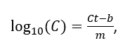 or
or
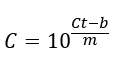
where C is genome copies per µL. - Average technical replicates and report final CME-AAV concentration as g.c./µL.
NOTE: Because heat denaturation during qPCR preparation disrupts both the enveloping membrane and AAV capsids, no separate DNA extraction is required. Do not perform DNase treatment on samples prior to heat lysis; DNase can access and degrade AAV genomes once capsids/membranes are disrupted, but before the sample reaches temperatures that inactivate DNase. Standards must be matched where possible: prepare a standard curve from a well-characterized AAV standard. If your samples or standards differ in genome configuration (single-stranded AAV vs self-complementary/double-stranded AAV), adjust reported values accordingly. Primer/probe design: primers may target any conserved AAV sequence; as an example, primers targeting the CMV promoter (upstream of the transgene) are commonly used.
5. Quantification of CME-AAV particle profiles
- Use Nanoparticle tracking analysis (NTA), tunable resistive pulse sensing (TRPS), or comparable particle analysis techniques to determine the size distribution and particle concentration of CME-AAV preparations.
- Thaw purified CME-AAV samples on ice and gently homogenize by pipetting prior to analysis. Perform measurements following standard laboratory practice and the manufacturer's instructions for the selected instrument, as described in previously published protocols12,27,28,29.
6. In vitro analysis of CME-AAV-mediated gene transfer
NOTE: This step describes the procedure to evaluate CME-AAV-mediated gene delivery and transgene expression in cultured cells. The assay is demonstrated using HEK293T cells and CME-AAV9.EGFP, but can be adapted to other cell types or reporter constructs. Conventional (non-membrane-enveloped) AAV9.EGFP serves as a positive control, while PBS or empty AAV particles are used as negative controls.
- Cell preparation
- Culture HEK293T cells under standard conditions (DMEM with 10% FBS, 1% penicillin-streptomycin) at 37 °C in 5% CO₂.
- Seed cells into 48-well or 96-well plates to reach approximately 70% confluency at the time of transduction (typically 24 h post-seeding). Adjust seeding density according to well size (e.g., ~5 × 10⁴ cells/well for 48-well plate or ~1 × 10⁴ cells/well for 96-well plate).
- Allow cells to attach and equilibrate for 24 h before adding vectors.
- Vector application
- Prepare CME-AAV9.EGFP and AAV9.EGFP working solutions in sterile PBS. Typical viral dose: 5 × 10⁸ g.c. per 48-well (adjust based on promoter strength, target gene, and experimental objectives).
- For each well, add equal genome copies (g.c.) of CME-AAV9.EGFP or AAV9.EGFP directly to the culture medium.
- Include negative control wells with an equivalent volume of PBS or empty AAV particles.
- Gently swirl the plate to ensure uniform distribution and return to the incubator.
- Continue incubation for 24-72 h, depending on promoter activity and transgene expression kinetics. For CMV-driven EGFP in HEK293T cells, strong fluorescence is typically observed within 48 h.
- Imaging and live-cell analysis
- When ready for imaging, gently aspirate the culture medium and wash cells twice with PBS to remove unbound particles. Avoid dislodging cells.
- Stain cell nuclei with Hoechst 33342 (live-cell compatible) following manufacturer's instructions. Do not use DAPI for live cells.
- Perform live-cell fluorescence imaging using an inverted fluorescence microscope equipped with appropriate filters (e.g., excitation 488 nm/emission 513 nm for EGFP; excitation 350 nm/emission 460 nm for Hoechst).
- Use 10× or 20× objectives for quantification of transduction efficiency and 63× objective for high-resolution subcellular localization if needed.
- Acquire at least five fields of view per well (e.g., four corners and one center) using identical imaging parameters (exposure time, gain, aperture, etc.).
- Save all images in raw and grayscale formats for quantitative analysis.
- Quantitative image analysis
- Open images in ImageJ (FIJI) or equivalent software.
- Measure mean fluorescence intensity for both the EGFP and Hoechst channels for each field.
- Determine background intensity by selecting a region without a fluorescent signal and averaging pixel values.
- Calculate corrected intensity for each channel:
Icorrected = Imean - Ibackground - Compute normalized intensity for each field as:
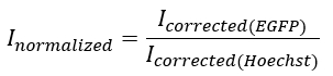
- Average normalized intensities from at least five fields per well, and report the mean ± SD across three independent biological replicates.
NOTE: CME-AAV typically demonstrates enhanced fluorescence intensity relative to conventional AAV when equal genome copies are applied, reflecting improved cellular uptake and/or protection from neutralizing antibodies. Optimal transduction efficiency depends on CME-AAV preparation quality, cell type, promoter strength, and viral titer. If weak signals are observed, increase the genome copy input, confirm vector integrity by qPCR, and verify microscope calibration. Other fluorescent reporters (e.g., mCherry, mNeonGreen, tdTomato) or luciferase constructs may be used depending on experimental needs. When working with primary or non-adherent cells, adjust seeding and imaging conditions accordingly (e.g., coating plates with poly-L-lysine or using chamber slides).
7. Evaluation of CME-AAV - mediated gene delivery in a murine model
NOTE: This step describes the evaluation of CME-AAV-mediated gene delivery in vivo using a murine model.
- Animal preparation
- Use wild-type mice such as C57BL/6. Ensure all animal procedures are approved by the institutional animal care and use committee and performed in accordance with relevant regulations.
- Select mice aged 4-6 weeks for optimal consistency across experiments.
- Maintain animals under standard housing conditions with ad libitum access to food and water prior to injection.
- Vector preparation and administration
- Select an AAV serotype according to the study purpose and target tissue. In this study, AAV9 and CME-AAV9 are used.
- Prepare viral vectors encoding a luciferase reporter gene (e.g., CME-AAV9.luciferase) for noninvasive imaging.
- Dilute the viral vectors in sterile saline to a final concentration of 1 × 10¹° genome copies (g.c.) per 50 µL for each mouse.
- Warm the tail gently using a heating pad or warm water to dilate the vein prior to injection.
- Load 50 µL of viral suspension into a 31-G insulin syringe and slowly inject into the lateral tail vein to minimize leakage and vein rupture.
- After injection, return the mice to their cages and monitor them until they are fully recovered.
- In vivo bioluminescence imaging
- Two weeks after administration, anesthetize the mice with isoflurane (according to institutionally approved protocols) and inject D-luciferin (150 µg/g body weight; Sigma) intraperitoneally in PBS.
- Place the anesthetized mice in a bioluminescence imaging system.
- Acquire luminescence images every 2 min post-injection until the signal reaches a plateau.
- Quantify bioluminescence using proper software. Define regions of interest (ROIs) around luminescent areas and calculate the total photon flux (photons/s) for each animal.
- Include AAV9.luc-treated and PBS-injected groups as positive and negative controls, respectively, for comparison.
- Ex vivo imaging
NOTE: This step is optional.- Immediately after in vivo imaging, euthanize the mice and collect major organs (liver, heart, spleen, kidney, and brain).
- Image the isolated organs under identical imaging parameters using the same IVIS system to determine tissue-specific luciferase expression.
NOTE: Alternative administration routes (e.g., intramuscular, intracerebral, or intraperitoneal injection) may be adopted depending on experimental goals. Bioluminescence intensity reflects both gene delivery efficiency and tissue biodistribution. CME-AAV-mediated luciferase expression typically exhibits earlier onset and enhanced peak expression compared to free AAV due to membrane-mediated endocytosis by up-taking cells. Ensure consistent luciferin dose, timing, and imaging settings across samples for accurate comparison. The described viral dose (1 × 10¹° g.c. per mouse) typically provides a detectable signal 2-4 weeks post-injection, but optimization may be required for different vectors or animal models.
Access restricted. Please log in or start a trial to view this content.
Results
MSCs used in this study were isolated from human umbilical cord tissue under protocols in accordance with relevant ethical guidelines. MSC membrane fractions were prepared according to the described protocol. AAV9.EGFP was produced in-house using a transient dual-plasmid transfection system in HEK293T cells, as previously reported12,26. For CME-AAV fabrication, membrane preparations, and AAV9.EGFP was co-incubated at a ratio of 150 µg of membrane protein per 1 × ...
Access restricted. Please log in or start a trial to view this content.
Discussion
Critical steps of the CME-AAV protocol
This protocol describes a method for generating MSC membrane-enveloped AAVs, and several steps are critical for successful CME-AAV formation. First, careful preprocessing of the cell membrane is essential. The goal of this step is to obtain membrane fragments that are suitable for extrusion while minimizing contamination from nuclear DNA, cytosolic proteins, and subcellular organelles. In addition, the size of membrane fragments is a key ...
Access restricted. Please log in or start a trial to view this content.
Disclosures
Dr. Daopin Wu is an employee of Guangdong Hengqin United Life Science Co., Ltd. The company did not provide funding for this study and had no role in study design, data collection, data analysis, manuscript preparation, or the decision to publish. The remaining authors declare no competing financial interests.
Acknowledgements
The work was supported by Guangdong S&T Program (2023B0303010002) to DM, and Guangdong Basic and Applied Basic Research Foundation (2025A1515011134) and BNU Faculty of Arts and Sciences Cross-disciplinary Research Project (12900-311324240581) to YL. This work was further supported by the instrumentation and technical expertise provided by the Instrumentation and Service Center for Science and Technology at Beijing Normal University.
Access restricted. Please log in or start a trial to view this content.
Materials
| Name | Company | Catalog Number | Comments |
|---|---|---|---|
| Bioluminescence imaging system | PerkinElmer | IVIS Spectrum optical imaging system (Lumina III) | |
| D-Luciferin potassium salt | Yeasen Biotechnology | 40902ES03 | |
| Fetal bovine serum | Yeasen Biotechnology | 40130ES76 | |
| Fluorescence microscope | Zeiss | Axio Observer 7 | |
| Formvar/carbon-coated copper grid 200 mesh | Millipore-Sigma | TEM-FCF400CU | |
| Glutaraldehyde | Millipore-Sigma | 354400 | |
| HBSS | Gibco | C14175500BT | |
| Hieff Unicon qPCR TaqMan Probe Master Mix | Yeasen Biotechnology | 11205ES08 | |
| High-glucose Dulbecco's modified Eagle's medium | Hyclone | SH30243 | |
| High-speed centrifuge | Beckman Coulter | Avanti J-E | |
| Hoechst 33342 | Beyotime | C1022 | |
| NTA instrument | Malvern Panalytical | NanoSight NS300 | |
| Open-top thinwall ultra-clear tube for ultracentrifuge | Beckman Coulter | 344059 | |
| OptiPrep iodixanol | Millipore-Sigma | D1556 | |
| Paraformaldehyde 4% | Beyotime | P0099 | |
| PBS | Biosharp | BL302A | |
| Penicillin-streptomycin | Millipore-Sigma | V900929 | |
| qPCR instrument | Applied Biosystems | QS6 real-time PCR system | |
| Rotor for high-speed centrifuge | Beckman Coulter | JA-25.50 | |
| SW 41 Ti Swing-bucket rotor for ultracentrifuge | Beckman Coulter | 331362 | |
| Transmission electron microscope | JEOL | JEM-2100F | |
| Ultracentrifuge | Beckman Coulter | Optima XE-100 | |
| Uranyl acetate | Electron Microscopy Sciences | NC1375332 | |
| Whatman Nuclepore polycarbonate membranes filters (200 nm) | Cytiva | 10417004 | |
| Whatman Nuclepore polycarbonate membranes filters (400 nm) | Cytiva | 10417104 |
References
- Wang, D., Tai, P. W. L., Gao, G. Adeno-associated virus vector as a platform for gene therapy delivery. Nat Rev Drug Discov. 18 (5), 358-378 (2019).
- Li, C., Samulski, R. J. Engineering adeno-associated virus vectors for gene therapy. Nat Rev Genet. 21 (4), 255-272 (2020).
- Mendell, J. R., et al. Current clinical applications of in vivo gene therapy with AAVs. Mol Ther. 29 (2), 464-488 (2021).
- Crunkhorn, S. Profiling aav tropism. Nat Rev Drug Discov. 22 (3), 183(2023).
- Kosaka, M., et al. Evaluation of the loading capacity and patterns of packaged DNA in AAV genomes of different sizes using long-read sequencing. Mol Ther Methods Clin Dev. 33 (2), 101474(2025).
- McCarty, D. M. Self-complementary AAV vectors; advances and applications. Mol Ther. 16 (10), 1648-1656 (2008).
- Lopez-Gordo, E., Chamberlain, K., Riyad, J. M., Kohlbrenner, E., Weber, T. Natural adeno-associated virus serotypes and engineered adeno-associated virus capsid variants: Tropism differences and mechanistic insights. Viruses. 16 (3), 442(2024).
- Louis Jeune, V., Joergensen, J. A., Hajjar, R. J., Weber, T. Pre-existing anti-adeno-associated virus antibodies as a challenge in AAV gene therapy. Hum Gene Ther Methods. 24 (2), 59-67 (2013).
- Wei, C. Prevalence of adeno-associated virus-9-neutralizing antibody in Chinese patients with Duchenne muscular dystrophy. Hum Gene Ther. 35 (1-2), 26-35 (2024).
- Directed evolution of AAV mutants for enhanced gene delivery. Schaffer, D. V., Maheshri, N. Conf Proc IEEE Eng Med Biol Soc, 2004, 3520-3523 (2004).
- Zhao, W., et al. Engineering sialylated N-glycans on adeno-associated virus capsids for targeted gene delivery and therapeutic applications. J Control Release. 380, 563-578 (2025).
- Liu, B., et al. Aav-containing exosomes as a novel vector for improved gene delivery to lung cancer cells. Front Cell Dev Biol. 9, 707607(2021).
- Li, X. Extracellular vesicle-encapsulated adeno-associated viruses for therapeutic gene delivery to the heart. Circulation. 148 (5), 405-425 (2023).
- Wang, W., et al. Intravitreal injection of an exosome-associated adeno-associated viral vector enhances retinoschisin 1 gene transduction in the mouse retina. Hum Gene Ther. 32 (13-14), 707-716 (2021).
- Breuer, C. B., et al. In vivo engineering of lymphocytes after systemic exosome-associated AAV delivery. Sci Rep. 10 (1), 4544(2020).
- Cheng, M., et al. Probing aspects of extracellular vesicle-associated AAV allows increased vector yield and insight into its transduction and immune-evasive properties. Mol Ther Methods Clin Dev. 33 (1), 101407(2025).
- Elmore, Z. C., et al. The membrane-associated accessory protein is an adeno-associated viral egress factor. Nat Commun. 12 (1), 6239(2021).
- Deshetty, U. M., Sil, S., Buch, S. Gene therapy for the heart: Encapsulated viruses to the rescue. Extracell Vesicles Circ Nucl Acids. 5 (1), 114-118 (2024).
- Fan, X., et al. Targeted repair of spinal cord injury based on miR-124-3p-loaded mesoporous silica camouflaged by stem cell membrane modified with rabies virus glycoprotein. Adv Sci (Weinh). 11 (21), e2309305(2024).
- Yao, C., et al. Self-assembly of stem cell membrane-camouflaged nanocomplex for microRNA-mediated repair of myocardial infarction injury. Biomaterials. , 257-120256 (2020).
- Tian, Y., et al. Biomimetic mesenchymal stem cell membrane-coated nanoparticle delivery of mkp5 inhibits hepatic fibrosis through the Ire/xbp1 pathway. J Nanobiotechnology. 22 (1), 741(2024).
- Kang, K., et al. Exosomes secreted from CXCR4 overexpressing mesenchymal stem cells promote cardioprotection via akt signaling pathway following myocardial infarction. Stem Cells Int. 2015, 659890(2015).
- Han, X., et al. Mesenchymal stem cells in treating human diseases: Molecular mechanisms and clinical studies. Signal Transduct Target Ther. 10 (1), 262(2025).
- Ong, S. G., Chitneni, M., Lee, K. S., Ming, L. C., Yuen, K. H. Evaluation of extrusion technique for nanosizing liposomes. Pharmaceutics. 8 (4), 36(2016).
- Tackenberg, M. W., Kleinebud, P. Encapsulation of liquids via extrusion--A review. Curr Pharm Des. 21 (40), 5815-5828 (2015).
- Bai, J. -J., et al. Dean-flow-coupled elasto-inertial focusing accelerates exosome purification to facilitate single vesicle profiling. Anal Chem. 95 (4), 2523-2531 (2023).
- He, S., et al. Biomimetic gene delivery system coupled with extracellular vesicle-encapsulated AAV for improving diabetic wound through promoting vascularization and remodeling of inflammatory microenvironment. J Nanobiotechnology. 23 (1), 242(2025).
- Smith, S. M., et al. Functional equivalence of stem cell and stem cell-derived extracellular vesicle transplantation to repair the irradiated brain. Stem Cells Transl Med. 9 (1), 93-105 (2020).
- Liang, Y., Lehrich, B. M., Zheng, S., Lu, M. Emerging methods in biomarker identification for extracellular vesicle-based liquid biopsy. J Extracell Vesicles. 10 (7), e12090(2021).
- Verma, S., et al. Seroprevalence of adeno-associated virus neutralizing antibodies in males with Duchenne muscular dystrophy. Hum Gene Ther. 34 (9-10), 430-438 (2023).
- Yan, B., Liang, Y. New therapeutics for extracellular vesicles: Delivering CRISPR for cancer treatment. Int J Mol Sci. 23 (24), 15758(2022).
- Hu, C. M. Erythrocyte membrane-camouflaged polymeric nanoparticles as a biomimetic delivery platform. Proc Natl Acad Sci U S A. 108 (27), 10980-10985 (2011).
- Aryal, S., et al. Erythrocyte membrane-cloaked polymeric nanoparticles for controlled drug loading and release. Nanomedicine (Lond). 8 (8), 1271-1280 (2013).
- Zhang, W., Huang, X. Stem cell membrane-camouflaged targeted delivery system in tumor. Mater Today Bio. 16, 100377(2022).
Access restricted. Please log in or start a trial to view this content.
Reprints and Permissions
