Here, we present a retrospective study (n=200) comparing anti-VEGF versus combination therapy, which showed faster early gains with combination therapy, but no sustained 6-month benefit and higher risks of elevated intraocular pressure and cataract progression.
Method Article
Clinical Outcomes of Anti-VEGF Monotherapy Versus Combination Therapy in Diabetic Macular Edema: A Retrospective Cohort Study
* These authors contributed equally
In This Article
Summary
Abstract
Diabetic macular edema (DME) is a leading cause of blindness involving vascular endothelial growth factor (VEGF) leakage and inflammation. This retrospective, single-center cohort study evaluated the efficacy and safety of intravitreal anti-VEGF monotherapy versus combination therapy with anti-VEGF plus dexamethasone implant in DME. A total of 200 patients (one eye per patient) treated between January 2021 and December 2024 were included, with 100 receiving anti-VEGF monotherapy and 100 receiving combination therapy. Best-corrected visual acuity (BCVA), central macular thickness (CMT), adverse events, and quality of life (NEI-VFQ-25) were assessed at baseline and at 1, 3, and 6 months. At 1 and 3 months, the combination therapy group showed faster improvement in BCVA (0.53±0.14 vs. 0.61±0.14 at 1 month; 0.46±0.13 vs. 0.53±0.12 at 3 months) and CMT (342±52 µm vs. 380±55 µm at 1 month; 315±48 µm vs. 345±50 µm at 3 months) compared with the monotherapy group (all P < 0.05), along with a higher rapid response rate (68.0% vs. 42.0%, P < 0.001). However, these differences were no longer significant at 6 months. The combination therapy group had higher rates of elevated intraocular pressure (18% vs. 6%, P < 0.05) and cataract progression (12% vs. 3%, P < 0.05). In conclusion, combination therapy was associated with faster early improvement in visual acuity and macular edema resolution within the first 3 months, but this early advantage was not sustained at 6 months and was accompanied by increased risks of elevated intraocular pressure and cataract progression.
Introduction
Diabetic macular edema (DME), as one of the most severe microvascular complications of diabetic retinopathy (DR), has become the leading cause of irreversible vision loss and legal blindness among working-age adults worldwide1. The persistent rise in global diabetes prevalence has led to an increased burden of DME, posing severe challenges to individual quality of life and public health systems. Epidemiological data confirm that the prevalence of DME reaches 10% among individuals with a 20-year history of diabetes. The risk of this condition is directly correlated with the level of glycemic, blood pressure, and lipid control, making systematic management crucial2.
The pathophysiology of DME involves a chronic, low-grade inflammatory process with multiple signaling pathways. Vascular endothelial growth factor (VEGF) plays a central role in the pathogenesis of DME, primarily by inducing a marked increase in vascular permeability and compromising the blood-retinal barrier through the disruption of inter-endothelial tight junctions3. In this, intravitreal injections of anti-VEGF drugs (such as ranibizumab, aflibercept, or bevacizumab) rapidly reduce vascular leakage by specifically neutralizing VEGF. Over the past decade, they have been established as the first-line standard treatment for DME with foveal involvement. Their efficacy in improving visual acuity has been demonstrated in multiple large randomized controlled clinical trials4,5,6.
However, limitations of anti-VEGF monotherapy have gradually become apparent. First, approximately 30–40% of patients continue to experience persistent or recurrent macular edema despite adequate anti-VEGF treatment, a phenomenon often termed “treatment resistance7. This suggests that mechanisms beyond the VEGF pathway, particularly inflammation, contribute to the disease. Elevated levels of inflammatory mediators such as interleukin-6 (IL-6), interleukin-8 (IL-8), and monocyte chemoattractant protein-1 (MCP-1) have been observed in the vitreous and aqueous humor of DME patients, and these factors can compromise blood-retinal barrier function. Therefore, for DME subtypes with a prominent inflammatory component, VEGF inhibition alone may be insufficient. Second, anti-VEGF treatment requires frequent injections and close follow-up, imposing significant time and financial burdens on patients and healthcare systems, which may reduce treatment adherence and affect long-term visual outcomes.
To overcome these challenges, researchers have turned their attention to corticosteroids (CS), which offer broad-spectrum anti-inflammatory effects and help stabilize the blood-retinal barrier8. Intravitreal dexamethasone implants provide sustained drug release for up to six months, potentially reducing treatment frequency. Despite their marked efficacy in reducing central macular thickness (CMT) and their inclusion in multiple treatment guidelines for DME, their clinical application remains constrained by two primary adverse effects: secondary intraocular pressure elevation9,10.
Against this backdrop, the concept of "combination therapy" has emerged as a potential strategy in DME treatment. The theoretical rationale is that anti-VEGF agents target the VEGF pathway to control acute leakage, while corticosteroids provide broader anti-inflammatory effects that may address non-VEGF-dependent pathways11,12. This approach has been hypothesized to offer faster anatomical resolution and potentially reduce treatment frequency13. However, it also introduces well-known corticosteroid-related risks, including elevated intraocular pressure and cataract progression. Given these trade-offs, the clinical value of combination therapy remains under investigation. This retrospective cohort study, therefore, aimed to compare the efficacy and safety of anti-VEGF monotherapy versus combination therapy with anti-VEGF plus dexamethasone implant in patients with DME, with a focus on early versus mid-term outcomes and the trade-off between therapeutic benefit and corticosteroid-related risks.
Protocol
This study was approved by the Institutional Review Board (IRB) of Guangdong Provincial People’s Hospital(Approval No.: GY--20200618). This clinical study complies with relevant ethical regulations, such as the Declaration of Helsinki14 and international research ethics standards. Informed consent was waived for this retrospective analysis of anonymized clinical data, with all identifiers removed to ensure privacy.
1. Patient selection and preoperative assessment (Figure 1)
Potential patients were screened for a diagnosis of center-involving DME, which was confirmed by optical coherence tomography (OCT) and defined as CMT > 300 µm. The diagnosis of type 2 diabetes mellitus was confirmed, and BCVA was verified to be between 0.05 and 0.5 on the Snellen chart (approximately 1.3 to 0.3 logarithm of the minimum angle of resolution (LogMAR)). For study purposes, BCVA was measured using the Early Treatment Diabetic Retinopathy Study (ETDRS) chart and converted to LogMAR units for analysis, with Snellen equivalents provided for clinical reference.

Figure 1: Research flowchart. Please click here to view a larger version of this figure.
The study cohort was established by applying predefined inclusion criteria15. Only patients with central macular edema (CMT > 300 µm) confirmed by OCT were included. Patients were required to have type 2 diabetes and baseline BCVA between 0.05 and 0.5 on the Snellen chart. Additionally, only patients with complete clinical data and a planned follow-up duration of ≥6 months were included.
Exclusion criteria were applied to ensure patient safety and protocol validity16,17. Patients with a history of intraocular surgery or laser treatment in the study eye within the past six months were excluded. Patients with co-existing ocular pathologies such as glaucoma, neovascular age-related macular degeneration, or retinal vascular occlusion in the study eye were also excluded. Individuals with known hypersensitivity to any component of ranibizumab, aflibercept, or dexamethasone were excluded, as were pregnant or lactating women.
A post-hoc sample size estimation was performed to assess the statistical adequacy of the retrospective dataset. Statistical software18 was used for the calculation, which was based on the primary efficacy measure (change in BCVA from baseline to 6 months), with a significance level (α) of 0.05 (two-tailed) and statistical power (1 – β) of 0.8, and assuming a large effect size (d=0.8) based on relevant literature19. The estimated minimum sample size was 58 patients per group (total 116). To account for a potential dropout rate of approximately 20% and to enable exploratory subgroup analyses, we enrolled 100 patients per group. This sample size provides adequate statistical power for the primary and secondary analyses.
A total of 200 patients were enrolled in this study, with 100 patients assigned to each treatment group. For patients with bilateral DME, only the study eye meeting the inclusion criteria was selected for analysis. If both eyes were eligible, the eye with the worse baseline BCVA was designated as the study eye to avoid within-patient correlation bias.
Follow-up and data completeness. All 200 enrolled patients completed the 6-month follow-up period. No patients were lost to follow-up, and no data were missing for the primary efficacy and safety endpoints. Therefore, no imputation methods were required.
Treatment group assignment
Treatment selection was determined by the attending ophthalmologist based on comprehensive clinical evaluation, including disease severity, inflammatory features (e.g., presence of hard exudates or cystoid edema on OCT), patient preference, and economic considerations. Patients with a history of glaucoma or significant lens opacity were more likely to receive anti-VEGF monotherapy to avoid corticosteroid-related adverse events. All treatment decisions were made prior to and independently of the study design.
Distribution of anti-VEGF agents. Ranibizumab and aflibercept were used in both groups. The distribution of these agents across groups is reported in the Results section (Additional treatment exposure data). The choice of anti-VEGF agent was not controlled in the analysis, and this heterogeneity is acknowledged as a limitation.
Treatment administration
All intravitreal injections were performed in a sterile operating room setting. The procedures were conducted by an experienced ophthalmologist. Preoperative topical anesthesia was administered, and routine antisepsis of the conjunctival sac was performed. Postoperative topical antibiotic eye drops were prescribed to prevent infection.
For patients assigned to the combination therapy group, the initial treatment consisted of same-day administration of both the anti-VEGF agent (ranibizumab 0.5 mg or aflibercept 2 mg) and the dexamethasone intravitreal implant (0.7 mg). The anti-VEGF agent was administered first via intravitreal injection, followed by administration of the dexamethasone implant in a separate quadrant using its dedicated injector system. Patients were classified as receiving combination therapy if they received at least one dexamethasone implant during the 6-month follow-up period. In this study, all patients in the combination group received the implant at baseline, with 32 patients (32.0%) receiving a second implant at month 3 due to recurrent edema. Unlike the monotherapy group, the combination therapy group did not receive a loading phase of three-monthly anti-VEGF injections, as the dexamethasone implant was expected to provide sustained anti-inflammatory coverage during the early treatment period. For subsequent treatments, anti-VEGF re-injection followed either a Treat-and-Extend (T&E) or Pro Re Nata (PRN) regimen (detailed criteria as described below). Re-implantation of the dexamethasone implant was considered based on its duration of efficacy (typically every 4–6 months) and clinical evidence of recurrent edema20.
Regarding the feasibility of this combined regimen, in China, the simultaneous administration of intravitreal anti-VEGF agents and dexamethasone implant on the same day is reimbursed under the national healthcare insurance system for patients meeting specific clinical criteria, including center-involving DME with documented inadequate response to monotherapy or presence of significant inflammatory features.
In the anti-VEGF monotherapy group, only intravitreal anti-VEGF injections (ranibizumab or aflibercept) were administered. Treatment was initiated with a loading phase of three-monthly injections. Thereafter, anti-VEGF re-injection followed the same T&E or PRN criteria as described for the combination therapy group. Specifically, in the T&E regimen, the injection interval was extended by 2-week increments in the absence of disease activity (CMT stable or decreased, no new fluid on OCT, and stable BCVA), up to a maximum of 12 weeks. In the PRN regimen, re-injection was performed upon meeting any of the following: (1) CMT increase ≥50 µm from the lowest recorded value, (2) new or persistent intraretinal or subretinal fluid on OCT, or (3) BCVA decline of ≥5 letters compared to the previous visit21.
Observation and measurement procedures
Primary efficacy endpoints were assessed at baseline, 1, 3, and 6 months post-treatment. BCVA was measured using a standard ETDRS chart or equivalent under standardized lighting conditions, and the results were converted to LogMAR units for recording and analysis22. CMT was measured using spectral-domain OCT, with the device's built-in software used to calculate the average retinal thickness within a 1 mm-diameter central subfield centered on the fovea. Treatment response was defined and evaluated as follows: "Rapid Response" was defined as a reduction in CMT of ≥100 µm at the 1-month visit compared to baseline, while "Effective Response" was defined as an improvement in BCVA of ≥5 letters (≈0.1 LogMAR) and a reduction in CMT of ≥20% at the 6-month visit compared to baseline23. Lens changes were quantified using anterior segment optical coherence tomography (AS-OCT) to measure lens thickness (LT) and lens density (LD) at each follow-up visit24. Cataract outcomes were analyzed only in phakic eyes at baseline (i.e., patients with prior cataract surgery or aphakia were excluded from lens-specific analyses). Baseline lens status was comparable between groups (Table 1), and no adjustment was made for lens status beyond this exclusion.
Safety indicators were monitored at each follow-up visit. Intraocular pressure (IOP) was measured using a calibrated tonometer, and any elevation defined as IOP > 25 mmHg or an increase of >10 mmHg from baseline was recorded25. The lens was assessed using slit-lamp biomicroscopy, and any new onset or progression of cataract was documented and graded according to the Lens Opacities Classification System III (LOCS III)26. All adverse events, including serious adverse reactions such as endophthalmitis, retinal detachment, or infectious keratitis, were documented27. Quality of life was evaluated using the validated Chinese version of the National Eye Institute Visual Function Questionnaire-25 (NEI-VFQ-25)27. The questionnaire was administered by trained research staff at baseline and at each follow-up visit (1, 3, and 6 months) in a standardized manner.
Data management and statistical analysis
Statistical analysis was performed using appropriate software. Quantitative data that were normally distributed were presented as mean ± standard deviation (x̄ ± s). Continuous variables between the two treatment groups at each time point were compared using independent samples t-tests. To evaluate the overall treatment effect over time and assess the interaction between treatment group and time, a two-way repeated-measures analysis of variance (ANOVA) with group (combination therapy vs. monotherapy) as the between-subjects factor and time (baseline, 1 month, 3 months, 6 months) as the within-subjects factor was performed. This model enabled formal testing of the group × time interaction, which is essential for determining whether the treatment effect differs across groups over the follow-up period. Categorical data were presented as counts and percentages (n, %), and comparisons between groups were performed using the Chi-square test or Fisher's exact test, as appropriate.
Results
Baseline characteristics
As shown in Table 1, baseline characteristics for the combination therapy group and the anti-VEGF monotherapy group (n = 100 each) are as follows. The two groups showed no significant differences in any of the demographic and clinical characteristics assessed, including age, gender, diabetes duration, baseline BCVA and CMT, lens status, and NEI-VFQ-25 scores (all P > 0.05). This indicates that the baseline homogeneity of patients in both groups was good and comparable.
| Variable | Combination Therapy Group (n=100) | Anti-VEGF Monotherapy Group (n=100) | Test | 95% CI | χ²/t | P-value |
| Age (years) | 58.6±7.2 | 59.3±6.8 | Independent-samples t-test | (-2.671, 1.251) | 0.706 | 0.688 |
| Gender(Male/Female) | 57/43 | 61/39 | Chi-Square Test | - | 0.331 | 0.565 |
| Duration of diabetes (years) | 12.3±4.5 | 11.8±4.2 | Independent-samples t-test | (-0.794 , 1.614) | 0.812 | 0.467 |
| Glycated hemoglobin(%) | 7.8±1.3 | 7.6±1.2 | Independent-samples t-test | (-0.225, 0.485) | 1.13 | 0.854 |
| History of hypertension(n,%) | 52(52.0) | 48(48.0) | Chi-Square Test | - | 0.32 | 0.571 |
| Baseline BCVA(LogMAR) | 0.68±0.15 | 0.69±0.14 | Independent-samples t-test | (-0.04, 0.038) | 0.487 | 0.421 |
| Baseline CMT(μm) | 492±68 | 485±72 | Independent-samples t-test | (-12.67, 26.65) | 0.706 | 0.938 |
| Lens Status (Natural Lens Eye/Artificial Lens Eye) | 82/18 | 79/21 | Chi-Square Test | - | 0.287 | 0.592 |
| Lens thickness(μm) | 4506.2±215.8 | 4498.5±208.4 | Independent-samples t-test | (-51.47, 66.85) | 0.258 | 0.921 |
| Lens Density (Gray Scale Units) | 25.3±4.2 | 24.9±4.0 | Independent-samples t-test | (-0.735, 1.535) | 0.689 | 0.737 |
| NEI-VFQ-25 | 52.3±8.6 | 51.8±9.2 | Independent-samples t-test | (-1.895, 3.075) | 0.397 | 0.108 |
Table 1: Group characteristics at baseline [x̄ ± s or n (%)]. Independent t-test for continuous variables; Chi-square test for categorical variables. Abbreviations: BCVA = best-corrected visual acuity; CMT = central macular thickness; LogMAR = logarithm of the minimum angle of resolution; NEI-VFQ-25 = National Eye Institute Visual Function Questionnaire-25; VEGF = vascular endothelial growth factor.
BCVA outcomes
Table 2 presents the BCVA values at each time point. The temporal trend is illustrated in Figure 2. Results showed that at 1 and 3 months, the combination therapy group achieved faster improvement in BCVA compared with the monotherapy group (1 month: 0.53 ± 0.14 vs. 0.61 ± 0.14; 3 months: 0.46 ± 0.13 vs. 0.53 ± 0.12, both P < 0.05). However, by 6 months, BCVA values were similar between groups (0.35 ± 0.14 vs 0.37 ± 0.13, P > 0.05), indicating that the early benefit was not sustained.
| Combination Therapy Group | Anti-VEGF Monotherapy Group | Test | 95% CI | χ²/t | P-value | |
| (n=100) | (n=100) | |||||
| Baseline | 0.68±0.15 | 0.69±0.14 | Independent-samples t-test | (-0.04, 0.038) | 0.487 | 0.421 |
| 1 month | 0.53±0.14 | 0.61±0.14 | Independent-samples t-test | (-1.226, -0.043) | 4.04 | <0.001 |
| 3 month | 0.46±0.13 | 0.53±0.12 | Independent-samples t-test | (-0.103, -0.035) | 3.96 | 0.001 |
| 6 month | 0.35±0.14 | 0.37±0.13 | Independent-samples t-test | (-0.056, 0.017) | 1.05 | 0.296 |
Table 2: Comparison in BCVA changes between the two groups before and after treatment (x̄±s). Independent t-test for between-group comparisons at each time point. Abbreviations: BCVA = best-corrected visual acuity; VEGF = vascular endothelial growth factor.
CMT outcomes
Table 3 presents the CMT values at each time point. Figure 3 illustrates the temporal trend Results indicate that the combination therapy group demonstrated faster resolution of macular edema. Combination therapy demonstrated significantly greater efficacy than anti-VEGF monotherapy in reducing CMT: at 1 month, the reduction was -150 ± 45 µm versus -105 ± 38 µm; at 3 months, it was -185 ± 52 µm versus -155 ± 48 µm (both P < 0.05). At the 6-month mark, there was no significant difference in CMT values between the two groups.
| Combination Therapy Group | Anti-VEGF Monotherapy Group | Test | 95% CI | χ²/t | P-value | |
| (n=100) | (n=100) | |||||
| Baseline | 492±68 | 485±72 | Independent-samples t-test | (-12.67, 26.65) | 0.706 | 0.938 |
| 1 month | 342±52 | 380±55 | Independent-samples t-test | (-53.92, -24.08) | 5.02 | <0.001 |
| 3 month | 307±48 | 330±51 | Independent-samples t-test | (-35.82, -8.19) | 3.28 | 0.001 |
| 6 month | 298±45 | 305±47 | Independent-samples t-test | (-19.82, 5.839) | 1.07 | 0.283 |
Table 3: Comparison of CMT changes in two groups of patients before and after treatment (x̄±s, µm). Independent t-test for between-group comparisons at each time point. Abbreviations: CMT = central macular thickness; VEGF = vascular endothelial growth factor.
Response rates
Table 4 compares the rapid response rates between the two groups post-treatment. Results indicate that the rapid response rate (CMT reduction ≥ 100 µm at 1 month) in the combination therapy group was 68.0% (68/100), significantly higher than the 42.0% (42/100) observed in the anti-VEGF monotherapy group (χ2 = 13.66, P < 0.001). No statistically significant difference in the effective response rate was observed between the combination therapy group (75.0%, 75/100) and the anti-VEGF monotherapy group (72.0%, 72/100) at 6 months (χ2 = 0.231, P = 0.631).
| Combination Therapy Group | Anti-VEGF Monotherapy Group | χ²/t | P-value | |
| (n=100) | (n=100) | |||
| Rapid Response Rate | 68(68.0%) | 42 (42.0%) | 13.66 | <0.001 |
| 6-month effective response rate | 75(75.0%) | 72 (72.0%) | 0.231 | 0.631 |
Table 4: Comparison of treatment response rates between two groups of patients [ n (%)]. Chi-square test. Abbreviation: VEGF = vascular endothelial growth factor.
Lens changes
Table 5 compares changes in lens thickness and density between the two patient groups. Results indicate that dexamethasone implants were associated with significant lens changes. Lens density in the combination therapy group increased markedly as early as 3 months post treatment, and lens thickness also increased significantly by 6 months, consistent with cataract progression. These findings support the known association between corticosteroid use and cataract development.
| Timing | Combination Therapy Group | Anti-VEGF Monotherapy Group | χ²/t | P-value | |
| (n=100) | (n=100) | ||||
| Lens Thickness (μm) | Baseline | 4506.2 ± 215.8 | 4498.5 ± 208.4 | 0.258 | 0.921 |
| 1 month | 4512.4 ± 208.1 | 4501.2 ± 206.3 | 0.382 | 0.703 | |
| 3 month | 4530.5 ± 222.1 | 4505.3 ± 210.7 | 0.823 | 0.414 | |
| 6 month | 4625.8 ± 235.6 | 4512.1 ± 209.5 | 3.606 | <0.001 | |
| Lens Density (Gray Scale Units) | Baseline | 25.3 ± 4.2 | 24.9 ± 4.0 | 0.689 | 0.737 |
| 1 month | 26.3 ± 4.3 | 25.0 ± 4.0 | 2.214 | 0.028 | |
| 3 month | 27.1 ± 4.5 | 25.2 ± 4.1 | 3.121 | 0.002 | |
| 6 month | 31.5 ± 5.1 | 25.5 ± 4.3 | 8.994 | <0.001 |
Table 5: Comparison of lens thickness and density changes between two patient groups (x̄ ± s). Independent t-test for between-group comparisons at each time point. Abbreviation: VEGF = vascular endothelial growth factor.
Adverse events
Table 6 compares adverse reactions between the two patient groups post-treatment. Results showed that the incidence of elevated intraocular pressure at 3 months and 6 months (15%, 18%) was significantly higher in the combination therapy group than in the monotherapy group (5%, 6%). Additionally, the cataract progression/incidence at 6 months in the combined treatment group (12%) was also significantly greater than in the monotherapy group (3%). The incidence of injection-related adverse reactions (e.g., injection site pain and subconjunctival hemorrhage) was comparable between the groups. The incidence of serious adverse events was extremely low and did not differ between groups. This indicates that while combination therapy offers therapeutic advantages, it does indeed increase specific risks associated with corticosteroids.
| Timing | Combination Therapy Group | Anti-VEGF Monotherapy Group | χ²/t | P-value | |
| (n=100) | (n=100) | ||||
| Elevated intraocular pressure | 1 month | 8 (8%) | 4 (4%) | 1.418 | 0.234 |
| 3 month | 15 (15%) | 5 (5%) | 5.556 | 0.018 | |
| 6 month | 18 (18%) | 6 (6%) | 6.818 | 0.009 | |
| Progression/New Onset of Cataracts | 1 month | 2 (2%) | 1 (1%) | - | 1.000* |
| 3 month | 5 (5%) | 2 (2%) | 0.592 | 0.442 | |
| 6 month | 12 (12%) | 3 (3%) | 5.838 | 0.016 | |
| Pain at the injection site | 1 month | 10 (10%) | 8 (8%) | 0.244 | 0.621 |
| 3 month | 12 (12%) | 10 (10%) | 0.204 | 0.651 | |
| 6 month | 15 (15%) | 12 (12%) | 0.385 | 0.535 | |
| Subconjunctival hemorrhage | 1 month | 15 (15%) | 12 (12%) | 0.385 | 0.535 |
| 3 month | 10 (10%) | 8 (8%) | 0.244 | 0.621 | |
| 6 month | 5 (5%) | 4 (4%) | - | 1.000* | |
| Serious adverse reactions | 1 month | 0 (0%) | 0 (0%) | - | - |
| 3 month | 1 (1%) | 0 (0%) | - | 1.000* | |
| 6 month | 2 (2%) | 1 (1%) | - | 1.000* |
Table 6: Comparison of adverse reaction incidence between two patient groups [ n (%)]. Chi-square test. Abbreviation: VEGF = vascular endothelial growth factor.
Quality of life outcomes
Based on NEI-VFQ-25 quality of life scores, the study findings indicate that at 1 and 3 months post-treatment, the combination therapy group demonstrated significantly greater improvements than the anti-VEGF monotherapy group in total scores and in the "Near Activities" and "Vision Dependence" dimensions, reflecting a marked early advantage in quality of life. However, by the sixth month, there were no statistically significant differences between the two groups in any of the scores, indicating that combination therapy has short-term benefits in accelerating quality-of-life recovery, while its long-term effectiveness is comparable to monotherapy. Table 7 presents the NEI-VFQ-25 scores across the two groups. Notably, despite the higher incidence of corticosteroid-related adverse events in the combination therapy group (Table 6), quality-of-life scores did not differ significantly between groups at any time point. This finding suggests that the early functional and anatomical improvements achieved with combination therapy may have a sustained positive effect on patients' perceived visual function, potentially offsetting the subjective burden of manageable IOP elevation and early cataract progression within the 6-month follow-up window.
| Timing | Combination Therapy Group | Anti-VEGF Monotherapy Group | χ²/t | P-value | |
| (n=100) | (n=100) | ||||
| Overall Score | Baseline | 52.3±8.6 | 51.8±9.2 | 0.397 | 0.108 |
| 1 month | 60.4±8.3 | 57.1±9.0 | 2.695 | 0.008 | |
| 3 month | 68.5±9.1 | 62.3±8.7 | 4.925 | <0.001 | |
| 6 month | 72.6±7.1 | 71.4±8.0 | 1.122 | 0.263 | |
| Close-range activities | 1 month | 62.0±10.3 | 57.0±9.8 | 3.517 | <0.001 |
| 3 month | 65.2±10.6 | 58.6±9.6 | 4.593 | <0.001 | |
| 6 month | 66.0±10.4 | 65.0±9.3 | 0.717 | 0.474 | |
| Visual dependency | 1 month | 66.3±11.2 | 60.0±10.5 | 4.104 | <0.001 |
| 3 month | 70.1±9.6 | 63.5±9.5 | 4.887 | <0.001 | |
| 6 month | 71.0±10.6 | 70.0±10.5 | 0.748 | 0.455 |
Table 7: Comparison of NEI-VFQ-25 scores before and after treatment in two patient groups [x̄ ± s , points]. Independent t-test for between-group comparisons at each time point. Abbreviations: VEGF = vascular endothelial growth factor; NEI-VFQ-25 = National Eye Institute Visual Function Questionnaire-25.
Treatment exposure data
Additional treatment exposure data. During the 6-month follow-up period, patients in the combination therapy group received a mean of 2.1 ± 0.8 anti-VEGF injections, with 68 patients (68.0%) receiving one dexamethasone implant and 32 patients (32.0%) receiving two implants (re-implantation at month 3 due to recurrent edema). In the anti-VEGF monotherapy group, patients received a mean of 4.5 ± 1.2 anti-VEGF injections, reflecting the loading phase (three monthly injections) followed by additional injections per T&E or PRN criteria. Regarding the choice of anti-VEGF agent, ranibizumab was used in 54 patients (54.0%) in the combination group and 52 patients (52.0%) in the monotherapy group, while aflibercept was used in 46 patients (46.0%) and 48 patients (48.0%), respectively, with no significant difference in distribution between groups (P = 0.78).
Data Availability:
All data generated or analyzed during this study are included in this article.
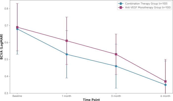
Figure 2: Temporal trends of best-corrected visual acuity (LogMAR) over 6 months in the anti-VEGF monotherapy and combination therapy groups. Data are presented as mean ± standard deviation. Abbreviations: BCVA = best-corrected visual acuity; VEGF = vascular epithelial growth factor; LogMAR = logarithm of the minimum angle of resolution. Please click here to view a larger version of this figure.
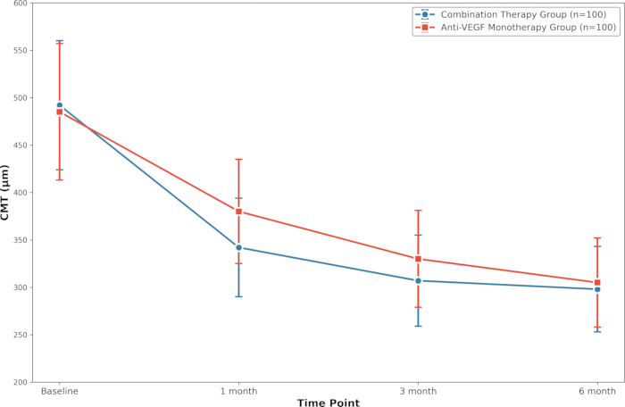
Figure 3: Temporal trends of central macular thickness (µm) over 6 months in the anti-VEGF monotherapy and combination therapy groups. Data are presented as mean ± standard deviation. Abbreviation: VEGF = vascular epithelial growth factor; CMT = central macular thickness. Please click here to view a larger version of this figure.
Discussion
This retrospective analysis of 200 DME patients over 6 months provides real-world evidence for understanding the clinical value of combined anti-VEGF and corticosteroid therapy. The combination therapy strategy was associated with faster early improvement in visual acuity and macular thickness within the first 3 months. However, this early advantage was not sustained at 6 months and was accompanied by higher rates of corticosteroid-related adverse events. These findings highlight a trade-off between short-term efficacy and safety risks, supporting individualized treatment selection rather than broad adoption of combination therapy.
The combined therapy was superior to anti-VEGF monotherapy in improving BCVA and reducing CMT at both the 1-month and 3-month time points. The combined therapy group showed statistically superior outcomes in both visual acuity (BCVA improvement: -0.08 vs. -0.07 LogMAR) and anatomic structure (CMT reduction: -150 µm vs. -185 µm) relative to the monotherapy group (all P < 0.05), consistent with prior literature. For example, the DRCR.net Protocol U study, despite its smaller sample size, also suggested that adding dexamethasone implants could yield further anatomical improvement in patients with a poor response to anti-VEGF28. More recently, Lin et al.29 reported that simultaneous intravitreal dexamethasone and aflibercept achieved sustained anatomical and functional improvement over 2 years in patients with DME. Study extends this observation to a broader initial treatment population and clarifies the temporal window for its "early" benefit. The clinical significance of this early advantage should not be underestimated. First, it lies in "rapid restoration." For patients experiencing sudden vision loss due to diffuse macular edema or large cystoid edema, a prolonged recovery process means sustained impairment in quality of life and psychological distress. Combination therapy can rapidly break the vicious cycle of edema, creating an optimal microenvironment for the recovery of retinal photoreceptor function9,30. This may positively influence the long-term prognosis of visual function. Second, it lies in the opportunity for "functional remodeling." Theoretical evidence suggests that prolonged chronic macular edema may lead to irreversible apoptosis and structural remodeling of retinal neurons (photoreceptor cells, bipolar cells, etc.)30. Therefore, the earlier and more thoroughly edema is eliminated, the greater the likelihood of protecting and salvaging neurons on the brink of death, thereby preserving more visual potential. For certain patients with specific needs—such as those requiring surgery on the contralateral eye, seeking driving qualifications, or pursuing visually demanding professions—this rapid visual improvement holds immense practical value.
The early advantages of combination therapy are likely attributable to complementary mechanisms. Anti-VEGF agents rapidly reduce vascular permeability by blocking VEGF signaling31, while corticosteroids exert broad-spectrum anti-inflammatory effects that help stabilize the blood-retinal barrier and reduce edema32,33. In DME, persistent hyperglycemia drives a chronic inflammatory network involving multiple cytokines (e.g., IL-6, IL-8) and transcription factors (e.g., NF-κB)34, which may not be fully addressed by anti-VEGF monotherapy. By targeting both VEGF and inflammatory pathways, combination therapy may achieve faster edema resolution, particularly in patients with a prominent inflammatory component.
The results of this study suggest that the incidence of adverse reactions was significantly higher in the combination therapy group than in the monotherapy group, mainly due to two inherent risks of corticosteroids: increased intraocular pressure (cumulative incidence 18% vs 6%) and cataract progression/new onset (12% vs 3%). This is consistent with the known pharmacological effects and safety profile of the drug. Regarding IOP elevation, the 18% incidence in the combination therapy group represented transient increases controllable with topical antihypertensive agents, with no cases requiring glaucoma surgery. This is related to the pharmacokinetics of the dexamethasone implant, which exhibits a high-to-low release curve pattern5. This underscores the critical importance of establishing a structured IOP monitoring regimen for patients receiving combination therapy (especially those on long-acting corticosteroid regimens), including scheduled follow-ups at 1 week, 1 month, and 2 months post-surgery. Prior to treatment, thorough screening and risk assessment for patients with glaucoma risk factors35 (such as high myopia, family history of glaucoma, or history of elevated IOP in the contralateral eye) constitute the first step in ensuring safety. Regarding cataracts, this study observed a 12% progression/incidence rate in the combined therapy group among patients with intact lenses, significantly higher than the 3% rate in the anti-VEGF monotherapy group. This represents a long-term consequence of receiving prolonged corticosteroid treatment. Therefore, when discussing treatment options with patients, it is essential to fully inform them that this is an anticipated adverse event, not an incidental occurrence. For older patients with existing signs of lens opacity, it is reasonable to anticipate that cataract surgery will likely be required within the next 1–2 years. However, for younger patients with intact lenses, this side effect warrants greater consideration in decision-making.
An additional observation worth discussing is the apparent discrepancy between the higher rates of cataract progression in the combination therapy group (12% vs. 3%) and the comparable NEI-VFQ-25 scores between groups throughout the follow-up period. Several factors may account for this finding. First, the early rapid improvement in visual acuity and anatomical outcomes observed at 1 and 3 months likely had a sustained positive impact on patients' perceived visual function, which may have outweighed the subjective effects of early-stage cataract changes within the 6-month follow-up window. Second, cataract progression in this study was defined based on objective measures (increased lens thickness and density), which may not yet have translated into clinically meaningful visual symptoms or functional impairment in daily activities within the relatively short follow-up period. Third, the NEI-VFQ-25 questionnaire assesses multiple dimensions of vision-related quality of life; early improvements in domains such as "Near Activities" and "Vision Dependence" may have compensated for any subtle decline related to early cataract formation. These findings highlight the importance of longer follow-up to fully capture the impact of corticosteroid-induced cataract progression on patient-reported outcomes.
Combination therapy may be considered as an option for selected patients rather than a universal first-line approach. Potential candidate groups include: First, patients requiring rapid visual recovery: As previously noted, individuals with urgent visual improvement needs due to occupational, lifestyle demands, or psychological factors; Second, DME patients with high inflammatory burden: Clinically, patients exhibiting vitreous inflammatory cells, extensive macular hard exudates, or optical coherence tomography angiography revealing widespread destruction of the retinal capillary network may have a heavier inflammatory component and represent ideal candidates for combination therapy36. Detection of inflammatory markers in aqueous humor or vitreous fluid may serve as biomarkers for precise subtyping. Third, patients with poor or inadequate response to anti-VEGF therapy: For "initial non-responders" showing less than 20% reduction in CMT or no visual acuity improvement after three loading-dose treatments, adding corticosteroids is a reasonable escalation strategy. Fourth, patients are unable to bear the burden of frequent injections: For those residing in remote areas or with poor follow-up compliance, the "burden-reducing" effect of combination therapy may outweigh its risks. Additionally, for patients with double-monotherapy-resistant chronic DME, combination therapy has shown promising results as a rescue strategy37. Notably, the difference in efficacy metrics between the two groups disappeared at 6 months, indicating that the early advantage of combination therapy did not persist beyond 6 months. Several factors may explain this convergence. First, the anti-VEGF monotherapy group achieved “therapeutic catch-up” through continued monthly injections (mean 4.5 vs. 2.1 injections in the combination group), gradually narrowing the initial gap. Second, the dexamethasone implant exhibits peak efficacy within the first 3 months with a gradual decline thereafter, contributing to the convergence of outcomes by month 6. Third, the higher rate of cataract progression in the combination therapy group (12% vs. 3%) may have partially offset early functional gains. Regarding longer-term outcomes, cataract surgery in combination therapy patients who develop clinically significant cataracts could restore lens clarity and eliminate cataract-related visual impairment, potentially allowing the full anatomical benefits of prior combination therapy to be realized. This potential for long-term benefit is supported by real-world evidence: Choi et al.38 reported in a 10-year study that patients receiving combination therapy maintained favorable visual outcomes with acceptable safety profiles. Future studies with extended follow-up are warranted to evaluate this hypothesis.
This single-center retrospective analysis has several noteworthy limitations. First, although the sample size (n=200) met the minimum statistical requirement, it remains relatively small. This may compromise the power of statistical tests, particularly in subgroup analyses where subtle yet clinically significant differences may be difficult to detect. Second, the non-randomized study design inevitably carries a risk of selection bias. Treatment allocation may have been influenced by multiple factors, including patient baseline characteristics, physician preferences, and patient financial status. Although baseline data indicated comparability between groups, potential confounding factors could still compromise the reliability of the results. Third, while the 6-month follow-up period allows assessment of early treatment efficacy and safety, it is clearly insufficient for evaluating long-term outcomes, visual acuity maintenance, and steroid-related late complications (such as cataract progression requiring surgical intervention and persistent high intraocular pressure risk). Fourth, the study did not perform stratified analysis based on morphological subtypes of DME (e.g., diffuse edema, cystoid edema), despite these subtypes potentially exhibiting differential treatment responses. This omission limits the study's applicability in precision medicine contexts. Fifth, the single-center design may limit the generalizability of our findings, as the treatment and management approaches may not fully reflect practices at other institutions. Sixth, the simultaneous administration of anti-VEGF and dexamethasone implant on the same day, while reimbursed in our healthcare system, may not be covered or feasible in other countries. This limits the generalizability of our findings to regions with different reimbursement policies, and the applicability of this combined regimen may vary depending on local healthcare regulations. Seventh, this study exclusively used the dexamethasone intravitreal implant (Ozurdex) as the corticosteroid component. Other corticosteroid formulations, such as intravitreal triamcinolone acetonide or subtenon triamcinolone injection, were not evaluated. Therefore, the findings may not be directly generalizable to other corticosteroid preparations, which may have different efficacy and safety profiles. Eighth, the study included both ranibizumab and aflibercept as anti-VEGF agents, and the distribution between groups was balanced but not adjusted in the analysis. This heterogeneity may influence outcomes and is acknowledged as a limitation. These limitations need to be addressed and validated in the future through more rigorous, larger-sample, longer-follow-up multicenter prospective randomized controlled trials. In summary, combination therapy was associated with faster early improvement in visual acuity and macular thickness within the first 3 months, but this early advantage was not sustained at 6 months and was accompanied by higher rates of elevated intraocular pressure and cataract progression. These findings suggest that combination therapy may offer short-term benefits for selected patients, with careful consideration of the trade-off between efficacy and safety. Future research should focus on optimizing patient selection, refining treatment protocols, and developing anti-inflammatory agents with improved safety profiles. As DME management evolves toward precision medicine, treatment decisions should be individualized based on patient characteristics and clinical needs.
Disclosures
The authors have no conflicts of interest to disclose.
Acknowledgements
The study received funding from The Research Funds of the State Key Laboratory of Ophthalmology (No.2025QNJS13) and Key Projects of Huizhou Science and Technology (No.2023CZ010009).
Materials
| Name | Company | Catalog Number | Comments |
|---|---|---|---|
| Aflibercept | Regeneron Pharmaceuticals / Bayer | 2 mg/0.05 mL | Anti-VEGF agent used in intravitreal injections. |
| Anterior Segment OCT (AS-OCT) | Zeiss | Visante OCT / Anterion | Used for measuring lens thickness and density. |
| Dexamethasone Intravitreal Implant (Ozurdex®) | Allergan (AbbVie) | 0.7 mg | Biodegradable sustained-release corticosteroid implant. |
| National Eye Institute Visual Function Questionnaire-25 (NEI-VFQ-25) | N/A | N/A | Patient-reported quality of life assessment tool. |
| Non-contact Tonometer | Suzhou Liuliu Vision Technology Co., Ltd. | YZ30 | Pneumatic tonometer for intraocular pressure (IOP) monitoring. |
| Optical Coherence Tomography (OCT) | Shenzhen Moting Medical Technology Co., Ltd. | Colombo IOL series | Used for measuring Central Macular Thickness (CMT). |
| Povidone-Iodine 5% Ophthalmic Solution | Alcon | 5% w/v sterile solution | Gold standard for pre-intravitreal injection antisepsis. Used for conjunctival sac and eyelid skin disinfection. Contact time ≥30 seconds, followed by irrigation with sterile BSS to minimize endophthalmitis risk. |
| Ranibizumab | Novartis | 0.5 mg/0.05 mL | Anti-VEGF agent used in intravitreal injections. |
| Sample Size Calculation Software | G*Power | Version 3.1 | Used for a priori power analysis. |
| Slit Lamp Microscope | Wuhan Yijiexun’an Trading Co., Ltd. / Shanghai Yimu Medical Equipment Co., Ltd. | YZ5G | For cataract assessment using LOCS III grading. |
| Statistical Analysis Software | IBM SPSS Statistics | Version 25.0 | Used for all data analysis. |
| Topical Antibiotic Eye Drops | Alcon | 0.5% Moxifloxacin | Broad-spectrum fluoroquinolone, common choice for post-operative prophylaxis.Frequently specified in clinical trial protocols, typically used QID for 3-7 days post-injection. |
| Topical Anesthetic Eye Drops | Akorn | 0.5% Proparacaine HCl | Most commonly used topical anesthetic.Rapid onset (~20 seconds). 1-2 drops are instilled pre-injection for adequate corneal and conjunctival anesthesia. |
| Visual Acuity Chart | Wenzhou Xingkang Medical Technology Co., Ltd. | XK100 | Low-vision chart for BCVA testing, converted to LogMAR. |
References
- Yau, J. W., et al. Global prevalence and major risk factors of diabetic retinopathy. Diabetes Care. 35 (3), 556-564 (2012).
- Funatsu, H., et al. Vitreous levels of interleukin-6 and vascular endothelial growth factor are related to diabetic macular edema. Ophthalmology. 110 (9), 1690-1696 (2003).
- Mitchell, P., et al. The RESTORE study: ranibizumab monotherapy or combined with laser versus laser monotherapy for diabetic macular edema. Ophthalmology. 118 (4), 615-625 (2011).
- Schmidt-Erfurth, U., et al. Guidelines for the management of diabetic macular edema by the European Society of Retina Specialists (EURETINA). Ophthalmologica. 237 (4), 185-222 (2017).
- Boyer, D. S., et al. Three-year, randomized, sham-controlled trial of dexamethasone intravitreal implant in patients with diabetic macular edema. Ophthalmology. 121 (10), 1904-1914 (2014).
- Maturi, R. K., et al. Effect of adding dexamethasone to continued ranibizumab treatment in patients with persistent diabetic macular edema: a DRCR Network phase 2 randomized clinical trial. JAMA Ophthalmol. 136 (1), 29-38 (2018).
- Wykoff, C. C., et al. Randomized trial of treat-and-extend versus monthly dosing for neovascular age-related macular degeneration: 2-year results of the TREX-AMD study. Ophthalmol Retina. 1 (4), 314-321 (2017).
- Barnes, P. J. How corticosteroids control inflammation: Quintiles Prize Lecture 2005. Br J Pharmacol. 148 (3), 245-254 (2006).
- Das, A., McGuire, P. G., Rangasamy, S. Diabetic macular edema: pathophysiology and novel therapeutic targets. Ophthalmology. 122 (7), 1375-1394 (2015).
- Jabbehdari, S., Yazdanpanah, G., Cantor, L. B., Hajrasouliha, A. R. A narrative review on the association of high intraocular pressure and glaucoma in patients with retinal vein occlusion. Ann Transl Med. 10 (19), 1072(2022).
- Gibran, S. K., Cullinane, A., Jungkim, S., Cleary, P. E. Intravitreal triamcinolone for diffuse diabetic macular oedema. Eye. 20 (6), 720-724 (2006).
- Massin, P., et al. Intravitreal triamcinolone acetonide for diabetic diffuse macular edema: preliminary results of a prospective controlled trial. Ophthalmology. 111 (2), 218-224 (2004).
- Cekiç, O., et al. Cataract progression after intravitreal triamcinolone injection. Am J Ophthalmol. 139 (6), 993-998 (2005).
- Serra, M. E. Participation and education: the Declaration of Helsinki in today's context. Arch Argent Pediatr. 123 (3), e202410585(2025).
- Gu, Z., Xi, T., Zhang, C., Yang, G. Optical coherence tomography classifications of diabetic macular edema and response to aflibercept: one-year follow-up outcomes in a Chinese population. Medicine (Baltimore). 102 (4), e32815(2023).
- Pessoa, B., et al. Intravitreal ranibizumab or aflibercept after bevacizumab in diabetic macular edema: exploratory retrospective analysis. Clin Ophthalmol. 15, 253-260 (2021).
- Chakraborty, D., et al. Transitioning from aflibercept to biosimilar ranibizumab in diabetic macular edema (DME): the TRANSFORM-DME trial, a multicenter observational study. Clin Ophthalmol. 18, 3449-3456 (2024).
- Kim, N., Fischer, A. H., Dyring-Andersen, B., Rosner, B., Okoye, G. A. Research techniques made simple: choosing appropriate statistical methods for clinical research. J Invest Dermatol. 137 (10), e173-e178 (2017).
- Esteban-Floría, O., et al. Efficacy and safety of faricimab in diabetic macular edema: real-world outcomes in treatment-naïve and previously treated eyes. J Clin Med. 14 (17), 6173(2025).
- Cai, X., Zhao, J., Dang, Y. Combination therapy with anti-VEGF and intravitreal dexamethasone implant for macular edema secondary to retinal vein occlusion. Curr Eye Res. 49 (8), 872-878 (2024).
- Patil, N. S., et al. Intravitreal steroids compared with anti-VEGF treatment for diabetic macular edema: a meta-analysis. Ophthalmol Retina. 7 (4), 289-299 (2023).
- Hussein, Z. R., et al. Efficacy of aflibercept as initial treatment for neovascular age-related macular degeneration in an Iraqi patient sample. J Med Life. 16 (2), 235-243 (2023).
- Cicinelli, M. V., et al. Factors associated with the response to fluocinolone acetonide 0.19 mg in diabetic macular oedema evaluated as the area-under-the-curve. Eye (Lond). 37 (2), 242-248 (2023).
- Gupta, A., et al. Age-related changes in geometry and transparency of human crystalline lens revealed by optical signal discontinuity zones in swept-source OCT images. Eye Vis (Lond). 10 (1), 46(2023).
- Maruyama-Inoue, M., et al. Incidence of elevated intraocular pressure after intravitreal injection in Japanese patients with age-related macular degeneration. Sci Rep. 11 (1), 12246(2021).
- Chylack, L. T. Jr, et al. The Lens Opacities Classification System III. The longitudinal study of cataract study group. Arch Ophthalmol. 111 (6), 831-836 (1993).
- Lee, J. S., Lin, K. K., Hou, C. H., Li, P. R., See, L. C. Chinese version of the vision-related quality of life (NEI-VFQ-25) among patients with various ocular disorders: a pilot study. Medicina (Kaunas). 58 (5), 602(2022).
- Lally, D. R., Shah, C. P., Heier, J. S. Vascular endothelial growth factor and diabetic macular edema. Surv Ophthalmol. 61 (6), 759-768 (2016).
- Lin, T. C., et al. Two-year treatment outcomes of simultaneous intravitreal dexamethasone and aflibercept on diabetic macular edema. Ophthalmologica. 249 (1), 70-78 (2026).
- Barber, A. J., et al. Neural apoptosis in the retina during experimental and human diabetes. Early onset and effect of insulin. J Clin Invest. 102 (4), 783-791 (1998).
- Wang, L., et al. A cyclic peptide epitope of an under-explored VEGF-B loop 1 demonstrated in vivo anti-angiogenic and anti-tumor activities. Front Pharmacol. 12, 734544(2021).
- Fujioka, S., et al. NF-kappaB and AP-1 connection: mechanism of NF-kappaB-dependent regulation of AP-1 activity. Mol Cell Biol. 24 (17), 7806-7819 (2004).
- Naser, A. N., Lu, Q., Chen, Y. H. Trans-compartmental regulation of tight junction barrier function. Tissue Barriers. 11 (4), 2133880(2023).
- Yang, P., Feng, J., Peng, Q., Liu, X., Fan, Z. Advanced glycation end products: potential mechanism and therapeutic target in cardiovascular complications under diabetes. Oxid Med Cell Longev. 2019, 9570616(2019).
- Lin, L. Y., et al. Ocular complications of SGLT-2 inhibitors, GLP-1 receptor agonists, and DPP-4 inhibitors in T2DM treatments: a retrospective real-world cohort study. Clin Pharmacol Ther. , (2025).
- Sakini, A. S. A., et al. Diabetic macular edema (DME): dissecting pathogenesis, prognostication, diagnostic modalities along with current and futuristic therapeutic insights. Int J Retina Vitreous. 10 (1), 83(2024).
- Heinke, A., et al. Combination intravitreal steroid and anti-VEGF therapy for double-monotherapy-resistant chronic diabetic macular edema. Ophthalmic Surg Lasers Imaging Retina. 56 (11), 664-671 (2025).
- Choi, S. W., Lee, S. H., Kim, M. S. Real-world outcomes of anti-VEGF and corticosteroid therapies for diabetic macular edema: up to 10 years of follow-up in Korean patients. Jpn J Ophthalmol. , (2025).