This protocol describes a head-mounted optically transparent skull (HOTS) window based on an optimized two-step skull-clearing procedure. This approach yields a highly transparent skull, enabling two-photon imaging of the adult mouse cerebral cortex to depths of up to ~800 µm.
Method Article
A Head-Mounted Optically Transparent Skull (HOTS) Window For Deep Transcranial Imaging of the Mouse Cortex
In This Article
Summary
Abstract
High-resolution two-photon imaging of the adult mouse cerebral cortex is severely limited by light scattering from the skull, which attenuates signals and restricts imaging depth in vivo. Although skull-clearing methods have been developed to provide optical access to the cortex through the intact skull, their practical performance is constrained by limited clearing time and suboptimal clearing cocktails. Here, we present a detailed protocol for implementing a head-mounted optically transparent skull (HOTS) window. In this approach, a head-mounted cap was used to maintain clearing solutions over the skull, thereby avoiding prolonged anesthesia or physical restraint and enabling extended skull clearing (several hours) in awake, freely behaving mice. Additionally, a two-step clearing procedure was performed using reagents (HOTS-S1: 10% wt/v EDTA, 15% wt/v D-mannose, 10% wt/v sulfolane, 0.5% wt/v Tween 20; HOTS-S2: 70% wt/v D-mannose, 5% wt/v sulfolane, 0.5% wt/v Tween 20) optimized through systematic chemical screening. We provide a step-by-step protocol that includes skull exposure and stabilization, creation and mounting of the head-mounted cap, delivery and refreshment of clearing reagents, and subsequent imaging preparation. In 6-week-old mice (~20 g), the HOTS protocol routinely produces a highly transparent skull that supports two-photon imaging of cortical structures to depths of up to ~800 µm below the pia, approaching the performance of open-skull windows. The HOTS window enables structural imaging in Thy1-GFP-M mice and functional calcium imaging in Thy1-GCaMP6s mice. We believe that, as a convenient and minimally invasive approach, the HOTS window will significantly facilitate deep transcranial imaging and optogenetic, photopharmacological, and other light-based manipulations in vivo.
Introduction
Intravital monitoring of cortical neurons is essential for understanding brain structure and function1. Two-photon microscopy (TPM) is a key tool for such in vivo studies2,3. However, its imaging quality and depth are severely constrained by strong light scattering from the opaque skull overlying the cortex4,5.
Open-skull6 and thinned-skull windows7,8 have greatly improved optical access to the cortex and are widely used, but both approaches have inherent limitations. Open-skull windows can induce acute brain injury and inflammatory responses9,10, whereas skull thinning is technically demanding and difficult to achieve uniform thickness11,12.
In vivo skull optical clearing techniques offer a minimally invasive alternative that enables two-photon imaging at axonal resolution in adult mice. However, the achievable imaging depth remains limited (typically 150–400 µm below the pia)12,13,14,15. In practice, skull clearing is usually restricted to a short period under anesthesia with rigid head fixation, and the systematic optimization of clearing cocktails for skull bone composition has remained underexplored12,13,14,15.
To overcome these limitations, we previously developed a head-mounted optically transparent skull (HOTS) window that supports prolonged, offline skull clearing in freely-behaving mice (Figure 1A)16. This approach employs a two-step clearing procedure using reagents (S1 and S2), which were optimized through systematic chemical screening in our previous work16. The systematic optimization of the clearing reagents and the characterization of clearing efficacy, reversibility, biosafety, and in vivo imaging performance of the HOTS window have been reported in our previous work16, which also demonstrated a favorable astroglial response following HOTS treatment. Here, we present a step-by-step protocol for establishing the HOTS window to enable deep transcranial two-photon imaging in adult mice. In representative preparations, imaging depths reach ~800 µm below the pia, nearly matching those achievable with open-skull windows. The HOTS window enables deep structural imaging in Thy1-GFP-M mice and functional calcium imaging in awake Thy1-GCaMP6s mice.
Protocol
All animal care and experimental procedures were approved by the Guangdong Provincial Animal Care and Use Committee and were carried out in accordance with the guidelines of the Animal Experimentation Ethics Committee of the Shenzhen Institute of Advanced Technology, Chinese Academy of Sciences. The reagents and equipment used in this protocol are listed in the Table of Materials.
NOTE: This protocol is optimized for 6-week-old mice (body weight ~20 g), which have a skull thickness of approximately 100 µm.
1. Preparation of skull-clearing solutions
- Prepare skull clearing solution S1 (10% wt/v EDTA, 15% wt/v D-mannose, 10% wt/v sulfolane, 0.5% wt/v Tween 20; RI: 1.38; final pH: 7.0-7.4)
- Weigh 0.35 g of sodium hydroxide (NaOH). Add it to 20 mL of deionized water in a beaker and stir until completely dissolved. This raises the pH of the solution, facilitating the subsequent dissolution of EDTA.
NOTE: NaOH is corrosive. Wear appropriate personal protective equipment (gloves, lab coat, eye protection) and handle in a chemical fume hood. - Add 4 g of ethylenediaminetetraacetic acid (EDTA) to the NaOH solution. Place the beaker in a 40 °C water bath and stir continuously until the EDTA is fully dissolved.
- Cool the mixture to room temperature. Add 6 g of D-mannose, 4 g of sulfolane, and 0.2 g of Tween 20 to the solution. Stir at room temperature until all components are completely dissolved.
- Add deionized water to bring the final volume to 40 mL. Mix thoroughly to obtain S1.
NOTE: S1 can be stored at room temperature for several months.
- Weigh 0.35 g of sodium hydroxide (NaOH). Add it to 20 mL of deionized water in a beaker and stir until completely dissolved. This raises the pH of the solution, facilitating the subsequent dissolution of EDTA.
- Prepare skull clearing solution S2 (70% wt/v D-mannose, 5% wt/v sulfolane, 0.5% wt/v Tween 20; final RI: 1.44; final pH: 7.0-7.4)
- Weigh 28 g of D-mannose, 2 g of sulfolane, and 0.2 g of Tween 20 and transfer them into a clean beaker.
- Add deionized water to approximately 30 mL and stir at room temperature until all components are completely dissolved.
- Add deionized water to bring the final volume to 40 mL and mix thoroughly to obtain S2.
NOTE: Gentle heating (25–30 °C) and continuous stirring accelerate the dissolution of D-mannose. S2 can be stored at room temperature for several months.
2. Preparation for surgery
- Autoclave all surgical instruments and thoroughly sterilize the workspace with 75% alcohol.
- Anesthetize the mouse by intraperitoneal injection of 1% (w/v) sodium pentobarbital (150–200 µL per 20 g of body weight).
NOTE: Other reversible anesthetics or appropriate anesthetic/analgesic regimens can be used as alternatives to sodium pentobarbital. - Monitor anesthesia depth using the toe-pinch reflex test and ensure that the mouse is fully anesthetized before starting the surgery.
- Place the mouse in a stereotaxic frame and secure the head with ear bars.
- Maintain body temperature at 37 °C using a heating pad.
- Apply ophthalmic ointment to both eyes to prevent corneal drying.
NOTE: Monitor respiration, anesthetic depth, and reflex responses continuously throughout the procedure. If signs of inadequate anesthetic depth are observed, pause the procedure and ensure an adequate anesthetic plane before continuing. Supplemental oxygen may be provided when indicated, particularly if respiratory depression is observed.
3. Surgery and mounting the headplate
- Skull exposure and cleaning (Figure 1Ba)
- Remove the scalp over the entire dorsal skull surface using fine forceps and surgical scissors. Trim the skin laterally to expose the edges of the temporal muscles and posteriorly toward the neck muscles.
- Gently remove the periosteum from the skull surface using fine forceps.
- Clean the skull surface with a moist cotton-tipped applicator and dry the skull with a gentle stream of air.
- If precise localization of functional areas of the cerebral cortex is required, mark 1 mm-spaced reference points on the skull surface using a stereotactic apparatus.
- Application of dental cement and head-plate (Figure 1Bb)
- Prepare the dental cement according to the manufacturer’s instructions. Dental cements with strong adhesion and good durability, suitable for long-term fixation, are recommended to ensure stable attachment.
- Apply a thin layer of dental cement to the exposed skull surface and surrounding tissues for protection, leaving the region designated for the clearing window uncoated.
NOTE: After mixing the powder and liquid components of the dental cement at a 1:1 volume ratio, wait approximately 10 s for the cement to adhere to the tip of a toothpick before application. This reduces its fluidity and improves handling. During application, apply the cement from the periphery toward the designated clearing window, and keep the window area completely free of cement. The cleared region is defined by the dental sealant (dental cement and soft liner material) boundary on the skull surface. Therefore, its size can be adjusted, but it is limited by the cranial sutures or the size of the cap. For common application in one hemisphere, the cleared region is usually ~3–5 mm in diameter. - Secure a small titanium headplate (for head fixation to a custom holder during subsequent imaging) to the skull using dental cement.
NOTE: Different headplate designs can be used for head fixation. In this study, both commercially available headplates and custom two-hole titanium plates were successfully used. Regardless of the design, the headplate should provide sufficient mechanical stability for subsequent imaging and should not obstruct the intended clearing/imaging window.
4. Creating a HOTS window
- Seal the skull with self-curing dental soft liner material (Figure 1Bc)
- After the dental cement has fully cured for approximately 5 min, cover the cement layer, excluding the clearing window region, with a thin layer of self-curing dental soft liner material to form a continuous seal over the skull.
NOTE: The soft liner material improves sealing and reduces leakage during prolonged exposure to clearing reagents. - Shape the self-curing dental soft liner material smoothly around the planned clearing window margin, creating a flat sealing surface for the cap.
- After the dental cement has fully cured for approximately 5 min, cover the cement layer, excluding the clearing window region, with a thin layer of self-curing dental soft liner material to form a continuous seal over the skull.
- Mounting the cap (Figure 1Bd)
- Cut the head of a disposable plastic pipette (polyethylene, PE) to create a small cylindrical cap with a closed top and open bottom. PE exhibits good chemical stability and shows no visible degradation or reaction with the S1 or S2 clearing solutions during the clearing procedure.
NOTE: The cap cut from the head of a 1 mL transfer pipette bulb is typically ~12 mm in diameter and ~10–14 mm in height. - Place the cap over the designated clearing window region and press it gently into the soft liner material.
NOTE: The cap is larger than the cleared region, and part of the skull underneath the cap that is not used for the cleared region can be applied with dental sealant to protect the skull from clearing. - Fix the cap in place using additional soft liner material around its base, ensuring that the junction between the cap and skull is fully sealed.
- Use a 1 mL syringe with a 26 G needle to pierce one or more small holes in the cap wall to serve as ports for injecting and withdrawing clearing solutions.
- Allow the soft liner material to be fully cured. Verify that the cap is firmly attached and that no visible gaps remain at the interface.
- Remove the mouse from the stereotaxic frame and place it in a warm cage until full recovery from anesthesia.
NOTE: During recovery from anesthesia, keep the mouse in a warmed recovery cage and monitor respiration, return of the righting reflex, spontaneous movement, and ambulation. If clinically indicated, 0.2 mL of sterile isotonic saline may be administered subcutaneously as supportive fluid therapy. Continue postoperative monitoring following recovery from anesthesia, and provide additional supportive care whenever clinically indicated. This is a suitable stopping point within the HOTS procedure. The clearing phase should be initiated only after the dental cement and soft liner material have fully cured and the mouse has adequately recovered from the initial anesthesia. In our routine practice, the HOTS window can be installed in the afternoon and the clearing phase initiated the following morning. Postoperative monitoring should continue throughout this period.
- Cut the head of a disposable plastic pipette (polyethylene, PE) to create a small cylindrical cap with a closed top and open bottom. PE exhibits good chemical stability and shows no visible degradation or reaction with the S1 or S2 clearing solutions during the clearing procedure.
5. Skull clearing in awake freely-behaving mice
NOTE: During the clearing phase, the mouse remains awake and freely behaving in its home cage. Use brief isoflurane anesthesia only for solution injection and exchange through the head-mounted cap.
- Initial loading of S1
- Place the mouse in an induction chamber and induce brief anesthesia with 3–4% isoflurane in air at a flow rate of 300–500 mL/min until the mouse becomes immobile. No maintenance anesthesia was applied.
- Gently restrain the mouse and stabilize the head-mounted cap by hand.
- Use a 1 mL syringe with a 26 G needle to slowly inject approximately 1 mL of S1 into the head-mounted cap.
- Return the mouse to a home cage and allow S1 to act on the skull for 2 h.
NOTE: During the S1 and S2 incubation, house the mouse individually in a standard cage.
- Refreshing S1
- At 2 h after the initial S1 loading, briefly anesthetize the mouse again with isoflurane.
- Aspirate S1 from the cap using a 1 mL syringe.
- Refill the cap with fresh S1 using a 1 mL syringe.
- Return the mouse to the cage and allow S1 to act for another 2 h.
- Repeat steps 5.2.1–5.2.4 at 4 h after the initial loading to perform a second S1 replacement.
- After the second replacement, allow S1 to continue acting until a total S1 exposure time of 6 h is reached.
NOTE: This protocol (1 mL S1 for 6 h) is optimized for 6-week-old mice (~20 g). For older animals with thicker skulls, S1 incubation time may be incrementally extended. Any adjustment must be accompanied by close monitoring of animal welfare and final imaging quality to avoid excessive skull softening.
- Application of S2
- After 6 h of S1 treatment, briefly anesthetize the mouse with isoflurane.
- Aspirate S1 from the cap with a 1 mL syringe and replace it with 0.7 mL of S2.
- Return the mouse to the cage and allow S2 to act on the skull for 1 h to further enhance transparency and provide refractive index matching.
NOTE: Under these conditions, the combined S1 and S2 clearing phase is typically completed within 7 h (6 h S1 + 1 h S2). Successful clearing is indicated by a visibly transparent skull surface without bleeding. The skull may become slightly softened after clearing; when gently touched with fine forceps, it should show mild compliance but remain structurally intact. Avoid excessive pressure on the skull surface.
6. Imaging preparation
- Anesthesia and head fixation for imaging
- At the end of the S2 incubation, anesthetize the mouse by intraperitoneal injection of 1% (w/v) sodium pentobarbital at 150–200 µL per 20 g of body weight.
- Confirm adequate anesthesia using the toe-pinch reflex test.
- Fix the mouse’s head by screwing the titanium headplate to a custom-designed head holder.
- Apply ophthalmic ointment to both eyes to prevent drying during imaging.
- Maintain body temperature at ~37 °C using a heating pad.
- Exposing the cleared skull surface (Figure 1Be,Bf)
- Aspirate the S2 solution from the cap using a syringe.
- Carefully cut the plastic cap with fine scissors and remove it using forceps, avoiding excessive force on the skull.
- Gently peel off the soft liner material surrounding the clearing window to fully expose the cleared skull region.
- Apply a small drop of S2 solution directly onto the cleared skull to serve as a refractive index-matching immersion medium during imaging.
NOTE: Standard water-immersion objectives were used in this study, including a 16×/NA 0.8 objective (N16XLWD-PF, Nikon) and a 25×/NA 1.1 objective (N25X-APO-MP, Nikon). When using S2 as the immersion medium, adjust the objective’s correction collar, if available, to the appropriate setting for the refractive index of S2 to reduce spherical aberrations. In the experiments here, the cleared skull maintained sufficient optical clarity for approximately 2 h of two-photon imaging, with no obvious degradation in image quality during dendritic spine observation. After about 2 days, re‑clearing was required for further imaging, and the re‑clearing time was substantially shorter than the initial clearing, allowing efficient repeated imaging from the same animal.
- Awake head-fixed imaging (optional)
NOTE: For awake imaging, habituate the mouse to head fixation before the experiment. Briefly anesthetize the mouse with isoflurane, then secure the implanted head-plate to the holder for 15–30 min per session, and repeat this for 2–3 days.- Use brief isoflurane anesthesia to facilitate handling. Fix only the implanted head-plate to the holder, allowing the body to move freely on the running wheel or platform.
- Follow steps 6.2.1–6.2.4 to expose the cleared skull surface and apply S2 before imaging.
7. Recovery
- After imaging, gently aspirate and remove any residual S2 solution from the skull surface.
- Rinse the cleared skull region with phosphate-buffered saline (PBS) several times to promote gradual restoration of the skull’s native opaque state.
- Gently dry residual fluid from the skull and surrounding tissue with sterile cotton swabs, avoiding mechanical damage to the bone.
- If the animal is kept alive for subsequent experiments, apply a layer of the biocompatible sealant Kwik-Cast (WPI) over the skull window to protect the bone during the survival period.
- Allow the protective layer to cure completely.
- Detach the mouse from the head holder and place it in a warm cage. Monitor the animal until it fully recovers from anesthesia, then return it to its home cage.
Results
Following the protocol, the head-mounted optically transparent skull (HOTS) window enables deep transcranial two-photon imaging in adult mice while preserving skull integrity. A successful preparation is indicated by two key outcomes: (1) the cortical vasculature becomes clearly visible through the skull upon clearing (Figure 1Bf), and (2) the cleared skull shows slight indentation under gentle pressure, indicating effective decalcification and collagen loosening. The HOTS window must be positioned over the intact skull and away from sutures to avoid capillary bleeding, a common cause of HOTS window failure. Another common failure mode is leakage of clearing reagents from an improperly sealed cap–soft liner material interface, leading to incomplete transparency.
To assess the optical clearing efficacy, we examined the progressive improvement in transparency both ex vivo and in vivo. Ex vivo imaging of skull pieces placed on a resolution target shows a stepwise increase in clarity after S1 and S2 treatment (Figure 2A). In vivo bright-field imaging reveals pial vasculature through the cleared skull (Figure 2B). Two-photon imaging through the HOTS window visualizes neuronal structures (Figure 2C–F). The HOTS window enables maximum imaging depth approaching ~800 µm below the pia. A direct before-and-after comparison of the same cortical region demonstrates that, whereas neurites become blurred at ~100–200 µm through the native skull, fine structures are clearly visible at ~100–200 µm after clearing, and neuronal somata can remain identifiable at 500–650 µm. These results confirm that the HOTS protocol reliably achieves the optical clarity necessary for deep cortical imaging.
Finally, to demonstrate the utility of the HOTS window for functional studies, we captured spontaneous calcium activity in awake mice. In awake Thy1-GCaMP6s mice, two-photon imaging at ~150 µm depth in the barrel cortex captured spontaneous calcium activity from multiple neurons through the cleared skull (Figure 3A,B), with ΔF/F0 traces of marked somata revealing heterogeneous activity patterns across cells (Figure 3C). Throughout the recording session, neuronal somata remained sharply defined with minimal motion artifacts. The HOTS window thus provides not only optical access but also the mechanical stability required for functional imaging in the awake state.
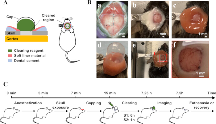
Figure 1: Preparation and workflow of the HOTS window. (A) Schematic illustration of the HOTS window setup. (B) Sequential photographs illustrating key steps in HOTS window preparation in an adult mouse. Scale bar: 1 mm. (a) Skull exposure and optional stereotaxic marking of the intended imaging region. (b) Application of dental cement and attachment of a small titanium headplate for head fixation. (c) Application of soft liner material. (d) Mounting of a cap to contain clearing reagents. (e, f) Appearance of the cleared skull after cap and soft liner material removal. (C) Experimental timeline of the HOTS window procedure. Please click here to view a larger version of this figure.
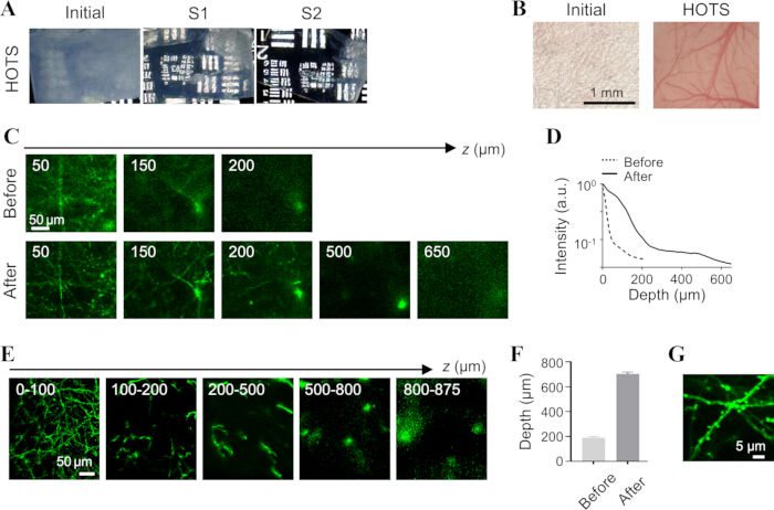
Figure 2: Optical transparency and imaging depth achieved with the HOTS window. (A) Ex vivo and (B) in vivo bright-field imaging of the skull before and after HOTS clearing. Scale bar: 1 mm. (C, D) Before-and-after comparison of the same cortical region through the native and cleared skull (C), with corresponding fluorescence intensity profiles versus depth (D). Scale bar in panel C: 50 µm. The fluorescence intensity at each depth was calculated by integrating signals from all neurons within the imaging field and normalized to the maximum value of the corresponding post-clearing dataset. (E) Maximum-intensity z-projections over different depth ranges (depth below the pia) of an adult Thy1-GFP-M mouse acquired through the HOTS window. Scale bar: 50 µm (F) In vivo two-photon imaging depths below the pia before and after HOTS clearing (n ≥ 3 mice). (G) The close-up views demonstrate that the HOTS window enabled clear visualization of fine dendritic spines. Please click here to view a larger version of this figure.
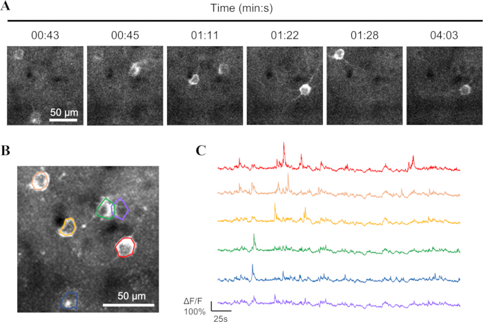
Figure 3: Representative spontaneous calcium activity in the barrel cortex of an awake mouse imaged through the HOTS window at a depth of 150 µm below the pia (n ≥ 3 mice). (A) Time-lapse images of a spontaneous calcium transient over a 4-minute period. Scale bar: 50 µm. (B) Maximum-intensity time projection of layer 2/3 neurons in an awake Thy1-GCaMP6s mouse, with active neurons indicated. Scale bar: 50 µm. (C) ΔF/F0 traces of the marked somata. Please click here to view a larger version of this figure.
Discussion
The HOTS method utilizes a head-mounted optically transparent skull (HOTS) window that combines a cap containing clearing reagents with a two-step skull-clearing procedure to enhance skull transparency in adult mice while preserving skull integrity. Under the standard protocol (6 h in S1 and 1 h in S2 for 6-week-old mice), the resulting window reliably supported deep transcranial two-photon imaging and allowed structures to be visualized to ~800 µm below the pia.
Several procedural steps are critical for obtaining a stable, transparent window. The skull must be clean and dry, with the scalp and periosteum carefully removed to ensure that the dental cement adheres firmly to the bone and anchors the head-plate securely during subsequent imaging. The cement layer should cover the exposed bone and surrounding tissue to provide a rigid base. A thin layer of self-curing dental soft liner material is then applied over the dental cement and around the window region to prevent later leakage of clearing reagents. The HOTS window should be positioned away from cranial sutures whenever possible, as clearing directly over sutures can induce capillary bleeding and often results in window failure. If the window must approach a suture, sealing the suture line with soft liner material can reduce this risk. During imaging, S2 is used as the immersion and index-matching medium, and the objective’s correction collar should be adjusted if available to compensate for refractive index mismatch.
The clearing parameters used here are optimized for 6-week-old mice with moderate skull thickness, but can be adjusted for other ages. For thicker skulls (e.g., older animals), the S1 incubation can be extended in small increments to improve transparency, whereas for very young mice, the S1 exposure can be shortened to avoid over-softening and fragility of the bone. After imaging, thoroughly rinse the skull surface with PBS to remove residual reagents and support the gradual recovery of the skull. Leakage from the head-mounted cap should be addressed immediately. If leakage is detected during the clearing phase, pause the clearing session, gently aspirate the leaked solution with a syringe, and apply additional soft liner material to seal any gaps before resuming clearing.
Compared with open-skull windows, the HOTS preparation retains the skull, thereby minimizing dural exposure and reducing the risk of meningeal injury or chronic inflammation while still granting optical access to deeper cortical layers. The head-mounted cap also allows the clearing reagents to act over several hours in awake, freely-behaving mice, rather than restricting skull clearing to short periods under anesthesia with rigid head fixation, as in previously reported in vivo skull optical clearing methods12,13,14,15. In 6-week-old mice, the HOTS window supports transcranial two-photon imaging of cortical structures, including spines and dendrites in superficial cortical layers and neuronal somata at greater depths. This approach also permits detection of functional signals, including spontaneous and whisker-evoked calcium transients, as well as cAMP dynamics16. Taken together, these features make the HOTS window a versatile, minimally invasive platform for deep cortical imaging. This minimally invasive transcranial access may be particularly useful for studying neuronal and non-neuronal interactions during neuroimmune responses, where reducing surgery-induced neuroinflammation is important. In addition, HOTS may also be useful for optical access to brain regions that are difficult to access surgically, such as the cerebellum. Photoacoustic imaging has recently gained increasing attention in brain imaging17,18,19, but the skull still causes optical attenuation and acoustic aberration, limiting high-resolution imaging at greater depths. Combining photoacoustic imaging with skull optical clearing may further enhance imaging depth and multimodal contrast in transcranial brain imaging. Optogenetic, photopharmacological, and other light-based manipulation techniques enable precise spatiotemporal control of neural activity and signaling20,21. Combining these approaches with skull optical clearing may further facilitate the study of brain function.
The HOTS window also has several practical limitations. The field of view with HOTS is constrained by both the diameter of the head-mounted cap and the need to avoid cranial sutures, limiting simultaneous coverage of widely separated cortical regions and making whole-skull clearing impractical. Although HOTS substantially improves skull transparency and enables deep transcranial imaging, the optical path still traverses a cleared skull with residual scattering. Therefore, spatial resolution and contrast in deep cortical layers remain lower than those achieved with open-skull windows. In addition, performing repeated clearing cycles at the same cranial site is technically challenging because the residual soft liner material hardens after use and becomes difficult to remove completely without disturbing the underlying cement and head-plate. Residual liner interferes with reapplying the soft liner and reconstructing a stable, leak-free HOTS setup. In practice, the current implementation is therefore optimized for one or two carefully planned imaging sessions at each window location.
Disclosures
The authors declare no conflicts of interest.
Acknowledgements
This work was supported by Science and Technology Innovation Key R&D Program of Chongqing (CSTB2024TIAD-STX0004), Shenzhen Medical Research Fund (D2404004), National Natural Science Foundation of China (62475278, 62305369, 62405351, 92359303); Basic and Applied Basic Research Foundation of Guangdong Province (2024A1515012517, 2020B121201010); Youth Innovation Promotion Association of the Chinese Academy of Sciences (2023377); Shenzhen Fundamental Research Program (RCJC20200714114433058, RCYX20210609104445093, JCYJ20241202124924033, ZDSY20130401165820357). We thank Prof. Yang Zhan (Brain Cognition and Brain Disease Institute, Shenzhen Institutes of Advanced Technology, Chinese Academy of Sciences, Shenzhen, China) for providing the Thy1-GFP-M mice.
Materials
| Name | Company | Catalog Number | Comments |
|---|---|---|---|
| 1 mL disposable pipette | Beijing Biotopped Science & Technology Co., Ltd. | N/A | |
| Dental cement | Shanghai Rongxiang Dental Materials Co., Ltd. | N/A | |
| D-mannose | Macklin | D813082 | |
| EDTA | Aladdin | El 16428 | |
| Isoflurane | RWD | N/A | |
| Kwik-Cast | WPI | N/A | A biocompatible sealant |
| NaOH | Macklin | S832169 | |
| Self-curing dental soft liner material | Yangmahu Biotechnology (Hebi) Co., Ltd. | N/A | |
| Sodium pentobarbital | Biopike | N/A | |
| Stereotaxic apparatus | RWD | N/A | |
| Sterile disposable syringe | Minank | N/A | 1 mL; 26 G needle |
| Sulfolane | Macklin | S817950 | |
| Thy1-GFP-M mice | N/A | Provided by Prof. Yang Zhan | |
| Thyl-GCaMP6s mice | Jackson Laboratory | ||
| Tween 20 | Sigma-Vetec | V900548 | |
| Water-immersion objective | Nikon | N25X-APO-MP | 25×/NA 1.1 objective |
| Water-immersion objective | Nikon | N16XLWD-PF | 16×/NA 0.8 objective |
References
- Lee, J., et al. Longitudinal intravital imaging of cerebral microinfarction reveals a dynamic astrocyte reaction leading to glial scar formation. Glia. 70 (5), 975-988 (2022).
- Hierro-Bujalance, C., Bacskai, B. J., Garcia-Alloza, M. In vivo imaging of microglia with multiphoton microscopy. Front Aging Neurosci. 10, 218 (2018).
- Fan, J. L., et al. High-speed volumetric two-photon fluorescence imaging of neurovascular dynamics. Nat Commun. 11 (1), 6020 (2020).
- Fan, X., Zheng, W., Singh, D. J. Light scattering and surface plasmons on small spherical particles. Light Sci Appl. 3 (6), e179 (2014).
- Kneipp, M., et al. Effects of the murine skull in optoacoustic brain microscopy. J Biophotonics. 9 (1-2), 117-123 (2016).
- Holtmaat, A., et al. Long-term, high-resolution imaging in the mouse neocortex through a chronic cranial window. Nat Protoc. 4 (8), 1128-1144 (2009).
- Drew, P. J., et al. Chronic optical access through a polished and reinforced thinned skull. Nat Methods. 7 (12), 981-984 (2010).
- Yang, G., et al. Thinned-skull cranial window technique for long-term imaging of the cortex in live mice. Nat Protoc. 5 (2), 201-208 (2010).
- Xu, H. T., Pan, F., Yang, G., Gan, W. B. Choice of cranial window type for in vivo imaging affects dendritic spine turnover in the cortex. Nat Neurosci. 10 (5), 549-551 (2007).
- Roth, T. L., et al. Transcranial amelioration of inflammation and cell death after brain injury. Nature. 505 (7482), 223-228 (2014).
- Dorand, R. D., et al. Comparison of intravital thinned skull and cranial window approaches to study CNS immunobiology in the mouse cortex. Intravital. 3 (2), e29728 (2014).
- Zhang, C., et al. A large, switchable optical clearing skull window for cerebrovascular imaging. Theranostics. 8 (10), 2696-2708 (2018).
- Zhao, Y. J., et al. Skull optical clearing window for in vivo imaging of the mouse cortex at synaptic resolution. Light Sci Appl. 7, 17153 (2018).
- Li, D. Y., et al. A through-intact-skull (TIS) chronic window technique for cortical structure and function observation in mice. eLight. 2 (1), 15 (2022).
- Liu, X., et al. SeeThrough: a rationally designed skull clearing technique for in vivo brain imaging. Nat Commun. 16 (1), 7584 (2025).
- Liu, L. N., et al. Structural and functional mouse brain imaging facilitated by a head-mounted optically transparent skull (HOTS) window allowing offline clearing. Optica. 12 (12), 1946-1960 (2025).
- Li, H., et al. Multidimensional dynamic optical imaging unveils anesthetic-driven hemispheric lateralization in blood-brain barrier homeostasis. Sci Adv. 11 (49), eady6275 (2025).
- Qin, W., et al. Amphiphilic hemicyanine molecular probes crossing the blood-brain barrier for intracranial optical imaging of glioblastoma. Sci Adv. 11 (3), eadq5816 (2025).
- Pang, W., et al. Direct monitoring of whole-brain electrodynamics via high-spatiotemporal-resolution photoacoustics with voltage-sensitive dye. Laser Photonics Rev. 18 (10), 2400165 (2024).
- Paoletti, P., Ellis-Davies, G. C. R., Mourot, A. Optical control of neuronal ion channels and receptors. Nat Rev Neurosci. 20 (9), 514-532 (2019).
- Deisseroth, K. Optogenetics: 10 years of microbial opsins in neuroscience. Nat Neurosci. 18 (9), 1213-1225 (2015).
Reprints and Permissions
This article has been published
Video Coming Soon