
Guest Editors
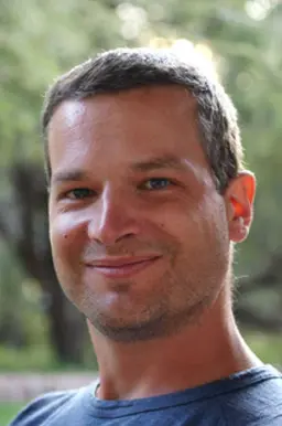
Patrick Herr
University of Sheffield
Dr. Patrick Herr is a Research Fellow at the University of Sheffield and the Weston Park Cancer Centre, Sheffield where he leads a multidisciplinary research project. The overall aim of his research is to understand fundamental processes in cancer biology and to translate those findings into clinically relevant applications. A central part of this work is to understand the mechanisms that control DNA damage, repair, nucleotide metabolism and the maintenance of nucleotide pool balance in cancer. This involves the molecular and functional characterization of novel proteins in these processes as well as the development and validation of small molecules to target these enzymes. Dr. Herr initiated this work during his time as a Postdoctoral Research Fellow and Project Leader at the Karolinska Institute in Stockholm in Sweden where he initially worked on the link between transcription and homologous recombination-mediated DNA repair. His interest in cancer biology originated from his earlier work on signaling pathways that control early embryogenesis. Dr. Herr received his PhD from the University of Zürich, Switzerland for unraveling new mechanisms that control WNT signaling molecule secretion and signal transduction in Drosophila and mammalian cells. He received his Diploma in Biology from the Albert-Ludwigs University and Max Planck Institute in Freiburg, Germany for the characterization of a novel regulator of TGF-beta signaling.

Vijay Menon
Yale University, Department of Therapeutic Radiology
Dr. Vijay Menon is an Associate Research Scientist in the Department of Therapeutic Radiology at Yale University. His current work involves the study of UV-induced rare mutations in sun-exposed skin using high-throughput duplex sequencing to be used as a metric to measure a patient’s past UV exposure and future skin cancer risk. Dr. Menon received his PhD in Pharmacology and Toxicology from Virginia Commonwealth University (VCU) in Richmond, Virginia, during which he investigated the mechanism of action of novel platinum compounds in cancer cells. This was followed by postdoctoral positions at VCU and Icahn School of Medicine. His research expertise mainly includes DNA damage response and repair pathways, erythropoiesis and hematopoiesis, mitochondrial dynamics and redox signaling, and Next Generation Sequencing (NGS).
Collection Overview
The study of DNA damage and repair in mammalian cells is a central part of cancer research. The molecular understanding of pathways and mechanisms of how cells cope with damaged DNA is key to developing new hypotheses that drive preclinical research.
This collection summarizes a variety of established methods in the field, also highlighting novel and emerging techniques. Many methods are microscopy-based and allow for a detailed look at processes happening directly at the site of DNA damage in fixed as well as live cells. The use of nuclei allows researchers to study the recruitment of repair proteins to defined DNA strand breaks in cells and is a standard to study (1) homologous recombination or (2) non-homologous end joining in the bulk of cells by FACS. High content imaging techniques allow researchers to not only study hundreds of cells at a time but also to correlate various markers simultaneously to identify repair factor recruitment (during specific cell cycle stages for example) with high statistical power. Laser micro-irradiation and FRAP combined with confocal microscopy enable the monitoring of real-time recruitment and dynamics of repair proteins in cells; various DNA and RNA labelling techniques combined with sequencing are used to study effects on a genome-wide scale. Post-translational protein modifications such as phosphorylation and ubiquitylation are studied using specific enrichment protocols and analyzed by mass spectrometry.
Examples of methods to be included in this collection are (1) DNA labelling and sequencing techniques, (2) fluorescence microscopy on live and fixed cells, (3) gel electrophoresis of cells, (4) FACS and (5) Mass-spectrometry.
Articles

Laser Microirradiation to Study In Vivo Cellular Responses to Simple and Complex DNA Damage
0 Views
•
Cited by 16
•
2018
•
Xiangduo Kong*1, Gladys M.S. Cruz*2, Bárbara A. Silva2, Nicole M. Wakida2, Nima Khatibzadeh2, Michael W. Berns2,3,4, Kyoko Yokomori1
1Department of Biological Chemistry, School of Medicine, University of California, Irvine, 2Beckman Laser Institute and Medical Clinic, University of California, Irvine, 3Department of Developmental and Cell Biology, School of Biological Sciences, University of California, Irvine, 4Department of Biomedical Engineering and Surgery, University of California, Irvine


Abstracts
MEASUREMENT OF DNA DAMAGE BY MASS SPECTROMETRY
Erdem Coskun*1
1PhD, ERT
DNA damage analysis using long-read sequencing data
Jun Kim*1
1Chungnam National University
Imaging and automatic detection of Sister Chromatid Exchanges using Centromere Chromosome Orientation Fluorescent In Situ Hybridization (Cen-CO-FISH) in Human cells
Simona Giunta*1
1University of Rome Sapienza
Comet assay as a method for DNA damage assessment
Taline Ramos Conde1,
Renata Calil Lemos1,
Helena Pereira da Silva Zamith*1
1Departamento de Farmacologia e Toxicologia, Instituto Nacional de Controle de Qualidade em Saúde, Fundação Oswaldo Cruz, Rio de Janeiro, RJ, Brazil