Overview of the roles, applications, and future prospects of exosomes in the treatment of chronic pain.
A subscription to JoVE is required to view this content. Sign in or start your free trial.
Review Article
Exosomes: Roles and Applications in Chronic Pain Treatment
* These authors contributed equally
In This Article
Summary
Abstract
Chronic pain is increasingly recognized as a maladaptive outcome of dysregulated intercellular communication across neural, immune, and stromal systems. Among emerging mediators, exosomes—nanoscale extracellular vesicles carrying proteins, lipids, and regulatory RNAs—have attracted considerable attention not only as biomarkers and drug carriers but also as active regulators of pain pathophysiology and potential targets for therapeutic intervention. This review aims to synthesize current knowledge on the roles and applications of exosomes in chronic pain treatment and to highlight their potential clinical relevance. Specifically, the review examines how exosomes contribute to chronic pain in representative conditions, including osteoarthritis, ovarian cancer, and hepatocellular carcinoma. Three major functional dimensions are discussed: mechanistic regulation of pain-related processes, therapeutic modulation through endogenous repair and exogenous interventions, and diagnostic utility based on disease- and stage-specific exosomal signatures. In addition, current challenges and future prospects for the clinical application of exosomes are considered. This review provides an overview of the emerging roles of exosomes in chronic pain and offers insights into their potential use in future pain management strategies.
Introduction
Chronic pain affects over 30% of the global population and represents a major unmet medical need, contributing to substantial personal suffering, functional disability, and socioeconomic burden1,2. Chronic nociceptive pain refers to pain that persists or recurs for more than three months and can be broadly classified as nociceptive (arising from tissue damage), neuropathic (resulting from nerve injury), or nociplastic (associated with altered nociception)3. Despite decades of pharmacological development, current therapies remain largely symptomatic, are often ineffective in refractory cases, and are limited by systemic toxicity and tolerance. Drug delivery routes for chronic pain treatment include oral, percutaneous, and parenteral administration4. Traditional intramuscular or intravenous administration is the most commonly used approach and offers relatively high drug utilization efficiency; however, chronic pain requires long-term continuous treatment, which indirectly increases the medical and economic burden on patients. Compared with other invasive drug delivery methods, such as intravenous, abdominal, and vaginal delivery, percutaneous drug delivery is considered one of the most favored approaches in clinical practice because of its non-invasive nature, although its drug utilization efficiency is relatively low5. To improve drug utilization, researchers have explored exosomes as a novel drug delivery vector. Exosomes can not only serve as drug delivery carriers for chronic pain treatment but also regulate inflammation and other biological processes that contribute to chronic pain.
Despite increasing interest in exosomes, the field lacks a consolidated framework linking exosome biology to the multisystem dysregulation underlying diverse chronic pain conditions. This review therefore synthesizes current evidence to define how exosomes contribute to chronic pain across distinct pathological contexts. A structured literature search was conducted using major scientific databases, including PubMed, Web of Science, and Embase, incorporating relevant keywords and Medical Subject Headings to identify studies published in recent years. Reference lists of retrieved articles were also screened to ensure comprehensive coverage. Based on this approach, studies relevant to the scope of this review were selected for synthesis and analysis. This review aims to provide a comprehensive overview of recent research findings and emerging directions, highlighting the roles, applications, and future potential of exosomes in chronic pain management.
Access restricted. Please log in or start a trial to view this content.
Review and Perspective
Composition, Function, and Formation Mechanisms of Exosomes
Exosomes are phospholipid nanovesicles with a characteristic cup-shaped morphology and diameters ranging from 30 to 150 nm. They are widely present in biological fluids such as blood, urine, and most eukaryotic extracellular environments, including cell culture media6. These vesicles are enriched with endosomal membrane markers, including cluster of differentiation 63 (CD63), CD9, and CD817, and contain lipid components such as cholesterol, sphingomyelin, glycosphingolipids, and phosphatidylserine8. Under both physiological and pathological conditions, most cell types are capable of secreting exosomes, highlighting their fundamental role in intercellular communication. Exosomes mediate this communication primarily through the transfer of bioactive molecules, including microRNA (miRNA), messenger RNA (mRNA), DNA, proteins, and even microorganisms such as viruses9,10.
The biogenesis of exosomes originates from late endosomes through inward budding of the limiting membrane of multivesicular bodies (MVBs), resulting in the formation of intraluminal vesicles (ILVs)11 (Figure 1). During this process, specific proteins are incorporated into the invaginating membrane, while cytoplasmic components are enclosed within ILVs. Upon fusion of MVBs with the plasma membrane, ILVs are released into the extracellular space as exosomes12,13. Two principal mechanisms govern ILV formation: endosomal sorting complexes required for transport (ESCRT)-dependent and ESCRT-independent pathways7. The ESCRT machinery consists of four protein complexes (ESCRT-0–ESCRT-III) that coordinate cargo recognition, membrane deformation, and vesicle scission14,15. In the ESCRT-dependent pathway, ESCRT-0 subunits bind ubiquitinated cargo, facilitating their targeting to endosomes, while ESCRT-I and ESCRT-II drive membrane invagination. ESCRT-III subsequently mediates vesicle detachment into MVBs15,16. In contrast, ESCRT-independent mechanisms involve lipid-mediated processes, in which ceramide production—regulated by neutral sphingomyelinase—promotes membrane invagination and ILV formation. Additionally, phospholipase D2 contributes to exosome biogenesis through the generation of phosphatidic acid, which facilitates membrane curvature and vesicle formation17,18.
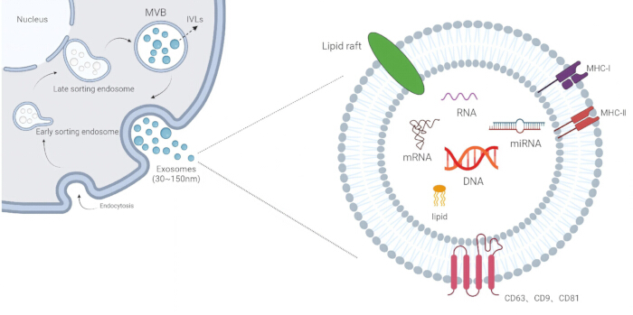
Figure 1. Biogenesis, composition, and secretion of exosomes. Exosome formation begins with the inward budding of the plasma membrane, leading to the generation of early sorting endosomes, which mature into late sorting endosomes and subsequently form multivesicular bodies (MVBs). Inward invagination of the endosomal membrane results in the formation of intraluminal vesicles (ILVs) within MVBs. Upon fusion of MVBs with the plasma membrane, ILVs are released into the extracellular space as exosomes with diameters ranging from 30 to 150 nm. Exosomes contain diverse biomolecules, including microRNA (miRNA), messenger RNA (mRNA), DNA, and lipids, and are enriched with membrane markers such as cluster of differentiation 63 (CD63), CD9, and CD81, as well as lipid rafts and major histocompatibility complex (MHC) molecules. Please click here to view a larger version of this figure.
Functionally, exosomes play a central role in intercellular communication and are involved in a wide range of physiological and pathological processes, including lactation and immune responses19, as well as the development of liver disease20, neurodegenerative disorders21, and cardiovascular disease22. Exosome-mediated signaling occurs through multiple mechanisms, including receptor–ligand interactions, direct membrane fusion, and endocytosis by recipient cells23. These processes enable both local and long-distance communication, facilitating the propagation of biological signals, including chemical and electrical cues24. Increasing evidence indicates that exosomes carry cell-specific molecular cargo and serve as an important mechanism for coordinated cellular regulation.
Exosomes interact with recipient cells through three primary pathways: binding to cell surface receptors, direct fusion with the plasma membrane, or internalization via endocytosis, which may occur through clathrin-dependent or clathrin-independent mechanisms25 (Figure 2). Through these pathways, exosomes regulate the biological activity of target cells by transferring functional biomolecules.
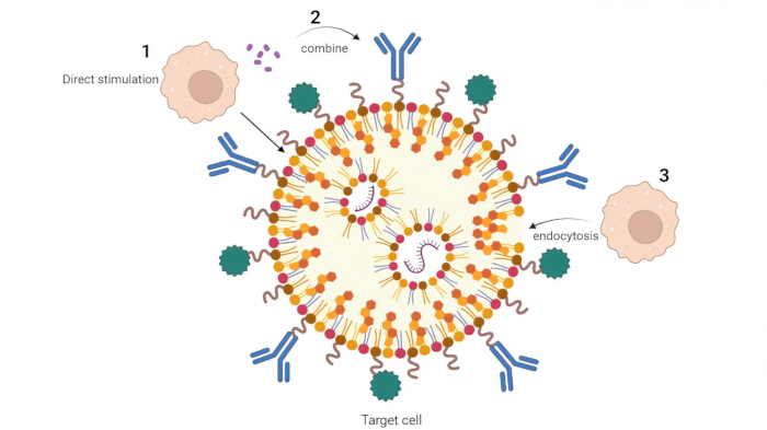
Figure 2. Mechanisms of exosome-mediated intercellular communication. This schematic illustrates three primary modes of interaction between exosomes and target cells: (1) direct stimulation, in which ligands on the exosome surface bind to receptors on the target cell and activate signaling pathways; (2) receptor-mediated binding, in which exosomes interact with specific receptors on the cell membrane to trigger downstream responses; and (3) endocytosis, in which exosomes are internalized by target cells. Through these mechanisms, exosomes facilitate intercellular communication and regulate biological processes, including inflammation, immune responses, and neural plasticity. Please click here to view a larger version of this figure.
In the context of immune regulation, activated T cells release exosomes containing T cell receptor (TCR)/CD3 complexes, Fas ligand (FasL), and Apolipoprotein 2 ligand, which contribute to immune modulation by inducing apoptosis of target cells. These exosomes also express CD4+ T-cell-associated molecules, including CD4, TCR, lymphocyte function associated antigen-1, CD25, and FasL, and have been shown to inhibit CD4+ T-cell proliferation in vitro26. Furthermore, exosomes derived from regulatory T cells carry microRNAs such as Lethal-7b (Let-7b), Let-7d, and miR-155, which suppress T-helper 1 immune responses and contribute to immunosuppressive signaling27.
Exosomes derived from mesenchymal stem cells (MSCs) have been extensively studied for their regenerative potential and are widely applied in tissue repair and wound healing. These exosomes have been shown to reduce myocardial infarction size and prevent adverse remodeling following ischemia–reperfusion injury28. In addition, they play important roles in skin wound healing29, acute kidney injury30, liver injury31, doxorubicin-induced cardiotoxicity32, uterine injury33, and protection of auditory hair cells from neomycin-induced damage34. Collectively, these findings highlight the diverse biological functions of exosomes and their potential therapeutic applications across a range of disease contexts.
Role of Exosomes in the Treatment of Chronic Pain
Chronic pain is increasingly recognized not merely as an abnormal sensory experience but as a complex systemic pathological condition characterized by dysregulation of neuro–immune–matrix interactions. Across diverse diseases, including osteoarthritis (OA), ovarian cancer (OC), and hepatocellular carcinoma (HCC), pain functions not only as a clinical symptom but also as a dynamic indicator of disease progression. In this context, exosomes have emerged as important mediators in the initiation, maintenance, and potential modulation of chronic pain. Rather than representing a single breakthrough, this evolving understanding reflects the progressive integration of knowledge related to the inflammatory microenvironment, neural plasticity, and tumor–host interactions. Collectively, current evidence suggests that exosomes may contribute to chronic pain through multiple biological pathways and may also provide novel opportunities for therapeutic intervention across different disease contexts.
Role of exosomes in OA
OA is a degenerative joint disease and a leading cause of chronic disability, particularly among older adults. It represents one of the most common joint disorders worldwide and contributes substantially to pain, functional limitation, and socioeconomic burden35. In 2020, approximately 654 million individuals globally were affected by knee OA36. OA is characterized as a chronic inflammatory condition involving progressive degeneration of articular cartilage, resulting in joint pain and stiffness37. Importantly, OA is not restricted to cartilage damage but also involves pathological changes in the synovium, subchondral bone, and surrounding ligaments38. Mechanistically, aberrant nerve ingrowth within joint tissues can generate pathological pain signals, directly activating dorsal root ganglia and contributing to neuronal injury. In addition, pro-inflammatory cytokines such as interleukin-1 (IL-1) and tumor necrosis factor (TNF), produced by immune and non-neuronal cells, further amplify pain signaling39,40.
Exosomes are increasingly recognized as key regulators of intercellular communication within the joint microenvironment and contribute to the maintenance of joint homeostasis by modulating cell proliferation, extracellular matrix (ECM) synthesis, and inflammatory responses41. MSC-derived exosomes, in particular, have been shown to exert therapeutic effects through the delivery of specific microRNAs. For example, MSC-derived exosomes can transport miR-204 into chondrocytes, where it modulates intracellular signaling pathways. This process increases the intracellular levels of miR-204 and contributes to pain alleviation by inhibiting transcription factor specificity protein 1–low-density lipoprotein receptor–related protein (SP1-LRP1) signaling and regulating neuro–cartilage interactions. Specifically, suppression of SP1 expression reduces LRP1 expression and associated transporter activity, thereby decreasing miR-204-mediated pain signaling. In parallel, modulation of this pathway reduces neuronal activation and limits nociceptor invasion at the synovium–cartilage interface, ultimately contributing to pain relief42. In addition, exosomes exhibit favorable biocompatibility and biodegradability, enabling protection of miRNAs from degradation and enhancing their therapeutic stability43. MSC-derived exosomes have also been shown to promote a shift in macrophage polarization toward an anti-inflammatory M2 phenotype while reducing pro-inflammatory M1 macrophages and cytokine production.
Beyond pain modulation, MSC-derived exosomes play a significant role in cartilage repair and regeneration. These vesicles promote tissue repair by enhancing new tissue formation and increasing ECM deposition, including sulfated glycosaminoglycans and type II collagen. Histologically, this is reflected in the formation of hyaline-like cartilage, increased chondrocyte presence, and elevated expression of cartilage-specific markers. In addition, regeneration of subchondral bone and structural integration with surrounding cartilage contribute to improved joint integrity. Through these mechanisms, MSC-derived exosomes facilitate dynamic cartilage remodeling by promoting cell proliferation and reducing apoptosis44.
Exosomes derived from synovial MSCs further contribute to cartilage protection and anti-inflammatory regulation. These exosomes inhibit the production of pro-inflammatory cytokines such as IL-6 and TNF-α and reduce IL-1β-induced ECM degradation. Moreover, they mediate the transfer of miR-485-3p into chondrocytes, enhancing its intracellular expression. Targeting of neuropilin-1 by miR-485-3p leads to inactivation of the phosphoinositide 3-kinase/protein kinase B signaling pathway, resulting in reduced apoptosis, decreased inflammatory cytokine release, and overall attenuation of cartilage injury45. Consistent with these findings, overexpression of miR-485-3p has also been shown to protect chondrocytes from OA-related damage46.
Notably, exosomes may also exert deleterious effects under certain conditions. They can promote the production of inflammatory mediators and cartilage-degrading enzymes through interactions with synovial cells and chondrocytes, thereby enhancing the expression of OA-related genes and contributing to disease progression41. These dual roles highlight the importance of carefully regulating exosome-based interventions to maximize therapeutic benefit while minimizing potential adverse effects. Overall, current evidence suggests that exosome-based therapies for OA may not function as direct substitutes for surgical intervention or conventional analgesics but instead offer a strategy to restore disrupted joint homeostasis. By modulating the underlying biological interactions within the joint, exosomes may enable pain to be addressed as a dynamic and potentially reversible pathological process rather than an irreversible outcome.
Role of exosomes in OC
When considering pain associated with malignant tumors, the role of exosomes becomes increasingly complex and multifaceted. OC has the highest mortality rate among gynecological malignancies, and most patients are diagnosed at advanced stages due to nonspecific or subtle early clinical symptoms. In this context, tumor progression and the associated alterations in the tumor microenvironment (TME) contribute not only to disease severity but also to the development and persistence of cancer-related pain.
Natural killer (NK) cells are key components of the innate immune system. Originating from the bone marrow, they represent a major lymphocyte population alongside T cells and B cells and are primarily distributed in peripheral blood, liver, and spleen. NK cells exert cytotoxic effects through the release of effector molecules such as perforin and granzyme, which contribute to their ability to eliminate tumor cells. Exosomes derived from activated NK cells (eNK-EXO) have been shown to enhance the sensitivity of OC cells to chemotherapeutic agents such as cisplatin, which exerts antitumor effects by binding to genomic and mitochondrial DNA and inhibiting DNA replication. In addition, eNK-EXO can restore impaired NK cell function within the TME, promoting the release of cytotoxic mediators such as perforin and TNF-α47.
Furthermore, eNK-EXO are preferentially internalized by OC cell lines, facilitating efficient and targeted activity. These exosomes have also demonstrated cytotoxic effects in breast cancer and melanoma cell lines48,49, while exhibiting minimal cytotoxicity toward normal ovarian epithelial cells, suggesting a favorable safety profile. From a broader perspective, these findings indicate that exosome-mediated modulation of tumor–immune interactions may contribute not only to tumor control but also to the indirect regulation of cancer-associated pain. Rather than solely masking pain signals, exosome-based strategies may help reprogram interactions within the TME, allowing pain to diminish as underlying disease processes are modulated.
Role of exosomes in HCC
HCC remains a major global health challenge and is among the most common malignancies worldwide. According to recent estimates, liver cancer accounts for more than 905,000 new cases annually, with China bearing a disproportionately high burden, contributing over 40% of global cases50. Chronic hepatitis B virus infection is a well-established etiological factor in liver cancer development51. In addition, cirrhosis of any origin represents a major risk factor for HCC, while other contributing factors include chronic alcohol consumption, metabolic disorders such as diabetes and obesity-related nonalcoholic fatty liver disease, and hepatitis C virus infection52. These conditions collectively contribute to tumor progression and the associated clinical manifestations, including cancer-related pain.
Cancer stem cells (CSCs), also referred to as tumor-initiating cells, play a critical role in tumor initiation and progression. These cells possess the capacity for self-renewal and multidirectional differentiation and exhibit strong tumorigenic potential53. Emerging evidence suggests that hepatic CSCs can influence the malignant behavior of surrounding HCC cells through the secretion of exosomes, thereby contributing to tumor progression and microenvironmental remodeling. Among the molecular cargo carried by these exosomes, circular RNAs (circRNAs) function as important regulatory mediators in intercellular communication.
Circular actin filament–associated protein 1 (circ-AFAP1) has been identified as a key circRNA involved in this process. Overexpression of circ-AFAP1 has been shown to significantly increase the expression of CD133, enhance epithelial–mesenchymal transition, and promote tumor progression and growth in HCC54. Through these mechanisms, exosome-mediated transfer of circRNAs contributes to the aggressive biological behavior of tumor cells. These findings suggest that the expression levels of specific circRNAs in exosomes may serve as potential indicators of disease progression and poor prognosis in patients with HCC. From a broader perspective, the involvement of exosomes in tumor progression highlights their potential relevance to cancer-associated pain. By modulating tumor–host interactions and influencing the tumor microenvironment, exosomes may indirectly affect the development and persistence of pain in HCC.
In summary, the growing recognition of exosomes in chronic pain research reflects a broader conceptual shift in pain medicine. Rather than focusing solely on the suppression of terminal pain signals, current approaches increasingly emphasize the regulation of upstream biological interactions, including neuro–immune–matrix communication. This shift represents a transition from symptomatic relief toward restoring underlying biological balance. In this context, exosomes offer a promising avenue for integrating pain management within a precision medicine framework, where pain is understood as part of a complex and dynamic disease process.
Role of Exosomes in the Diagnosis of Chronic Pain
Recent studies have shown that exosomes can also serve as novel diagnostic indicators for chronic pain. By measuring the expression levels of specific proteins or molecules, such as RNA contained within exosomes, the occurrence and extent of chronic pain may be assessed. For example, exosomal miRNAs have been investigated as potential biomarkers of cardiovascular risk in children. MiRNAs participate in almost all biological processes in eukaryotic cells and are stably present in body fluids through extracellular vesicles, where they can exert regulatory effects at distant sites55,56,57. Cardiovascular disease is a major source of morbidity and mortality worldwide, and obesity is widely recognized as an independent risk factor for cardiovascular disease58. In children, obesity is associated with increased metabolic and cardiovascular dysfunction, including endothelial dysfunction (ED)59,60,61. Overweight children are more likely to develop early ED, high blood pressure, and type 2 diabetes62. In addition, even a brief period of overweight or obesity in childhood has been associated with earlier mortality in adulthood63. Therefore, early disease detection and personalized intervention are crucial for children with cardiovascular disease. In this context, exosomes have been explored as a means of predicting pediatric cardiovascular risk64,65,66. More broadly, miRNAs contained in exosomes have been identified as biomarkers for a variety of diseases, owing to several advantages: they are stable in circulation and can survive in harsh environments; most miRNA sequences are conserved across species; circulating miRNA levels vary across diseases and pathological stages; and miRNA levels can be measured using relatively simple methods67,68,69,70.
MiRNAs may also serve as potential biomarkers for ulcerative colitis (UC). UC is a chronic, nonspecific inflammatory bowel disease that commonly presents with abdominal pain, diarrhea, and bloody stool. Colonoscopy is currently the gold standard for diagnosis, while blood tests and biomarker analysis are used as adjunctive approaches. However, colonoscopy is invasive, imposes substantial physical and financial burden on patients, and may be associated with serious complications such as bowel perforation and death71,72. As important post-transcriptional regulatory factors, some miRNAs are abnormally expressed during the carcinogenic progression of UC and may therefore be useful in the early detection of disease-related changes. MiR-21, one of the most extensively studied miRNAs in UC, is upregulated in the blood73, feces74, and colon tissues75 of patients with UC and promotes intestinal inflammation. In addition, the nuclear factor kappa-light-chain-enhancer of activated B cells, signal transducer and activator of transcription 3, and B-cell lymphoma-2 signaling pathways can be activated through targeting programmed cell death factor 4, thereby reducing tumor cell apoptosis76. Beyond their diagnostic potential, miRNAs may also participate in the regulation of UC and influence disease onset and progression.
Although the examples above are not specific to chronic pain, they illustrate the broader diagnostic relevance of exosomal biomarkers in disease detection and monitoring. Because many chronic pain conditions involve inflammatory and molecular changes, these findings may indirectly support the potential use of exosomal miRNAs as minimally invasive biomarkers in chronic pain assessment. Further studies are needed to define pain-specific exosomal signatures and clarify their diagnostic utility in clinical settings.
Exosomes as a New Drug Carrier
In recent years, exosomes have attracted considerable attention as potential drug delivery carriers, particularly in the context of cancer therapy. Their unique biological properties, including biocompatibility, stability, targeting capability, and controllable drug release, make them promising candidates for therapeutic applications. The favorable biocompatibility of exosomes is largely attributed to their endogenous origin, which reduces the likelihood of immune rejection. In addition, their lipid bilayer structure provides structural stability and protects encapsulated bioactive molecules from degradation and inactivation.
Exosomes also exhibit effective targeting capabilities, as surface proteins and glycoproteins can interact with specific receptors on target cells, facilitating selective delivery. Furthermore, their capacity for controlled drug release is enhanced through genetic engineering and surface modification techniques, enabling the loading of specific therapeutic agents, including drugs and genes. Based on these advantages, exosomes have been widely explored as drug carriers in cancer treatment77.
To further enhance their therapeutic potential, exosomes can be combined with various antitumor strategies, enabling a transition from single-modality treatment to combination therapy. Such approaches are particularly valuable in addressing tumor heterogeneity through synergistic effects. Combination strategies may involve engineered exosomes used in conjunction with chemotherapy drugs, therapeutic nucleic acids, photosensitizers, immunotherapies, and phytochemicals78.
For example, engineering modifications of exosomes can improve drug delivery efficiency, enhance in vivo tissue penetration, and strengthen targeting specificity79. These modifications may also extend circulation time and improve tissue distribution, thereby increasing therapeutic efficacy80. Despite these advances, challenges remain in optimizing exosome-based drug delivery systems for clinical application. Further investigation is required to determine the most effective delivery methods and strategies to improve drug delivery efficiency and enhance absorption across different target tissues.
Problems in the Clinical Application of Exosomes
Exosomes have demonstrated substantial potential in clinical applications, playing important roles in disease diagnosis, treatment, and prevention, as well as in monitoring and evaluating disease prognosis. In addition, they contribute significantly to tissue regeneration and repair and have broad applicability in regenerative medicine. Through their involvement in intercellular communication, exosomes provide valuable insights into disease pathogenesis and progression and offer new strategies for diagnosis, treatment, and prognostic assessment. Despite these promising applications, several challenges remain that limit their widespread clinical use.
One major challenge is the safety of exosome-based therapies. Exosomes derived from different cellular sources can exert distinct biological effects. For example, exosomes derived from MSCs have demonstrated protective effects in pulmonary arterial hypertension81, whereas exosomes derived from tumor stem cells may promote tumor initiation and metastasis82,83. Moreover, exosomes themselves may exert either beneficial or adverse effects depending on their origin and cargo. Therefore, improving the safety profile of exosomes is essential to ensure their reliable use in clinical diagnosis and therapy.
A second challenge relates to the selection and sourcing of exosomes. Different diseases may require exosomes derived from specific cell types, which increases the complexity of research and places additional demands on resources, including time, labor, and cost. Identifying broadly applicable or standardized exosome sources that can achieve consistent therapeutic effects across multiple disease contexts would help simplify clinical translation and improve feasibility.
A third limitation is the variability in the quantity and quality of exosomes. The yield of exosomes during in vitro culture is often inconsistent, and insufficient exosome production may reduce therapeutic efficacy. In addition, variations in exosome quality can influence treatment outcomes and may even lead to undesirable effects. Therefore, optimizing exosome production, improving cargo loading capacity, and ensuring the quality of source cells are critical factors for effective clinical application.
Another important obstacle is the lack of efficient and standardized exosome culture and production systems. Current methods typically involve isolating cells from target tissues and culturing them in vitro, which can be time-consuming and may result in unpredictable yields. Developing more efficient, scalable, and reproducible production systems is necessary to support clinical implementation.
Finally, the scope of exosome applications remains relatively limited. To date, most advances have been concentrated in areas such as inflammation, metabolism, and oncology. Expanding the application of exosomes to a broader range of diseases may further enhance their clinical value and therapeutic potential.
Overall, exosomes serve as important mediators of intercellular communication and play significant roles in regulating the tumor microenvironment, immune responses, neurodegenerative disease progression, tissue repair, and biomarker discovery for liquid biopsy. Their intrinsic lipid bilayer structure contributes to their biocompatibility and targeting capabilities, making them attractive candidates for advanced drug delivery systems. Given their broad biological functions and translational potential, exosomes represent a promising direction for future research in regenerative medicine, vaccine development, and targeted therapy.
Access restricted. Please log in or start a trial to view this content.
Conclusions
In summary, this review highlights the multifaceted roles of exosomes in the development, progression, and potential management of chronic pain across diverse pathological conditions, including OA, OC, and HCC. Exosomes contribute to chronic pain through their involvement in intercellular communication, regulation of inflammatory responses, and modulation of neural and microenvironmental interactions. In addition to their mechanistic roles, exosomes demonstrate significant potential as diagnostic biomarkers and therapeut...
Access restricted. Please log in or start a trial to view this content.
Disclosures
The authors declare no competing financial interests.
Acknowledgements
The authors have no acknowledgments or funding sources to declare.
Access restricted. Please log in or start a trial to view this content.