检测氧化性细胞环境的一种筛选方法是测定CM-H2DCFDA的氧化程度。CM-H2DCFDA在细胞内被氧化后,会从无荧光状态转变为荧光化合物。这种荧光变化可通过流式细胞术进行检测,从而反映处于氧化环境中的细胞数量。
需要JoVE订阅才能观看此内容。 请登录或开始免费试用
本文内容
摘要
摘要
病毒诱导神经元损伤的常见机制之一是氧化应激。星形胶质细胞在调控中枢神经系统(CNS)氧化应激中发挥重要作用。星形胶质细胞不仅有助于为神经元维持稳态环境,还能保护神经元免受活性氧(ROS)的损害。CM-H2DCFDA 是一种可穿透细胞膜的活性氧指示剂。CM-H2DCFDA 以无荧光的化合物形式进入细胞,在细胞内酯酶去除乙酸基团并发生氧化反应后,转变为荧光化合物。通过流式细胞术检测到的绿色荧光细胞数量,可反映处于氧化状态的细胞数量。CM-H2DCFDA 可被多种不同的活性氧氧化,这种对具体何种活性氧的非特异性使其成为病理发生早期研究中的有用试剂,因为该检测方法可用于筛查氧化性细胞环境,而无需预先确定导致细胞状态的具体氧自由基或活性氧组合。一旦通过 CM-H2DCFDA 的氧化证实活性氧的存在,即可进一步开展实验,以明确参与特定病理过程的具体活性氧种类或其组合。本研究结果表明,与生理盐水对照组相比,加入过氧化氢后检测到 CM-H2DCFDA 荧光显著增强,表明该检测方法可用于有效识别 G355-5 细胞(一种猫源星形胶质细胞系)内的氧化环境。
方案
1. 猫星形胶质细胞的培养及H处理2O2
- 将G355-5细胞系在含DMEM培养基(添加营养物质)的75 cm2培养瓶中,于37°C、5% CO2条件下培养至60%融合(约5天)。每1-2天或根据需要更换培养基,以维持细胞健康生长。
- 移除培养基,加入2 ml 0.25%胰蛋白酶。室温(RT)下吹打45秒至1分钟,使细胞脱落。
- 迅速将细胞悬液转移至含有10 ml培养基的15 ml锥形离心管中,轻轻混匀。
- 在23°C下以300 × g离心3分钟,收集细胞沉淀。弃去上清液,注意不要扰动沉淀。
- 加入6-7 ml新鲜培养基,重悬细胞。
- 向6孔板的每个孔中加入约1 ml细胞悬液,每孔再添加2-3 ml培养基,孵育至细胞融合度达80-90%。
- 在培养基中配制100 μM的过氧化氢(H2O2)溶液。
- 吸去各孔中的培养基,分别替换为2 ml新鲜培养基(对照组)、2 ml H2O2溶液(处理组)或2 ml DPBS。于37°C、5% CO2条件下孵育3小时。
- 移除溶液,每孔加入1 ml胰蛋白酶消化细胞。将细胞转移至含有0.5 ml DPBS的1.7 ml离心管中。此步骤应逐孔操作,以避免因胰蛋白酶长时间作用导致细胞死亡。
- 以300 × g离心3分钟,弃去上清液,用1 ml DPBS重悬细胞沉淀。
2. 活性氧染色
- 染色的所有步骤均应在尽量减少光照的条件下进行,以防止荧光淬灭。
- 向阳性对照组加入 1 μl 的 50 mM CCCP,于 37°C、5% CO2 条件下孵育 5 分钟。
- 向 H2O2 组加入 5 μl 的 10 mM CM-H2DCFDA 溶液,于 37°C、5% CO2 条件下孵育 15–30 分钟。生理盐水样本中不添加染料。
- 用 2 ml DPBS 洗涤样品,以去除残留染料。
- 以 300 × g 离心 3 分钟,获得细胞沉淀。
- 将细胞重悬于 0.5 ml DPBS 中。除生理盐水组外,向每个样品中加入 1 μl PI。
- 将样品转移至 5 ml 流式细胞仪检测管中。
- 上机进行流式细胞仪检测。
3. 流式细胞术
- 使用配备488 nm氩激光器及以下带通滤光片(±20 nm)的Beckman Coulter FC500(或同等设备)运行样品:525 nm、775 nm和620 nm。
- 准备适当的对照:未染色细胞、仅CM-H2DCFDA染色、仅PI染色、双染色(阴性对照)。
- 建立包含以下散点图的实验方案:FS vs. SS、PI vs. CM-H2DCFDA;同时应包含以下直方图:细胞数量 vs. FL1、FL2和FL3,分别对应相应的染料信号。
- 运行样品前,检查仪器、流体系统和废液容器,并将仪器预热至少20分钟。
- 清洁仪器,并根据制造商标准使用适当的校准微球进行校准。
- 选择实验方案并运行样品。
- 按照之前的方法清洁仪器。
- 根据制造商标准清洁真空管路。
- 关闭流路,并清洁探头/真空管路。
- 关闭软件和计算机。
4. 代表性结果:
使用 FlowJo 7.6 分析流式细胞术结果,并利用染色对照客观设定门控。根据该方法,健康细胞在直方图左侧显示,而活性氧(氧化应激)则表现为细胞向右侧偏移。图1展示了过氧化氢对健康猫星形胶质细胞作用效果的结果。采用FL1通道数据测量CM-H2DCFDA的荧光强度,以指示活性氧的存在。正如预期,经H2O2处理的样本中检测到更高水平的活性氧。在直方图上表现为荧光强度从左向右的偏移。比较经DMEM与DPBS处理的健康细胞时,活性氧水平无显著变化(图2)。
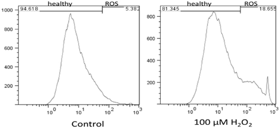
图1. 健康猫星形胶质细胞中氧化应激水平的流式细胞术检测结果,以及H2O2对健康细胞的影响。结果表明,与未处理细胞(DMEM)相比,H2O2可增加健康细胞中活性氧(ROS)的水平。
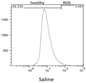
图 2. 健康细胞在 DPBS 中孵育 3 小时后检测到的活性氧水平的流式细胞术结果。
访问受限。请登录或开始试用以查看此内容。
讨论
病毒引起神经元损伤的常见机制之一是氧化应激1, 4, 7, 9, 13, 15, 16, 18-22, 27, 31。基本观点认为,病毒感染可导致胶质细胞(星形胶质细胞和小胶质细胞)释放活性氧自由基,如羟自由基、超氧阴离子、一氧化氮和/或过氧化氢,这些物质对神经元具有毒性。类似地,氧化应激也被认为是甲基苯丙胺诱导神经毒性的主要病理机制之一2, 6, 17。由于本研究团队关注病毒和滥用药物对中枢神经系统(CNS)的共同影响,我们选择采用一种快速筛选方法来检测星形胶质细胞中是否存在过量的活性氧(ROS)。大多数神经元内的谷胱甘肽由星形胶质细胞合成,随后输送给神经元5, 8, 10-12, 24, 26, 29, 30。因此,星形胶质细胞在氧化应激过程中发挥重要作用,不仅有助于维持神经元的稳态环境,还能保护神经元免受活性氧的损伤,这也是本研究选用星形胶质细胞培养体系的原因。
CM-H2DCFDA 也被称为 2',7'-二氯荧光素(2',7'-dichlorofluorescein)和 H2DCF。CM...
访问受限。请登录或开始试用以查看此内容。
披露
我们感谢 ReadiSorb Products 对这些研究的大力支持。
致谢
我们感谢 ReadiSorb Products 对这些研究的大力支持。
访问受限。请登录或开始试用以查看此内容。
材料
| 姓名 | 公司 | 目录编号 | 评论 |
|---|---|---|---|
| DMEM | Cellgro | 15-013-CV | |
| MEM 维生素 | Cellgro | 25-020-cl | 1% (v/v) |
| L-谷氨酰胺 | Hyclone | 17-605E | 1% (v/v) |
| 非必需氨基酸 | Hyclone | 25-025-cl | 1% (v/v) |
| FBS | Hyclone | SH30071.03 | 20% (v/v) |
| 必需氨基酸 | Cellgro | 25-030-cl | 0.2% (v/v) |
| 500 ml 真空过滤系统 | VWR international | 87006-076 | |
| 15 ml 锥形管 | Falcon BD | 352097 | |
| 75 cm2 组织培养瓶 | Falcon BD | 353136 | |
| 6 孔组织培养板 | Falcon BD | 353224 | |
| CM-H2DCFDA | Invitrogen | C6827 | |
| 碘化丙啶(PI) | Sigma-Aldrich | P4170-10mg | |
| 胰蛋白酶 | Lonza Inc. | 17-160F | |
| H2O2 | Sigma-Aldrich | H6520 | |
| HyQ 抗生素 | Hyclone | SV30079.01 | 0.1% (v/v) |
| G355-5 细胞 | ATCC | CRL-2033 | 正常猫脑 |
参考文献
- Bukrinsky, M. I., Nottet, H. S., Schmidtmayerova, H. L., Dubrovsky, H., Flanagan, C. R., Mullins, M. E., Lipton, S. A., Gendelman, H. E. Regulation of nitric oxide synthase activity in human immunodeficiency virus type 1 (HIV-1)-infected monocytes: implications for HIV-associated neurological disease. J Exp Med. 181, 735-745 (1995).
- Cadet, J. L., Ali, S., Epstein, C. Involvement of oxygen-based radicals in methamphetamine-induced neurotoxicity: evidence from the use of CuZnSOD transgenic mice. Ann N Y Acad Sci. 738, 388-3891 (1994).
- Cathcart, R., Schwiers, E., Ames, B. N. Detection of picomole levels of hydroperoxides using a fluorescent dichlorofluorescein assay. Anal Biochem. 134, 111-116 (1983).
- Chao, C. C., Hu, S., Peterson, P. K. Glia: the not so innocent bystanders. J Neurovirol. 2, 234-239 (1996).
- Cooper, A. J., Kristal, B. S. Multiple roles of glutathione in the central nervous system. Biol Chem. 378, 793-802 (1997).
- Cubells, J. F., Rayport, S., Rajendran, G., Sulzer, D. Methamphetamine neurotoxicity involves vacuolation of endocytic organelles and dopamine-dependent intracellular oxidative stress. J Neurosci. 14, 2260-2271 (1994).
- Dawson, V. L., Dawson, T. M., Uhl, G. R., Snyder, S. H. Human immunodeficiency virus type 1 coat protein neurotoxicity mediated by nitric oxide in primary cortical cultures. Proc Natl Acad Sci U S A. 90, 3256-329 (1993).
- Desagher, S., Glowinski, J., Premont, J. Astrocytes protect neurons from hydrogen peroxide toxicity. J Neurosci. 16, 2553-2562 (1996).
- Dewhurst, S., Gelbard, H. A., Fine, S. M. Neuropathogenesis of AIDS. Mol Med Today. 2, 16-23 (1996).
- Dringen, R. Metabolism and functions of glutathione in brain. Prog Neurobiol. 62, 649-671 (2000).
- Dringen, R., Hamprecht, B. Glutathione restoration as indicator for cellular metabolism of astroglial cells. Dev Neurosci. 20, 401-407 (1998).
- Dringen, R., Pfeiffer, B., Hamprecht, B. Synthesis of the Antioxidant Glutathione in Neurons: Supply by Astrocytes of CysGly as Precursor for Neuronal Glutathione. J Neurosci. 19, 562-569 (1999).
- Everall, I. P. H. udson, L, R. W. K. erwin Decreased absolute levels of ascorbic acid and unaltered vasoactive intestinal polypeptide receptor binding in the frontal cortex in acquired immunodeficiency syndrome. Neurosci Lett. 224, 119-1122 (1997).
- Gabriel, C., Camins, A., Sureda, F. X., Aquirre, L., Escubedo, E., Pallas, M., Camarasa, J. Determination of nitric oxide generation in mammalian neurons using dichlorofluorescin diacetate and flow cytometry. J Pharmacol Toxicol Methods. 38, 93-938 (1997).
- Gendelman, H. E., Genis, P., Jett, M., Zhai, Q. H., Nottet, H. S. An experimental model system for HIV-1-induced brain injury. Adv Neuroimmunol. 4, 189-1893 (1994).
- Hayman, M., Arbuthnott, G., Harkiss, G., Brace, H., Filippi, P., Philippon, V., Thomson, D., Vigne, R., Wright, A. Neurotoxicity of peptide analogues of the transactivating protein tat from Maedi-Visna virus and human immunodeficiency virus. Neuroscience. 53, 1-6 (1993).
- Hirata, H., Ladenheim, B., Rothman, R. B., Epstein, C., Cadet, J. L. Methamphetamine-induced serotonin neurotoxicity is mediated by superoxide radicals. Brain Res. , 677-6345 (1995).
- Koka, P., He, K., Zack, J. A., Kitchen, S., Peacock, W., Fried, I., Tran, T., Yashar, S. S., Merrill, J. E. Human immunodeficiency virus 1 envelope proteins induce interleukin 1, tumor necrosis factor alpha, and nitric oxide in glial cultures derived from fetal, neonatal, and adult human brain. J Exp Med. 182, 941-951 (1995).
- Kong, L. Y., Wilson, B. C., McMillian, M. K., Bing, G., Hudson, P. M., Hong, J. S. The effects of the HIV-1 envelope protein gp120 on the production of nitric oxide and proinflammatory cytokines in mixed glial cell cultures. Cell Immunol. 172, 77-83 (1996).
- Koutsilieri, E., Gotz, M. E., Sopper, S., Sauer, U., Demuth, M., ter Meulen, V., Riederer, P. Regulation of glutathione and cell toxicity following exposure to neurotropic substances and human immunodeficiency virus-1 in vitro. J Neurovirol. 3, 342-349 (1997).
- Lipton, S. A. Similarity of neuronal cell injury and death in AIDS dementia and focal cerebral ischemia: potential treatment with NMDA open-channel blockers and nitric oxide-related species. Brain Pathol. 6, 507-517 (1996).
- Lipton, S. A., Yeh, M., Dreyer, E. B. Update on current models of HIV-related neuronal injury: platelet-activating factor, arachidonic acid and nitric oxide. Adv Neuroimmunol. 4, 181-188 (1994).
- Mills, E. M., Takeda, K., Yu, Z. X., Ferrans, V., Katagiri, Y., Jiang, H., Lavigne, M. C., Leto, T. L., Guroff, G. Nerve growth factor treatment prevents the increase in superoxide produced by epidermal growth factor in PC12 cells. J Biol Chem. 273, 22165-22168 (1998).
- Peuchen, S., Duchen, M. R., Clark, J. B. Modulation of the glutathione redox state in adult astrocytes. Biochem Soc Trans. 24, 449S-449S (1996).
- Possel, H., Noack, H., Augustin, W., Keilhoff, G., Wolf, G. 2,7-Dihydrodichlorofluorescein diacetate as a fluorescent marker for peroxynitrite formation. FEBS Lett. 416, 175-178 (1997).
- Sagara, J., Makino, N., Bannai, S. Glutathione efflux from cultured astrocytes. J Neurochem. 66, 1876-1881 (1996).
- Stefano, G. B., Smith, E. M., Paemen, L. R., Hughes, T. K. Jr HIV gp120 associated neurological deficits: a potential role for nitric oxide and other signal molecules. Advances in Neuroimmunology. 3, 47-57 (1993).
- Sundaresan, M., Yu, Z. X., Ferrans, V. J., Irani, K., Finkel, T. Requirement for generation of H2O2 for platelet-derived growth factor signal transduction. Science. 270, 296-299 (1995).
- Wang, X. F., Cynader, M. S. Astrocytes provide cysteine to neurons by releasing glutathione. J Neurochem. 74, 1434-1442 (2000).
- Wilson, J. X. Antioxidant defense of the brain: a role for astrocytes. Can J Physiol Pharmacol. 75, 1149-1163 (1997).
- Zenger, E., Collisson, E. W., Barhoumi, R., Burghardt, R. C., Danave, I. R., Tiffany-Castiglioni, E. Laser cytometric analysis of FIV-induced injury in astroglia. Glia. 13, 92-100 (1995).
访问受限。请登录或开始试用以查看此内容。
重印与许可
