This article provides a streamlined protocol for the dissection and dissociation of murine olfactory epithelium for the purpose of single-nucleus RNA sequencing.
A subscription to JoVE is required to view this content. Sign in or start your free trial.
Method Article
Rapid Dissection and Dissociation of the Mouse Olfactory Epithelium for Single-Nucleus Suspensions
In This Article
Summary
Abstract
The murine olfactory epithelium is the initial entry point of the olfactory system, housing various cell types that include olfactory sensory neurons, their regenerating progenitors, and support cells. Olfactory sensory neurons transduce chemical odorants into neural signals, yet the mechanisms underlying how these cells develop and turnover, create synapses with the olfactory bulb, and regulate their odorant receptors remain areas of intense study. Located on the dorsal aspect of the nasal cavity, the olfactory epithelium adheres to intricate bony structures known as turbinates. This anatomy poses unique challenges for its extraction and dissociation, especially in the context of preparing viable single-cell suspensions. Because single-cell suspension protocols often involve preparatory steps (e.g., papain dissociation, FACS) that can stress cell viability and/or delay library preparation, minimizing tissue extraction time is crucial. This article presents a streamlined method for the rapid dissection of the olfactory epithelium and a protocol for generating high-quality single-nucleus suspensions.
Introduction
The murine olfactory epithelium (OE) is an essential component of the olfactory system and is responsible for detecting and transducing odorant signals into the central nervous system. Its most superficial layer contains the odorant-detecting cilia of the olfactory sensory neurons, which are covered in a layer of mucous and surrounded by the supporting sustentacular and microvillar cells1,2. OSNs are bipolar, extending their cilia apically to form a lattice within the mucus layer and their axons basally through the bony cribriform plate to coalesce into glomeruli within the olfactory bulb. These glomeruli are the site of synapses between OSNs and downstream neurons. Underlying OSNs, neuronal progenitor cells called globose basal cells constantly replenish OSNs as they die3 following direct exposure to environmental insults like airborne toxins, pathogens, and physical trauma. Globose basal cells are derived from a reserve pool of multipotent horizontal basal cells4,5, located most basally and adherent to the basal lamina of bony protrusions called nasal turbinates. The nasal turbinates are tortuous, rendering the OE difficult to separate and homogenize. Furthermore, the OE is surrounded by bones of the skull, making access to the area difficult and traditionally requiring a time-intensive procedure.
Prior studies have relied on an extended papain incubation to release the olfactory epithelium from the bone6,7,8,9,10, which can further delay downstream processing of the single-cell suspension. Because shearing of OSN axons triggers apoptosis11, minimizing the amount of time from tissue extraction to cDNA library preparation is imperative. Furthermore, the architecture of the OE and its projections to the olfactory bulb complicate the extraction of intact cells for successful single-cell RNA sequencing. Fortunately, single-nucleus RNA sequencing (snRNA-seq) has emerged as a powerful tool for profiling cells while preserving cell type distribution, particularly in tissues where single-cell dissociation is difficult or impractical12,13.
While a number of studies6,7,8,10,14,15 have performed single-cell sequencing of the mouse OE, none offer a detailed dissection procedure optimized for consistent and expedient tissue collection, which is necessary given the OE's fragility and encasement by various bones of the skull. Additionally, existing dissection guides16 are focused on preserving anatomical structure rather than speed, leading to potentially lengthy dissection times. Because expedient tissue preparation is essential to minimize RNA degradation and maximize the quality of prepared nuclei17,18, an optimized protocol for the rapid dissection and homogenization of murine OE into a single-nucleus suspension for use in various downstream applications is warranted.
This protocol (Figure 1) presents a streamlined method designed to overcome these challenges, enabling the rapid dissection of the murine OE and efficient dissociation of the tissue for the purpose of isolating nuclei. This approach facilitates high-quality single-nucleus sequencing19, providing a reliable technique for investigating the molecular underpinnings of olfactory biology.
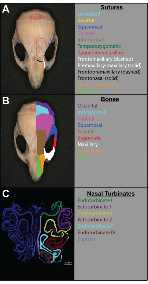
Figure 1: Anatomy of the relevant sutures and bones of C57BL/6 mouse skull and representative cross-section of the olfactory epithelium. (A) Sutures of the skull are color-coded. Sutures are color-coded on one side of the skull, while the other side is left unmarked for reference (left). Names of the sutures color-coded accordingly (right). (B) Bones of the skull color-coded. Bones are color-coded on one side of the skull, while the other side is left unmarked for reference (left). Names of the bones color-coded accordingly (right). (C) Cross section of the central portion of the OE (dissected using the method described in this article) with turbinates color-coded on one side and the over left unmarked for reference (left). Names of the visualized turbinates (right). Please click here to view a larger version of this figure.
Access restricted. Please log in or start a trial to view this content.
Protocol
All animal experiments in this study were approved by and conducted in compliance with the Baylor College of Medicine IACUC. Female C57BL/6J (JAX:000664) mice were used. Details of the reagents and equipment are listed in the Table of Materials.
1. Olfactory epithelium dissection
- Begin by deeply anesthetizing the mouse using any preferred method (following institutionally approved protocols). Confirm surgical plane of anesthesia by firm toe pinch and proceed with transcardiac perfusion of at least 10 mL of ice-cold 1x PBS. Remove the skin to expose the skull (Figure 2A).
NOTE: Removal of the head from the body is optional. If one decides to remove the head, it is recommended to remove the skin and other soft tissue around the skull prior to decapitation. - Using a pair of fine, sharp-tipped scissors, make two incisions to break the zygomatic arches from the squamosal bone bilaterally by placing one scissor tip in the posterior orbit and the other adjacent to the auditory canal (green lines in Figure 2B).
- Make a shallow incision from orbit to orbit along the most anterior aspect of the frontal bone. Incise only the dorsal surface of the frontal bone (~1-2 mm deep), being careful not to cut any underlying structures or bones that comprise the orbit (blue line in Figure 2B).
NOTE: To achieve a shallow incision between the orbits, hold the scissors perpendicular to the skull and use only the sharp tips to cut. - Cut along the midline of the skull from the foramen magnum, through the sagittal and interfrontal sutures, until the interorbital incision made in the frontal bone in step 1.3 is reached (red line in Figure 2B).
- Insert a pair of forceps into the incision made along the midline of the skull and firmly pull one hemisphere of the cranial vault laterally. This action will also remove the bones of the orbit (frontal, maxillary, and lacrimal), exposing the lateral aspects of the olfactory turbinates (Figure 2D).
NOTE: Take care not to inadvertently damage the OE from this point forward. - Holding the nasal bone and incisor teeth, break away the contralateral cranial vault using forceps. Allow the brain (including the olfactory bulbs) to be removed with the hemisphere of bone as it is torn away (Figure 1E). The only visible bony structures that should remain after this step are the base of the skull and the bones surrounding the OE (Figure 2F).
- Using a pair of bone nippers or rongeurs, remove any remaining frontal bone and premaxillary (incisive) bone up to the base of the incisor teeth (Figure 2G).
- Use bone nippers to cut along the frontonasal suture to release the nasal bone from the cribriform plate, taking extreme caution not to damage the underlying exposed OE or fracture the cribriform plate.
- Lift the nasal bone off the cribriform plate using forceps to expose the OE (Figure 2H). If residual premaxillary bone obscures the dorsal OE, remove it before proceeding (black circles in Figure 1I).
- Using fine-tipped forceps, gently secure the dorsal aspect of the nasal septum at its junction with the cribriform plate and pull posteriorly (Figure 2J,K). With minimal resistance, the main olfactory epithelium will break free as a wholly intact structure (Figure 2L).
- Proceed immediately with OE dissociation or snap-freeze the tissue for later processing.
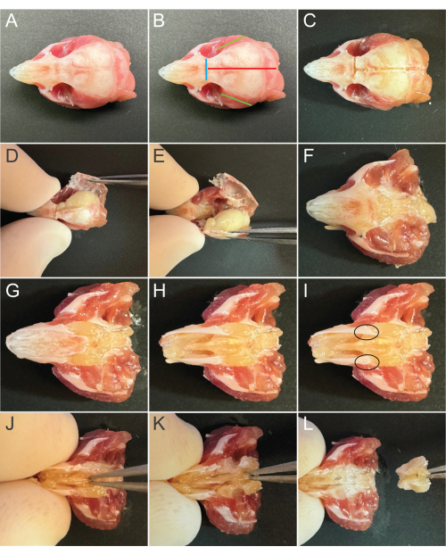
Figure 2: Technique for rapid dissection of the olfactory epithelium. (A) The mouse head following the removal of the surrounding soft tissue. (B) Color-coded guides for cranial vault incisions. (C) Appearance following cranial vault incisions. (D) Removal of the first hemisphere of the cranial vault along with the portion of the orbit adjacent to the olfactory epithelium. (E) Removal of the contralateral portion of the cranial vault along with the brain. (F) The head following removal of the brain and cranial vault. (G) Removal of the anterior-most frontal bone and posterior premaxilla, following incision at the frontomaxillary suture. (H) Removal of the nasal bones. (I) Black circles highlight areas of residual premaxillary bone that can be optionally removed. (J) Placement of forceps for removal of intact olfactory epithelium. (K) Pull posteriorly to release the olfactory epithelium. (L) Total release of the olfactory epithelium. Please click here to view a larger version of this figure.
2. Olfactory epithelium dissociation and single-nucleus isolation
- Place one C-tube and two 15 mL conical tubes per sample on ice.
- Prepare lysis buffer by adding RNAse inhibitor (to a final concentration of 0.2 U/µL) to pre-made nuclei extraction buffer.
- Add 4 mL of 1% BSA to each 15 mL conical tube, vortex briefly, and discard excess. This coats each of the 15 mL conical tubes with BSA to minimize nuclei adhesion to the tube.
- For one whole OE, add 2 mL of ice-cold lysis buffer to each C-tube.
- Add OE samples to the C-tubes. Using #2 forceps, hold each sample over its respective C-tube and cut it into small pieces using a pair of fine-tip scissors.
- Place the C-tubes containing each sample upside-down onto the automated tissue dissociator blocks. On the dissociator screen, the square corresponding to each block will change from Free to Selected.
- Press the folder icon to cycle into the Miltenyi folder. Using the arrows, scroll to the 4C_nuclei_1 program (a pre-loaded program within the dissociator). Press OK to apply the program to each block, and then press Start to begin the cycle. Remove the C-tubes from the dissociator immediately after cycle completion.
NOTE: If possible, keep the C-tubes ice-cold during the dissociation cycle. It is important to remove the samples from the dissociator immediately after cycle completion and proceed with the next steps of the protocol to avoid over-lysis of the nuclei. - With a pipette, aspirate the homogenized suspension from the C-tube and filter it through a 70 µm strainer into the first BSA-coated conical tube. Avoid aspirating the bone fragments that have accumulated at the bottom of the C-tube.
- Rinse the strainer with 1 mL of ice-cold 1x PBS to ensure complete filtration.
- Centrifuge the filtered suspension at 500 × g for 5 min at 4 °C in a swinging-bucket centrifuge.
- Gently aspirate and discard the supernatant, taking care to avoid disturbing the pellet. Completely resuspend the nuclei pellet in 1 mL of ice-cold 1x PBS by gentle pipetting.
- Pass the resuspended nuclei through a 30 µm strainer into the second BSA-coated conical tube. The concentration of nuclei can be quantified using a hemocytometer or an automated cell counter.
- Proceed immediately with the chosen downstream protocol (with or without FACS) to preserve sample integrity and prevent degradation (Figure 3).
NOTE: It is highly recommended that users perform practice runs of this entire protocol to ensure seamless and timely completion.
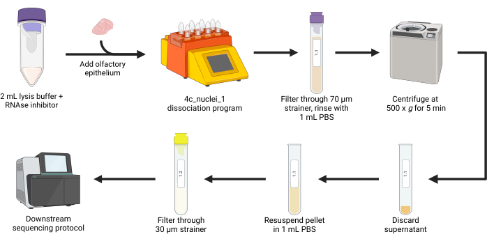
Figure 3: Olfactory epithelium dissociation and single-nucleus isolation workflow. Following dissection, the olfactory epithelium is mechanically and chemically dissociated. The lysate is then filtered through a 70 µm strainer, rinsed with 1x PBS, and centrifuged at 500 × g for 5 min. The pellet is resuspended in 1x PBS before being filtered through a 30 µm strainer. The resulting nuclei suspension is available for various downstream sequencing applications. Please click here to view a larger version of this figure.
Access restricted. Please log in or start a trial to view this content.
Results
To provide anatomical context for dissection and analysis, the sutures and bones of the C57BL/6 mouse skull, as well as the central portion of the olfactory epithelium (OE), were visualized and color-coded (Figure 1). One side of each image was annotated to highlight relevant sutures, bones, and turbinates, while the other side was left unmarked for reference. The OE cross-section was prepared following established protocols20, and turbinate identification was based o...
Access restricted. Please log in or start a trial to view this content.
Discussion
The procedure described here enables the consistent removal of an intact OE following intracardiac 1x PBS perfusion. This represents a notable improvement in speed compared to previous methods16 and is of particular importance given that OSNs begin to degenerate following axotomy2, which happens during brain removal.
Careful removal of the frontal, premaxillary, and nasal bones is crucial for consistent extraction of an intact OE. Additionally, d...
Access restricted. Please log in or start a trial to view this content.
Disclosures
The authors declare that this research was conducted in the absence of any commercial or financial relationships that could be construed as a potential conflict of interest.
Acknowledgements
The authors would like to acknowledge Baylor College of Medicine's Single Cell Genomics Core. This project was supported by the Cytometry and Cell Sorting Core at Baylor College of Medicine with funding from the NIH (P30 AI036211, P30 CA125123, and SS10 RR024574) and the expert assistance of Joel M. Sederstrom. This project was funded in part by the NIH/NICHD [P50HD103555], the NIH/NINDS [5R01NS078294], and the NIH/NIA [5F30AG076265]. Figure 3 is created in BioRender. https://BioRender.com/5418352.
Access restricted. Please log in or start a trial to view this content.
Materials
| Name | Company | Catalog Number | Comments |
|---|---|---|---|
| 15 mL Centrifuge Tube | Celltreat | 229411 | |
| Bone Nipper | Fine Science Tools | 16102-11 | |
| Bonn Miniature Iris Scissors | Miltex | 18-1392 | |
| Bovine Serum Albumin Solution | Sigma-Aldrich | A1595 | |
| Dumont #3 Forceps | Fine Science Tools | 11231-30 | |
| Female C57BL/6J mice | The Jackson Laboratory | JAX:000664 | |
| gentleMACS C tubes | Miltenyi Biotec | 130-093-237 | |
| gentleMACS Octo Dissociator | Miltenyi Biotec | 130-096-427 | |
| MACS SmartStrainers 30 um | Miltenyi Biotec | 130-098-458 | |
| MACS SmartStrainers 70 um | Miltenyi Biotec | 130-098-462 | |
| Micro Friedman-Pearson Rongeurs | Fine Science Tools | 16220-14 | |
| Nuclei Extraction Buffer | Miltenyi Biotec | 130-128-024 | |
| Phosphate-Buffered Saline (PBS), 1x without calcium and magnesium, PH 7.4 ± 0.1 | Corning | 21-040-CV | |
| Protector RNAse Inhibitor | Millipore Sigma | 3335402001 | |
| Sorvall Legend XTR Refrigerated Centrifuge | Thermo Scientific | 50119927-5 |
References
- Graziadei, P. P. C., Graziadei, G. A. M. Neurogenesis and neuron regeneration in the olfactory system of mammals. I. Morphological aspects of differentiation and structural organization of the olfactory sensory neurons. J Neurocytol. 8 (1), 1-18 (1979).
- Monti Graziadei, G. A., Graziadei, P. P. C. Neurogenesis and neuron regeneration in the olfactory system of mammals. II. Degeneration and reconstitution of the olfactory sensory neurons after axotomy. J Neurocytol. 8 (2), 197-213 (1979).
- Caggiano, M., Kauer, J. S., Hunter, D. D. Globose basal cells are neuronal progenitors in the olfactory epithelium: a lineage analysis using a replication-incompetent retrovirus. Neuron. 13 (2), 339-352 (1994).
- Iwai, N., Zhou, Z., Roop, D. R., Behringer, R. R. Horizontal basal cells are multipotent progenitors in normal and injured adult olfactory epithelium. Stem Cells. 26 (5), 1298-1306 (2008).
- Schwob, J. E., et al. Stem and progenitor cells of the mammalian olfactory epithelium: Taking poietic license. J Comp Neurol. 525 (4), 1034-1054 (2017).
- Fletcher, R. B., et al. Deconstructing olfactory stem cell trajectories at single-cell resolution. Cell Stem Cell. 20 (6), 817-830.e8 (2017).
- Gadye, L., et al. Injury activates transient olfactory stem cell states with diverse lineage capacities. Cell Stem Cell. 21 (6), 775-790.e9 (2017).
- Saraiva, L. R., et al. Hierarchical deconstruction of mouse olfactory sensory neurons: from whole mucosa to single-cell RNA-seq. Sci Rep. 5 (1), 18178(2015).
- Fletcher, R. B., et al. p63 regulates olfactory stem cell self-renewal and differentiation. Neuron. 72 (5), 748-759 (2011).
- Brann, D. H., et al. Non-neuronal expression of SARS-CoV-2 entry genes in the olfactory system suggests mechanisms underlying COVID-19-associated anosmia. Sci Adv. 6 (31), eabc5801(2020).
- Carter, L. A., MacDonald, J. L., Roskams, A. J. Olfactory horizontal basal cells demonstrate a conserved multipotent progenitor phenotype. J Neurosci. 24 (25), 5670-5683 (2004).
- Lake, B. B., et al. A comparative strategy for single-nucleus and single-cell transcriptomes confirms accuracy in predicted cell-type expression from nuclear RNA. Sci Rep. 7 (1), 6031(2017).
- Bakken, T. E., et al. Single-nucleus and single-cell transcriptomes compared in matched cortical cell types. PLoS One. 13 (12), e0209648(2018).
- Ruiz Tejada Segura, M. L., et al. A 3D transcriptomics atlas of the mouse nose sheds light on the anatomical logic of smell. Cell Rep. 38 (12), 110547(2022).
- Li, W., et al. A single-cell transcriptomic census of mammalian olfactory epithelium aging. Dev Cell. 59 (22), 3043-3058.e8 (2024).
- Dunston, D., Ashby, S., Krosnowski, K., Ogura, T., Lin, W. An effective manual deboning method to prepare intact mouse nasal tissue with preserved anatomical organization. J Vis Exp. (78), e50538(2013).
- Gurumurthy, R. K., Kumar, N., Chumduri, C. Optimized protocol for isolation of high-quality single cells from the female mouse reproductive tract tissues for single-cell RNA sequencing. STAR Protoc. 2 (4), 100970(2021).
- Nguyen, Q. H., Pervolarakis, N., Nee, K., Kessenbrock, K. Experimental considerations for single-cell RNA sequencing approaches. Front Cell Dev Biol. 6, 108(2018).
- Belfort, B. D. W., et al. Comparative analysis of AAV serotypes for transduction of olfactory sensory neurons. Front Neurosci. 19, 1531122(2025).
- Huang, J. S., et al. Immature olfactory sensory neurons provide behaviourally relevant sensory input to the olfactory bulb. Nat Commun. 13 (1), 6194(2022).
- Barrios, A. W., Nunez, G., Sanchez Quinteiro, P., Salazar, I. Anatomy, histochemistry, and immunohistochemistry of the olfactory subsystems in mice. Front Neuroanat. 8, 63(2014).
- Antunes, M. B., et al. Murine nasal septa for respiratory epithelial air-liquid interface cultures. Biotechniques. 43 (2), 195-204 (2007).
- Wu, J., Cai, Y., Wu, X., Ying, Y., Tai, Y., He, M. Transcardiac perfusion of the mouse for brain tissue dissection and fixation. Bio Protoc. 11 (5), e3988(2021).
- Bakulski, K. M., et al. Single-cell analysis of the gene expression effects of developmental lead (Pb) exposure on the mouse hippocampus. Toxicol Sci. 176 (2), 396-409 (2020).
- Chang, C. -S., et al. Single-cell RNA sequencing uncovers the individual alteration of intestinal mucosal immunocytes in Dusp6 knockout mice. iScience. 25 (2), 103738(2022).
- Denisenko, E., et al. Systematic assessment of tissue dissociation and storage biases in single-cell and single-nucleus RNA-seq workflows. Genome Biol. 21 (1), 130(2020).
- Koenitzer, J. R., Wu, H., Atkinson, J. J., Brody, S. L., Humphreys, B. D. Single-nucleus RNA-sequencing profiling of mouse lung. Reduced dissociation bias and improved rare cell-type detection compared with single-cell RNA sequencing. Am J Respir Cell Mol Biol. 63 (6), 739-747 (2020).
- Xiong, A., Zhang, J., Chen, Y., Zhang, Y., Yang, F. Integrated single-cell transcriptomic analyses reveal that GPNMB-high macrophages promote PN-MES transition and impede T cell activation in GBM. eBioMedicine. 83, 104239(2022).
Access restricted. Please log in or start a trial to view this content.
Reprints and Permissions
