
Guest Editors
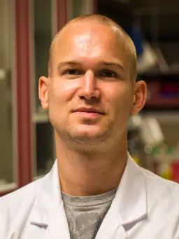
Koen Galenkamp
Sanford Burnham Prebys Medical Discovery Institute, NCI-designated Cancer Center
Dr. Koen Galenkamp completed his PhD in Biochemistry, Molecular Biology, and Biomedicine in 2015 at the Autonomous University of Barcelona (Spain), where he studied the possibilities of targeting death receptor signaling and the NF-κB pathway in neuroblastoma. In 2016, he joined the lab of Dr. Commisso at the Sanford Burnham Prebys Medical Discovery Institute’s NCI-designated Cancer Center as a post-doctoral associate and was later awarded a post-doctoral fellowship by the Tobacco-Related Disease Research Program (TRDRP) and a Fishman Fund Career Development Award. Here, his research focuses on the role of sodium-hydrogen exchange in the regulation of pH homeostasis in pancreatic cancer and its implications in cell fitness, actin remodeling, and macropinocytosis.
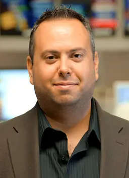
Cosimo Commisso
Sanford Burnham Prebys Medical Discovery Institute, NCI-designated Cancer Center
Dr. Cosimo Commisso earned his PhD in 2008 at the University of Toronto in the Department of Molecular Genetics and completed his Postdoctoral training at New York University’s Langone Medical Center in 2014. He is currently an Associate Professor in the Sanford Burnham Prebys Medical Discovery Institute NCI-designated Cancer Center. Dr. Commisso made a seminal contribution to the field of cancer research by identifying the mechanism by which Ras-mutated tumors augment their glutamine supply through boosting macropinocytosis-dependent protein catabolism. In his follow up studies, it was established that human pancreatic tumors scavenge extracellular protein via macropinocytosis. The Commisso laboratory has developed extensive protocols to evaluate macropinocytosis in tumor cells and tissue, a methodology that could be utilized to stratify patient samples. Current research in Dr. Commisso’s lab is centered on identifying the underlying mechanisms that drive macropinocytosis in Ras-mutated tumors and understanding the role of nutrient stress in tumor progression. Dr. Commisso’s work has garnered several awards, including a Natural Sciences and Engineering Research Council of Canada award, an Ontario Scholar Award, two Canadian Institutes of Health Research awards, two AACR Pancreatic Cancer Action Network awards, a Cancer Centers Council Pedal the Cause Team Science Award and most recently a Career Development Award from the Department of Defense.
Yijuan Zhang
Sanford Burnham Prebys Medical Discovery Institute, NCI-designated Cancer Center
Dr. Yijuan Zhang is a Postdoctoral Associate in Dr. Cosimo Commisso's laboratory at Sanford Burnham Prebys Medical Discovery Institute’s NCI-designated Cancer Center. Her current research focuses on studying the mechanism and function of macropinocytosis in pancreatic cancer-associated fibroblasts. She received her PhD in Microbiology from the University of Wuhan and then worked as a research assistant on nanoscale material-based drug delivery for cancer chemotherapy and immunotherapy in Dr. Yifan Ma's group at Shenzhen Institute of Advanced Technology, Chinese Academy of Sciences. Before her current position, she conducted her first postdoctoral studies in Dr. Xiaoting Zhang's laboratory at the University of Cincinnati, exploiting pRNA nanoparticle-delivered MED1 shRNAs to overcome tamoxifen resistance of breast cancer.
Collection Overview
Macropinocytosis is an endocytic mechanism of fluid-phase uptake that functions in a variety of cells to internalize extracellular fluid and any molecules or particles found therein. Macropinocytosis is important in many biological processes, including cancer metabolism, immune surveillance, and pathogen invasion. Moreover, macropinocytic uptake has emerged as a drug delivery mechanism for nanoscale therapeutics and bioactive molecules.
Macropinocytosis is a non-selective form of uptake and depends on actin-dependent membrane ruffles and cytoskeletal dynamics to form the nascent vesicular structure, termed a macropinosome. Once formed, the macropinosome compartment then matures and depending on the cellular context, internalized cargo can be degraded via the lysosome, recycled back to the cell surface, or trafficked to other cellular locations.
In this Methods Collection, we aim to explore state-of-the-art methods utilized in the study of macropinocytosis. These include approaches to (1) dynamically visualize macropinocytosis in vitro, (2) quantify macropinocytosis using medium/high-throughput microscopy or flow cytometry, (3) examine macropinosomes in live tissue in vivo or ex vivo, and (4) investigate macropinocytic dependency of functional read-outs such as nutrient production, antigen presentation, pathogen invasion or drug delivery.
Articles

Visualizing Membrane Ruffle Formation using Scanning Electron Microscopy
0 Views
•
Cited by 3
•
2021
•


Fluorescence Microscopy for ATP Internalization Mediated by Macropinocytosis in Human Tumor Cells and Tumor-xenografted Mice
0 Views
•
Cited by 3
•
2021
•
Corinne M. Nielsen*1,2,3, Yanrong Qian*4, Subhodip Adhicary1,5, Yunsheng Li4, Pratik Shriwas1,2,4, Xuan Wang1,2,4, Lindsey Bachmann6, Xiaozhuo Chen1,2,4,7
1Department of Biological Sciences, Ohio University, 2Molecular and Cellular Biology Program, Ohio University, 3Neuroscience Program, Ohio University, 4Edison Biotechnology Institute, Ohio University, 5Translational Biomedical Sciences Program, Ohio University, 6Honors Tutorial College, Ohio University, 7Department of Biomedical Sciences, Heritage College of Osteopathic Medicine, Ohio University

Quantitative Assessment of Macropinocytosis in mTORC1-Hyperactive Cells using Flow Cytometry
0 Views
•
Cited by 3
•
2021
•
Abstracts
Measuring the pH and redox chemistries of macropinosomes by dual-fluorophore ratiometric microscopy.
Johnathan Canton*1
1The University of Calgary
Automated quantification of individual macropinosomes using FIJI.
Adam Wall1,
Yvette Koh1,
Jennifer Stow*1,
Nicholas Condon1
1The University of Queensland
A click-chemistry based flux assay to measure the anabolic contributions of macropinocytosis
Aimee Edinger *1,
Vaishali Jayashankar 1
1University Of California Irvine