The lacrimal gland (LG) is a branching organ that produces the aqueous components of tears necessary for maintaining vision and ocular health. Here we describe murine LG dissection and ex vivo culture techniques to decipher signaling pathways involved in LG development.
The lacrimal gland (LG) secretes aqueous tears necessary for maintaining the structure and function of the cornea, a transparent tissue essential for vision. In the human a single LG resides in the orbit above the lateral end of each eye delivering tears to the ocular surface through 3 – 5 ducts. The mouse has three pairs of major ocular glands, the most studied of which is the exorbital lacrimal gland (LG) located anterior and ventral to the ear. Similar to other glandular organs, the LG develops through the process of epithelial branching morphogenesis in which a single epithelial bud within a condensed mesenchyme undergoes multiple rounds of bud and duct formation to form an intricate interconnected network of secretory acini and ducts. This elaborate process has been well documented in many other epithelial organs such as the pancreas and salivary gland. However, the LG has been much less explored and the mechanisms controlling morphogenesis are poorly understood. We suspect that this under-representation as a model system is a consequence of the difficulties associated with finding, dissecting and culturing the LG. Thus, here we describe dissection techniques for harvesting embryonic and post-natal LG and methods for ex vivo culture of the tissue.
The lacrimal gland (LG) is responsible for aqueous tear secretion critical for visual acuity and the health, maintenance, and protection of the cells of the ocular surface. LG dysfunction results in one of the most common and debilitating ocular disorders: aqueous deficient Dry Eye Disease, which is characterized by ocular irritation, light sensitivity and decreased vision1. In the human the LG resides in the orbit above the lateral end of the eye where 3 – 5 excretory ducts deposit tears onto the ocular surface. The mouse has three pairs of major ocular glands, the most studied of which is the lacrimal gland (LG) located anterior and ventral to the ear (exorbital) with tears traveling to the eye via a single excretory duct. Similar to other glandular organs, the LG develops through the process of epithelial branching morphogenesis in which a single epithelial bud within a condensed mesenchyme undergoes multiple rounds of bud and duct formation to form an intricate interconnected network of secretory acini and ducts (Figure 1)2. During development the epithelium becomes vascularized as well as heavily innervated by the parasympathetic nerves of the pterygopalatine ganglion and to a lesser extent by sympathetic nerves from the superior cervical ganglion3. Interactions between each of these cells types i.e. neuronal, epithelial, endothelial and mesenchymal cells, are essential to the function and maintenance of the adult tissue. However, the underlying molecular mechanisms coordinating LG development and regeneration as well as how inter-cell type communication guides these processes remains unclear.
The advent of embryonic ex vivo culture techniques has allowed the identification of developmental and regenerative pathways in multiple branching organs4. Culturing ex vivo gives the researcher the ability to manipulate the organ (mechanical, genetic or chemical) under defined conditions as well as to characterize organ development and cell-cell interactions in real time. The exorbital LG of the mouse is highly amenable to this technique and recent studies have defined signaling systems that regulate its development2,5. However, despite the need to understand molecular cues underpinning LG development and regeneration, it currently remains understudied, likely due to the technical difficulties in isolating the organ. In this paper, we describe how to isolate and perform ex vivo culture of the embryonic murine LG to define developmental programs.
All animal work was performed under strict accordance with the recommendations in the Guide for the Care and Use of Laboratory Animals of the National Institutes of Health. The protocol was approved by the Institutional Animal Care and Use Committee (IACUC) of the University of California, San Francisco.
1. Mouse Embryonic Lacrimal Glands (LG): Harvesting and Microdissection
2. Prepare Plates for Ex vivo Culture
This procedure is based on that employed for the developing salivary gland7,8.
3. Mouse Postnatal and Adult Lacrimal Glands (LG): Dissection
The LG develops through the process of epithelial branching morphogenesis. Brightfield images of embryonic LGs dissected at e14, e15, e16, e17, and P2 illustrate this event.
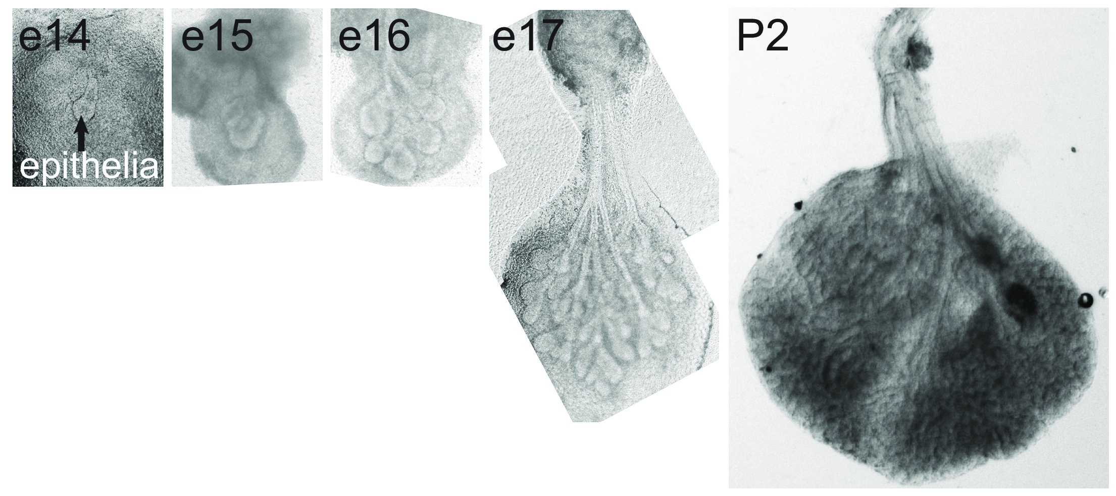
Figure 1. The LG develops through the process of epithelial branching morphogenesis. Brightfield images of embryonic LGs freshly dissected at e14, e15, e16, e17 and P2. Please click here to view a larger version of this figure.
Note that as the epithelium increases in size, the amount of mesenchyme decreases. The experimental steps for microdissection of the embryonic LGs are depicted in Figure 2.
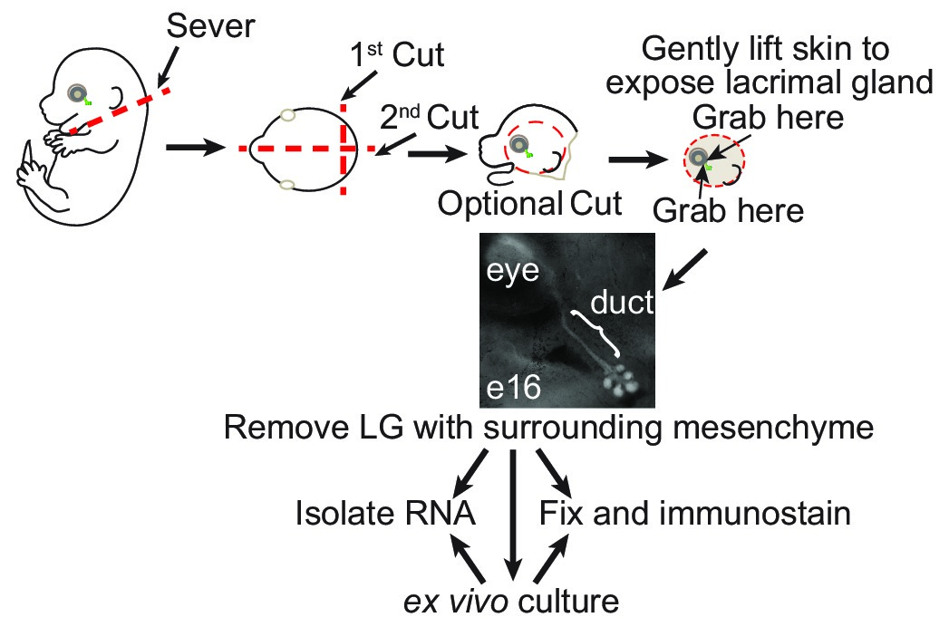
Figure 2. Schematic of steps involved in LG microdissection from mouse embryos. Schematic diagram of the dissection and removal of the LG from the embryonic mouse. Please click here to view a larger version of this figure.
For LG culture we routinely utilize e16 embryos due to the consistency in ex vivo growth. As shown in Figure 3, LG in ex vivo culture conditions develop in a similar manner to the in vivo gland (compare to Figure 1).
Figure 3. Ex vivo culture of e16 LGs recapitulates in vivo morphogenesis. e16 LGs were derived from Pax6-Cre, GFP embryos, where GFP is expressed by the epithelium. Consecutive fluorescent images were taken of this single gland at 0, 3, 6, 12, 24, and 48 hr. Please click here to view a larger version of this figure.
Here we employed Pax6-Cre, GFP (also called Le-Cre5) embryos to highlight epithelial cells, allowing us to take consecutive fluorescent images of the branching epithelium over the 48 hr culture period. Mice harboring the Pax6-Cre, GFP transgene can be obtained from JAX: Tg(Pax6-Cre, GFP)1Pgr but are not necessary for gland dissection and culture.
Cultures or freshly dissected LG can then be fixed for immunofluorescent analysis. Figure 4 shows the visualization of 4 cellular compartments, mesenchyme, epithelium (EpCAM), nerves (Tubb3) and blood vessels (PECAM), in an e16 LG from a wild type (CD1) embryo.
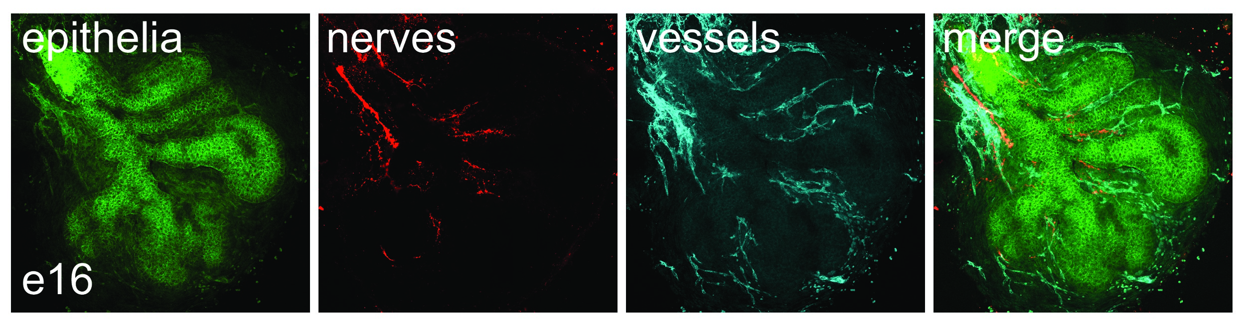
Figure 4.The LG is composed of multiple cell types including epithelial, neuronal and endothelial cells. e16 LG was immunostained for epithelial cells (Epcam, green), neurons (Tubb3, red) and endothelial cells (Pecam, cyan). Please click here to view a larger version of this figure.
For freshly dissected tissue it is easier to first embed the LG in laminin, as done for culture, so as to immobilize the gland for fixation and subsequent handling. Figure 5 shows a schematic for dissection of the postnatal/adult LG.
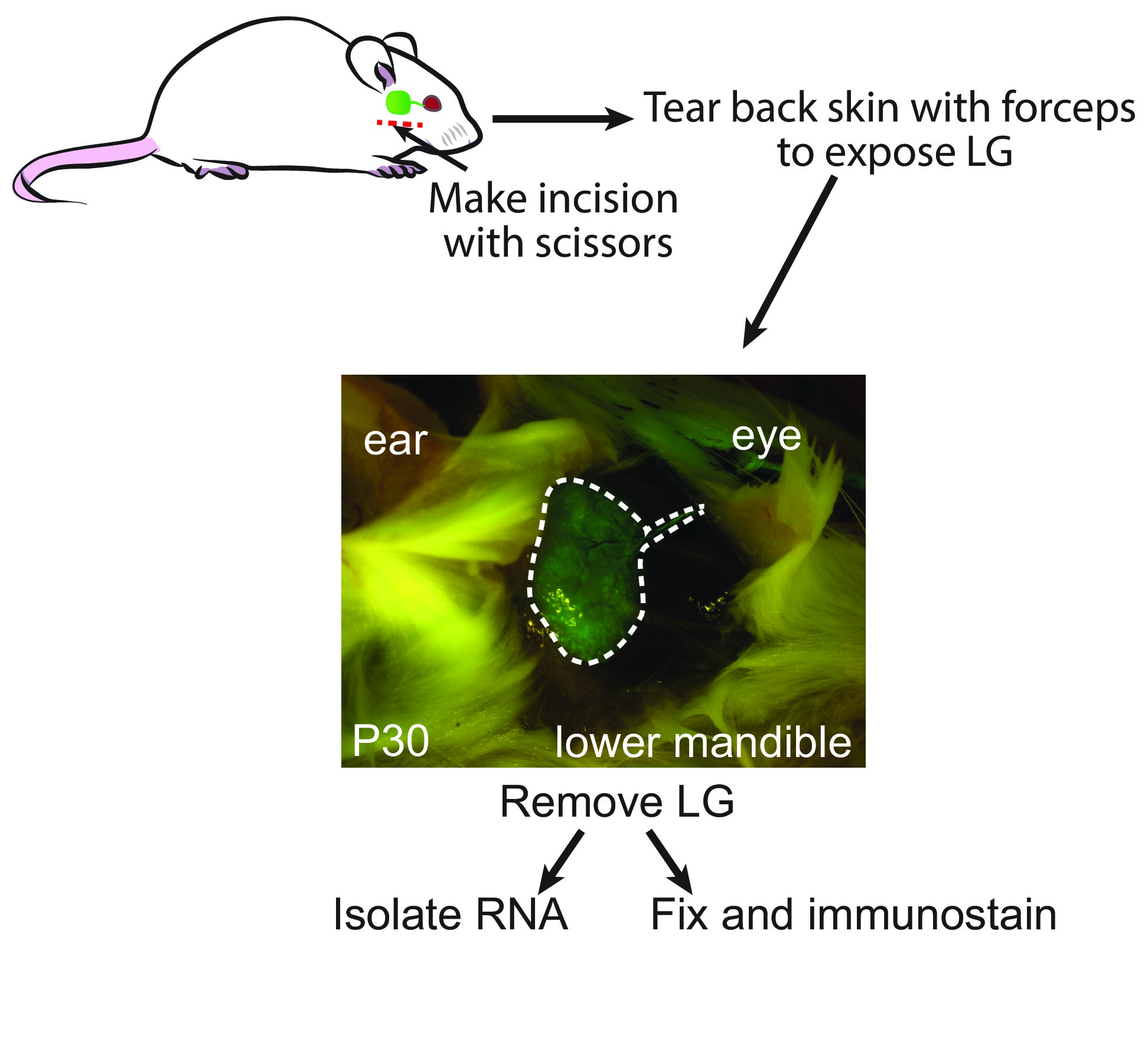
Figure 5. Schematic of steps involved in LG microdissection from post-natal and adult mouse. Schematic diagram of the dissection and removal of the LG from either post-natal or adult mouse. A Pax6-Cre,GFP post-natal day 30 mouse was used to visualize the position of the LG and associated duct. Please click here to view a larger version of this figure.
The position of the post-natal/adult LG has been visualized by using the Pax6-Cre, GFP mouse.
The versatility to culture and manipulate the LG ex vivo provides significant advantages for studying its development. This includes the speed at which the researcher is able to test hypotheses and the multitude of perturbations that can be performed to assess how epithelial, neuronal, endothelial and mesencyhmal cells interact to form the organ. However, there are a number of caveats when utilizing this model. First, by virtue of its isolation the gland is no longer connected to the peripheral vasculature or nervous system, which may affect the signaling pathways being tested if they work in conjunction with signals delivered by the nerves or blood vessels9,10. Second, the surrounding non-LG tissues may provide factors that regulate LG development4. To avoid this other non-mesenchymal tissue components must be removed prior to culture. Third, at present we are not able to provide the conditions necessary for complete cytodifferentiation into a fully functional tissue. This is the case for all organs cultured ex vivo and is likely due to multiple reasons including the absence of functional blood vessels and nerves and/or mechanical forces from surrounding tissues. Despite these caveats, mechanisms discovered in ex vivo culture have shown to be recapitulated in subsequent in vivo experiments, making this a robust system for testing new hypotheses8,11.
It has been shown that developmental and regenerative pathways significantly overlap, indicating that the ex vivo culture system can also be employed to study organ regeneration12. Although we have not demonstrated this aspect here we, and others, have previously shown the embryonic salivary gland can be used to model the effects of therapeutic radiation on organ damage and regeneration13. Future studies are needed to explore the use of the LG ex vivo system as a model for tissue regeneration.
The authors have nothing to disclose.
The authors would like to acknowledge funding provided by the UCSF Resource Allocation Program.
Name | Catalog #. | Company | Comments/Description |
DMEM/F12 without HEPES | SH3027101 | Thermo Scientific | |
Penicillin-Streptomycin | P0781 | Sigma-Aldrich | |
Albumin solution from bovine serum | A9576 | Sigma-Aldrich | |
Paraformaldehyde 16% Solution | 15710 | Electron Microscopy Sciences | Diluted to 4% in 1X PBS |
Phosphate Buffered Saline 10X | BP665-1 | Fisher Scientific | |
Phosphate Buffered Saline 1X | Prepared from 10X stock | ||
holo-Transferrin bovine | T1283 | Sigma-Aldrich | 25 mg/ml stock solution in water. Freeze single-use aliquots at -20C. Add to DMEM/F12 media to a final concentration of 50 ug/mL. |
L- Ascorbic acid (Vitamin C) | A4544 | Sigma-Aldrich | 25 mg/ml stock solution in water. Freeze single-use aliquots at -20C. Add to DMEM/F12 media to a final concentration of 50 ug/mL. |
BioLite 100mm Tissue Culture Treated Dishes | 130182 | Thermo Scientific | Non-tissue culture-treated plates can also be used. |
Stereo Microscope with substage iluminator | Stemi 2000 | Carl Zeiss | Any stereo dissecting microscope can be used that has a transmitted light base. |
Substage Illuminator Base for Stereo Microscope | TLB 4000 | Diagnostics Instruments, Inc. | |
Falcon 35mm Tissue Culture Treated Dishes | 353001 | Corning | Non-tissue culture-treated plates can also be used. |
50mm uncoated glass bottom dishes | P50G-1.5-14-F | MatTek Corporation | |
Widefield fluorescence microscope | Axio Observer Z1 | Carl Zeiss | Any fluorescence microscope (upright, inverted or stereo dissecting microscope) can be used to monitor GFP expression at low magnification with an attached digital camera. |
Confocal Microscope | TCS SP5 | Leica Microsystems | Confocal microscopy is necessary to see detailed cell structures. Any confocal microscope can be used. |
Ideal for harvesting glands from embryos | |||
SWISS Micro-Fine Forceps, #5 (11.2cm) | 17-305X | Integra LifeSciences Corporation | Fine tips are required for removing mesenchyme from epithelium. Tungsten needles can also be used. |
Dumont Standard Tip Forceps, #5 (11cm) | 91150-20 | Fine Science Tools (USA), Inc. | Ideal for harvesting glands from embryos |
Reusable Plastic Surgical Knife Handles, style no. 3. | 4-30 | Integra LifeSciences Corporation | |
Stainless Steel Sterile Surgical Blades, no. 10 | 4-310 | Integra LifeSciences Corporation | |
RNAqueous-Micro Total RNA Isolation Kit | AM1931 | Life Technologies | |
Cultrex 3D Culture Matrix Laminin I | 3446-005-01 | Trevigen | 6 mg/ml stock diluted 1:1 with DMEM/F12 |
Timed-pregnant Crl:CD1(ICR) mice | Charles River Labs | Embryos are harvested on day 14 (with day of plug discovery designated as day 0). | |
Nucleopore Track-Etched Hydrophilic Membranes, 0.1 um pre size, 13mm | 110405 | Whatman | |
Timed-pregnant Pax6-Cre,GFP mice | Tg(Pax-Cre, GFP)1Pgr | The Jackson Laboratory | Optional mice for learning how to locate the LG. |