Targeted manipulations to cause directed stress or death in individual cells have been relatively difficult to accomplish. Here, a single-cell-resolution ablation approach to selectively stress and kill individual cells in cell culture and living animals is described based on a standard confocal UV laser.
Method Article
Triggering Cell Stress and Death Using Conventional UV Laser Confocal Microscopy
In This Article
Summary
Abstract
Using a standard confocal setup, a UV ablation method can be utilized to selectively induce cellular injury and to visualize single-cell responses and cell-cell interactions in the CNS in real-time. Previously, studying these cell-specific responses after injury often required complicated setups or the transfer of cells or animals into different, non-physiological environments, confounding immediate and short-term analysis. For example, drug-mediated ablation approaches often lack the specificity that is required to study single-cell responses and immediate cell-cell interactions. Similarly, while high-power pulsed laser ablation approaches provide very good control and tissue penetration, they require specialized equipment that can complicate real-time visualization of cellular responses. The refined UV laser ablation approach described here allows researchers to stress or kill an individual cell in a dose- and time-dependent manner using a conventional confocal microscope equipped with a 405-nm laser. The method was applied to selectively ablate a single neuron within a dense network of surrounding cells in the zebrafish spinal cord. This approach revealed a dose-dependent response of the ablated neurons, causing the fragmentation of cellular bodies and anterograde degeneration along the axon within minutes to hours. This method allows researchers to study the fate of an individual dying cell and, importantly, the instant response of cells-such as microglia and astrocytes-surrounding the ablation site.
Introduction
Fluorescence microscopy has long been used to study the effects of transgenes in the zebrafish CNS, particularly their effects on development1. High-resolution microscopy has allowed a detailed mapping of the cellular processes involved in brain development, muscle generation, and many other developmental events2. Studying the death of an individual cell has been more challenging, mainly due to the technical difficulties of inducing selective cell death during standard imaging procedures. However, the combination of single-cell resolution imaging and highly targeted ablation techniques allows the investigation of immediate cellular responses to stress and injury, as well as of the consequent cell-cell interactions. Understanding these processes is critical, particularly for neurodegenerative diseases such as motor neuron disease (MND), where neuron-glia interactions have been shown to contribute to the progression of the disease3.
MND, or amyotrophic lateral sclerosis (ALS), is a devastating neurodegenerative disease that affects motor neurons in the brainstem, motor cortex, and spinal cord. Loss of these neurons leads to muscle loss, and patients die within 3 - 5 years of diagnosis4. Motor neurons in the spinal cord link to the muscle fibers and play an essential role in facilitating muscle contraction. Failure of this communication or death of these neurons gradually weakens the muscles and affects the patient's ability to swallow, walk, speak, and breathe. Visualizing the death of a motor neuron and the short-term consequences in a living animal provides an excellent opportunity to better understand the dynamic processes involved in normal cell homeostasis and disease.
Zebrafish have emerged as an attractive model system to study neurodegenerative diseases1. This is due to the advantages offered by this model organism, such as external fertilization, short developmental time, optical access to the nervous system, and ease of transgenesis. In addition, the ability to easily generate compound transgenic zebrafish allows for multiple labeling strategies of different cell types. Genetic ablation approaches to kill specific cell types allow rather broad disturbance, but lack the fine control of targeting individual cells5. Laser-assisted techniques, on the other hand, provide fine temporal and spatial control and have been used for different animal models. While most approaches use specialized equipment, such as pulsed lasers6,7,8,9,10,11,12 or two-photon set-ups13, other research groups have recently taken advantage of a UV laser in conventional confocal microscopes14.
The technique described here combines high-resolution confocal microscopy with a UV laser-mediated approach to cause cellular stress or death in a dose-dependent way in selected motor neurons. It relies on the use of the commonly installed 405-nm laser, has been tested successfully in cell culture and in living animals, and allows the detailed characterization of cellular interactions, such as microglial clearance after neuronal death.
Protocol
NOTE: Design, conduct, and reporting of animal experiments must take account of current guidelines15. Such work must be approved in advance by the local animal welfare authority (in our case, the Animal Ethics Committee of Macquarie University).
1. Prepare the Zebrafish for Mounting and UV Cell Ablation
- Generate zebrafish (Danio rerio) expressing fluorescent proteins.
- To express fluorescent proteins of interest in zebrafish, perform plasmid injections into the one-cell stage of the zebrafish egg (as described elsewhere16) or use fluorescent transgenic lines. To label multiple cell types, create compound transgenic zebrafish lines by crossing established transgenic lines pertinent to the question of interest. Place one male and one female zebrafish on each side of a false-bottom pair mating tank in the evening and remove the divider with the onset of light the next morning (as detailed elsewhere17). Keep the zebrafish at 28 °C and handle them according to the established protocols17,18.
- Collect the embryos after successful spawning by straining the tank water containing the embryos through a plastic tea strainer. Rinse the eggs with system water and transfer them into egg water in a Petri dish.
- Examine them under a light microscope to determine fertilization. Store fertilized eggs in a Petri dish and place them in an incubator at 28 °C18.
- Optional: Perform a microinjection to label specific cell populations.
NOTE: This is an alternative method that allows for the expression and visualization of proteins, without the need to raise stable transgenic lines. This method is also advantageous when the protein of interest is toxic and prohibits the generation of a stable transgenic lines.- Inject the plasmid constructs into the one-cell stage of zebrafish embryos, as described elsewhere19,20,21.
NOTE: This method results in the mosaic expression of the protein of interest. The protein of interest is driven from a promoter of choice (e.g., islet122, -3mnx123,24, met25, or mpeg126) flanked by Tol2 inverted repeats20.
- Inject the plasmid constructs into the one-cell stage of zebrafish embryos, as described elsewhere19,20,21.
- Age the fish to the desired size.
- Raise the fish to 3 - 5 days post fertilization (dpf) and place them under a fluorescent compound microscope. Screen the animals for appropriate fluorophore expression and select the brightly labeled fish. Separate the appropriate larvae into another dish with egg water for embedding later on (store in a 28 °C incubator).
Optional: Embryos can be placed into a 0.2 mM 1-phenyl-2-thioures (PTU) Ringers solution at 24 h post fertilization (hpf) to inhibit the formation of pigmentation. Care must be taken with PTU, as it is toxic and can have adverse physiological, genetic, or morphological effects. - For studies at an early developmental stage (< 2 dpf), dechorionate the embryos manually using sharp forceps. Dechorionate large numbers of embryos enzymatically by adding pronase (2 mg/mL) to the egg water and incubating them for 10 min at 28 °C.
- Pass the embryos periodically through a plastic Pasteur pipette to ease dechorionation. Terminate the process when the majority of embryos have emerged from their chorions by washing them several times with egg water.
- Raise the fish to 3 - 5 days post fertilization (dpf) and place them under a fluorescent compound microscope. Screen the animals for appropriate fluorophore expression and select the brightly labeled fish. Separate the appropriate larvae into another dish with egg water for embedding later on (store in a 28 °C incubator).
- Prepare solutions for zebrafish embedding in agarose.
- Prepare an anesthesia solution by adding 4 g/L MS222 (tricaine stock solution, pH 7.0) dropwise to a Petri dish containing egg water. A dose of 50 mg/L is a recommended starting point (Figure 1A).
- Prepare a stock of low-melting agarose (0.8 - 1.5%) in egg water and aliquot it into 1.5-mL microcentrifuge tubes. Place an aliquot into a pre-heated heat block (38 - 40 °C) and let it equilibrate to the set temperature (~ 30 min; Figure 1B).
- Optional: For longer-term imaging (> 4 h), prepare a little agarose circle within the 35-mm glass-bottom Petri dish and allow it to set (Supplementary Figure 1).
NOTE: This extra step was effective in avoiding any movement of the whole agarose drop with the zebrafish over longer time frames.- To do so, place ~ 300 µL of agarose along the inner circle of the glass-bottom dish to prepare a doughnut-shaped circle with a little opening in the middle in which to place the fish (step 1.5.3; Supplementary Figure 1).
- Mount the zebrafish in agarose for microscopy.
- Select 1 - 3 of the pre-screened fish for ablation and anesthetize the larvae by transferring them (using a transfer pipette) into a dish with the anesthesia solution (step 1.4.1; Figure 1C; approximately 5 min).
NOTE: The fish are anesthetized when they show a shallow opercular movement and a decreased heart rate and no longer display a touch-evoked escape response (TEER; failure to swim away after gently touching their tail with a brush). Ensure appropriate anesthesia for the ethical treatment of the fish and to prevent twitching upon transfer into agarose or exposure to fluorescent light. - After the anesthesia is confirmed, suck up a larva using an adjustable pipette (with a cut-off 200-µL tip set to ~ 30 µL) and let it sink to the bottom of the tip. Transfer the larva into preheated agarose (step 1.4.2) by releasing a drop of the liquid with the larva into the agarose (try to minimize the amount of egg water going into the agarose; Figure 1D).
- Suck up the fish surrounded by agarose. Dispense it quickly into the previously prepared glass-bottom 35-mm dish.
- Use a dissection microscope and a standard paint brush (long liner, size 1) to position the animal within the agarose on the side (head to the left) so that body and tail are flat (Figure 1E). If working with multiple fish, align all the fish in the dish so that they are easily located using the confocal microscope later on.
NOTE: Quickly perform this procedure of positioning and aligning (it may require some practice, as the agarose starts to set immediately after exposure to colder temperatures). - Leave the agarose-embedded fish for 10 - 15 min until the agarose is set firmly. Carefully top up the 35-mm Petri dish with ~ 2 mL of egg water containing tricaine (Figure 1F).
- Select 1 - 3 of the pre-screened fish for ablation and anesthetize the larvae by transferring them (using a transfer pipette) into a dish with the anesthesia solution (step 1.4.1; Figure 1C; approximately 5 min).
2. Set up the Confocal Microscope and Imaging Parameters
- Place the Petri dish with the embedded larva on the confocal microscope stage and focus on the dorsal side of the animal spinal cord (using bright field). Examine the animal under the appropriate magnification (40X) and fluorescent setting and visualize the structure of interest (e.g., fluorescence intensity of the labeled neurons or microglial movement) to confirm that all imaging parameters are as needed for subsequent ablation (Figure 2). We routinely use the 40X objective to perform our time lapse studies.
- Optional: To perform a time-lapse study for several hours, it is advisable to record a single or a few time-points prior to ablation to establish the unperturbed physiological response of the cell and its environment (e.g., microglial movement to establish baseline speed and motility).
- Determine the thickness of the structure of interest for the UV laser ablation.
- Using the z-drive, verify the top and bottom of the structure of interest (e.g., the cell soma) by manually focusing up and down. Note down the z-plane that will be ablated (e.g., the center of the cell).
NOTE: From experience, this method was most effective by targeting spinal cord neurons that were brightly labeled (a high signal-to-noise ratio that allows easy time-lapse visualization after ablation; e.g., Figure 4) and by ablating the middle of the cell soma. Cell nucleus fluorescence can be of advantage to assure correct targeting and high ablation efficiency.
- Using the z-drive, verify the top and bottom of the structure of interest (e.g., the cell soma) by manually focusing up and down. Note down the z-plane that will be ablated (e.g., the center of the cell).
3. Perform Targeted Laser Ablation of Individual Cells in the Zebrafish Spinal Cord
NOTE: For this ablation and visualization approach, a confocal microscope (Leica SP5) was used. The ablation procedure using a 405-nm diode for cell-specific destruction is detailed according to the software (Leica Application Suite, v2.7.3.9723). However, any conventional confocal microscope that is equipped with a 405-nm laser and a FRAP (fluorescence recovery after photobleaching) or bleach module will allow the performance of the same cell manipulations, but potentially with slightly different settings, parameters, and names.
- Start the FRAP wizard by clicking on the dropdown menu at the top of the software menu (Figure 3A, 1 and 2). Observe a new window with different steps that allows the set up of the specific parameters for the laser ablation (Figure 3B, 3).
- Determine the image parameters for the ablation approach by selecting the format, scan speed (Figure 3B, 4), and averaging (Figure 3B, 5). An image format of 1,024 x 1,024 at a scan speed of 400 Hz and a line average of 4 was most applicable.
NOTE: There is generally no need to change the spectral detection (such as the excitation or emission parameters), as they have been determined in the previous acquisition.- If the z-plane for ablation hasn't already been selected (as described in step 2.4.), press the "Live" button and focus through the specimen until the fluorescent structure or the desired z-plane that is going to be ablated is in focus.
- Once the general image parameters are set, access the "Bleach" step (Figure 3C, 6) to control the specific ablation components.
NOTE: A combination of the laser intensity (Figure 3C, 8), the scan speed, and the averaging that has been set in step 3.2 (Figure 3B, 4 and 5), as well as the number of repetitions that will be set in step 3.5 (Figure 3E, 12), will determine the overall dwell time of the UV laser at the ROI, and therefore, the bleaching efficiency.- Engage the 405-nm laser by activating it for the bleaching procedure (Figure 3C, 8).
NOTE: Most success with the aforementioned settings was achieved with 405-nm laser intensities between 60 - 80% in our experimental setup. Be aware that this laser power output is instrument-specific and will differ for every confocal setup. - Use the "zoom in" option (Figure 3C, 7) to maximize the bleaching intensity at the selected ROI by reducing the scan field, therefore maximizing dwell time. Alternatively, use the "Bleach point" option of the software of choice for this process.
- Engage the 405-nm laser by activating it for the bleaching procedure (Figure 3C, 8).
- Select one or multiple ROIs (Figure 3D, 10) for the ablation by using any of the drawing tools in the image acquisition window (Figure 3D, 9). Target the axon hillock, for example, with the circular drawing tool of approximately 4 - 8 µm.
NOTE: The ablation area is adjustable from a single pixel to a larger area, depending on the application. - After establishing the ROI, select the "Time Course" button (Figure 3E, 11) and confirm the number of cycles the ROIs will be scanned/ablated (Figure 3E, 12). Choose the "Pre-Bleach" and "Post-Bleach" frames as desired to permit an overview of the whole image just before and immediately after the bleaching process.
- After establishing all the necessary ablation parameters, press "Run Experiment" (Figure 3E, 13) and monitor the efficiency of the ablation.
NOTE: In our FRAP setup, a single image will be taken before and after the FRAP cycle with the appropriate laser excitation (e.g., 488-nm excitation for EGFP-expressing cells). These pre- and post-ablation pictures allow a quick judgment of how satisfactorily the ROI was bleached and how effective the chosen ablation parameters were. - Repeat the process by adjusting the laser intensity (Figure 3C, 8), scan speed and averaging (Figure 3B, 4 and 5), and repetitions (Figure 3E, 12) in case the selected ROI still shows high fluorescence intensity after completion of the FRAP cycle.
4. Perform the Follow-up Procedure, Including Fish "Rescue" or Disposal
- If the experiment is terminal, euthanize the animal with an overdose of tricaine. Remove the egg water and replace it with anesthesia stock solution for 10 min. To ensure euthanasia, check under the microscope for the cessation of the heartbeat.
- Optional: If the experiment is not terminal, remove the fish carefully from the agarose with fine forceps and a brush. Place the fish in fresh egg water and allow it to recover under observation for 15 min. If normal swimming behavior returns, return the fish to the incubator.
- Dispose transgenic animals according to the institution's approved GMO waste stream.
Results
The method described here allows the ablation of motor neurons in the zebrafish spinal cord using the FRAP module of a commercial confocal microscope. Transgenic zebrafish lines that express a green fluorescent protein in neurons under the control of specific promoters, such as -3mnx1, islet1, or met, were used. The expression of GFP driven by the motor neuron promoter (such as -3mnx1 or met) allows high-resolution visualization of the cell bodies, the main axons, and the peripheral branches extending to the muscles (Figure 4 and Video 1).
Neurons in the spinal cord of 3- to 5-day-old fish have been successfully ablated, with an overall dwell time of 60 - 80 s at a laser power of ~ 70% and the general settings described in step 3. Successful ablation is achieved when the fluorescence fades immediately after ablation and never resumes (Figure 5, C and D). Attempts at ablation with other laser lines (such as the 488-nm laser line) did not result in permanent fading, and fluorescence was restored within short time frames. Importantly, this technique demonstrated characteristic features of apoptotic cell death in the UV-ablated neurons, such as the presence of Annexin V, consistent morphological changes of somal degeneration, and axonal blebbing of the ablated neuron27.
The specificity of this approach is confirmed in the experiments using the photoconvertible fluorophore Kaede (that switches its emission from green to red after exposure to UV light), where a single targeted neuron was converted (Figure 5, A and B) without signs of cellular destruction over several hours. Usage of a higher laser power instead leads to extinction of the targeted neuron (no photoconversion or reappearance of fluorescence) and photoconversion (without death) of the cells in close proximity (~ 20 µm) to the ablation site (Figure 5, C and D).
One important advantage of this laser-induced ablation technique is the dose dependency of the approach. To target cells with different intensities, multiple layers of fine-tuning are available by adjusting the laser power (Figure 3C, 8), the scan speed and line averaging (Figure 3B, 4 and 5), the size of the ROI to be ablated (Figure 3D, 10), and the repetitions (Figure 3E, 12). Notably, this approach can also be utilized to apply cellular stress to individual cells instead of inducing cell death. For example, fine-tuning has been quite valuable to assess cellular processes during the death of a neuron. Motor neurons with long axonal projections that were ablated with lower UV laser intensities revealed characteristic "blebbing" (the formation and fragmentation of cellular vesicles), which commenced at the targeted soma and continued along the axon over time (40 - 90 min; Figure 4; 3D rendered movie of this ablation in Video 1). Consequently, modulating the different laser ablation parameters and therefore the level of induced cellular stress and the time course of death allows researchers a high level of experimental flexibility.
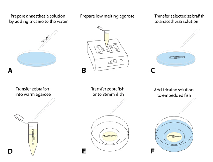
Figure 1: Embedding of zebrafish for live imaging. (A-F) Embedding procedure for live imaging: (A) Tricaine is added to egg water to anaesthetize the zebrafish at a starting dose rate of 50 mg/L. (B) Low-melting agarose (0.8 - 1.5%) is prepared and warmed up to 38 - 40 °C. (C) Using a transfer pipette, the screened and selected zebrafish are transferred into a dish with tricaine solution. After successful sedation (shallow opercular movement, decreased heart rate, lack of a touch-evoked response), a fish is transferred into the preheated agarose (D). Minimize the amount of egg water that is transferred into the agarose to prevent subsequent dilution. (E) Transfer a drop of agarose (~ 30 - 50 µL) containing the zebrafish onto a glass-bottom 35-mm dish. Perform this under a dissection microscope and use a brush to gently align the zebrafish to its preferred orientation. Wait 10 - 15 min, until the agarose is set, and add ~ 2 mL of tricaine solution to the dish (F). Please click here to view a larger version of this figure.
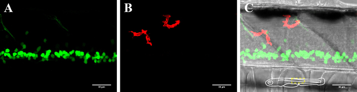
Figure 2: Visualization of neurons and microglia in the spinal cord of a 3-dpf zebrafish. Visualization of microglia and neurons in the spinal cord of a 3-day-old transgenic zebrafish expressing (A) GFP-positive neurons (islet1:GFP) and (B) mCherry-positive microglia (mpeg1:GAL4,UAS:mCherry). (C) Composite image of the neuron and microglia channel together with the bright-field image. The schematic insert in (C) depicts the orientation of the fish and outlines the presented area. Scale bar = 30 µm. Please click here to view a larger version of this figure.
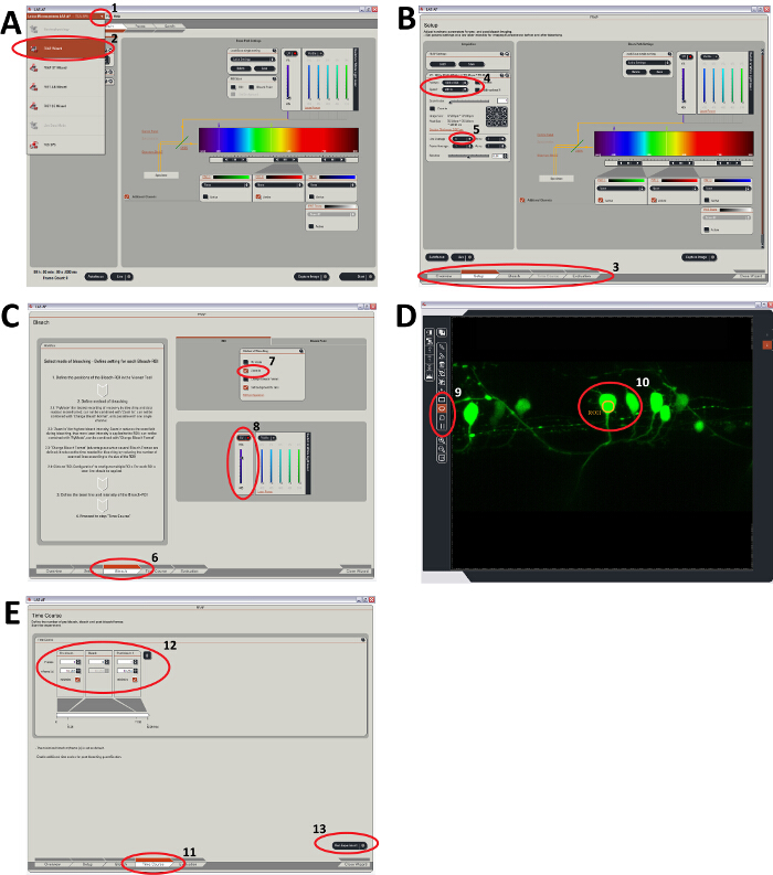
Figure 3: Steps in the process of UV laser ablation (as outlined in the protocol, step 3). Steps to control the FRAP software module in the confocal software (Leica Application Suite). (A) Starting the FRAP module as a tool to perform UV laser ablation. (B) Setting up the z-plane for ablation and other FRAP settings like format, speed, and averaging, which will determine the dwell time of the laser. (C) Control of the laser intensity and the "zoom in" option to maximize bleaching efficiency. (D) Selection of one or multiple regions of interest (ROI) that will be ablated. (E) Setting the time course of bleaching determines the bleach cycles and the overall laser dwell time at the ROIs. Please click here to view a larger version of this figure.
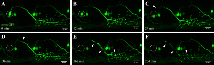
Figure 4: Anterograde degeneration of a UV-ablated neuron. Time-lapse imaging of the neurodegeneration of a UV-ablated spinal neuron. (A-F) UV-irradiation of a single spinal neuron (met:GAL4,UAS:EGFP; A; circle) resulted in the soma of the neuron shrinking and rounding up over time (A-C), followed by axonal fragmentation (C-F; arrowheads). The axonal degeneration started at the soma (site of ablation) and progressed anterograde toward the distal end of the axon until finally, the fluorescence in the soma disappeared and the entire axon showed "blebbing" (D-F). Scale bars = 20 µm. The 3D-rendered time-lapse movie of this ablation is shown in Video 1. Please click here to view a larger version of this figure.

Figure 5: Confirmation of the effect of single-cell UV irradiation using a photoconvertible fluorophore (Kaede) in a motor neuron. Validation of single-cell UV irradiation through the activation of the photoconvertible fluorophore Kaede in a neuron. (A-D) UV irradiation of neurons labeled with Kaede. (A) Photoconversion of a single neuron (circle) with a laser power of 30% for 10 s led to photoconversion of Kaede (from green to red) in only the targeted individual neuron (B). Note that the converted cell survived for several hours and showed no visual signs of deterioration, such as blebbing or rounding up. Ablation of a single neuron (C; circle) with a higher laser power (95% for 10 s) resulted in immediate disappearance of that neuron (D) and subsequent photoconversion of Kaede in a small number of surrounding neurons within a radius of approximately 20 µm. Scale bars = 20 µm. Please click here to view a larger version of this figure.
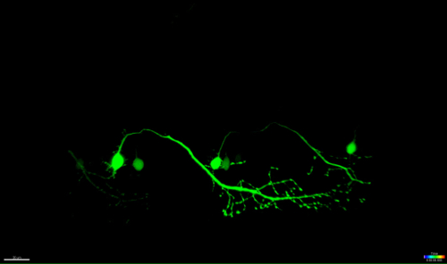
Video 1: 3D surface rendering (Imaris) of the UV ablated neuron illustrated in Figure 4.
The time-lapse video of the neuron depicted in Figure 4 is surface rendered using a visualization software (Imaris, Bitplane). It highlights the shrinking process of the ablated soma, followed by axonal fragmentation anterogradely towards the distal end of the cell. Please click here to download this file.
Supplementary Figure 1: Optional agarose cast for long-time imaging.
To avoid movement of the fish and the agarose during long term acquisition prepare a donut shaped circle of agarose along the edges in the middle of the glass-bottom 35 mm dish (A). Let the agarose set for ~10 min and transferred fish into the inner circle with a drop of agarose (B). Try to minimize the amount of agarose for the embedding (brush excess agarose to the outside after orientating the fish). Please click here to download this file.
Discussion
Laser Ablation Approaches
Laser-assisted ablation techniques allow the precise targeting of individual or small groups of cells. Combining this technique with high-resolution microscopy and genetic manipulations in animal models such as zebrafish allows researchers to systematically study the fate of an individual cell and the interactions after injury.
The UV (405 nm) laser ablation protocol described here outlines how individual cells can be stressed or killed selectively (in a dose-dependent manner), while neighboring neurons, glia, and axons are left unharmed. We have successfully utilized this approach in cell culture experiments and describe here the detailed approach for the zebrafish spinal cord. We show the implementation of this approach in the zebrafish spinal cord by selectively stressing an individual neuron within a network of other cells (Figure 5, A and B), or by killing a single neuron immediately and without recovery (Figure 5, C and D).
Previously, specialized laser systems, such as pulsed-nitrogen laser or two-photon laser systems, were required to induce tissue damage and motor nerve transections10,11,12,13. These laser systems have been successfully utilized to cause cell damage, such as thrombosis in arteries and veins6, acute kidney injury7, cardiac injury8, and to study calcium waves and microglial response after brain injury9. Furthermore, Soustelle and colleagues used a conventional confocal setup (351-nm and 364-nm UV lasers) to induce damage to epithelial and glial cells in Drosophila14.
Relevance of Zebrafish Models for Understanding ALS (and Other Human Diseases)
Zebrafish are a widely used model organism, especially for developmental studies28,29,30. While they have certain limitations, their potential to model human disease and give an understanding of pathogenic molecular mechanisms is enormous. Zebrafish models have been well-established for the study of MND and have led to important molecular insights31,32,33,34. Transgenic zebrafish lines can be rapidly generated (4 - 5 months) and allow the selective tracking of a specific cell type, features that make them a valuable addition to current animal models of ALS. Zebrafish embryos/larvae are optically transparent and offer unique experimental advantages that allow long-term live-imaging at the single-cell level in the brain or the spinal cord, which cannot be readily achieved in rodent models (or in humans). When combined with molecular techniques, such as single-cell ablation, this provides a unique experimental platform for studying precise molecular mechanisms in vivo.
Motor Neurons Can Be Selectively Targeted Using UV Laser Ablation
Spinal neurons in zebrafish start to develop within 10 h after birth and are established after approximately 48 h35,36. This rapid development allows the visualization of these neurons in short time frames and with high throughput. Motor neurons provide the essential link between brain and muscles and, in ALS, are affected in the motor cortex (upper motor neurons), the brainstem, and the spinal cord (lower motor neurons). Loss of these neurons inevitably leads to muscle atrophy and weakness. Motor neurons in the spinal cord of zebrafish can be identified by their distinct projections and by the usage of motor-neuron specific promoters like -3MNX1. Targeting the cell soma of such projecting neurons revealed the anterograde degeneration along the axonal projection over time (Figure 4 and Video 1). Single-cell resolution imaging of spinal motor neurons additionally confirmed phosphatidylserine translocation and consequent Annexin V-labeling after laser ablation (see Figure 4 and Supplementary Video 3 in Reference 27). Although we report the activation of Annexin V in dying neurons after our UV laser ablation approach, we cannot be certain that the cascade of death that is triggered during this accelerated process exactly matches the neuronal death that occurs during neurodegeneration or normal cell homeostasis.
While this ablation approach is highly reproducible and specific, different embedding strategies might also affect the efficiency of the UV ablation. In our experience, it was most successful to minimize the layer of agarose we embedded our fish in. Thicker layers of embedding medium with an additional layer of egg water may reduce the UV power ultimately received by the cell due to attenuation and scattering effects that occur along the beam path.
In the future, the crossing of different transgenic fish lines will allow for the visualization of the immediate and short-term (up to 12 h) responses of other affected cells, such as glia, to the laser-induced cell destruction. For example, astrocyte and non-cell autonomous toxicity in neurodegenerative disorders such as ALS have been in the research spotlight and are heavily implicated in the pathogenicity of sporadic and familial ALS37,38. However, the mechanisms underlying glial toxicity and selectivity toward motor neurons remain unclear. We and others recently took advantage of this approach to study the engulfment of dying neurons by microglia and visualized the clearance of neuronal remnants27,39,40.
Combining the ablation technique with high-resolution microscopy and markers for neuroinflammation will allow researchers in the future to expand the understanding of single-cell function and interconnected cell systems. Characterization of these processes in an in vivo setting is critical not only in developmental settings but also in models of neurodegenerative diseases, including MND, where cellular interactions may be impaired3,41.
Disclosures
The authors declare that they have no competing financial interests.
Acknowledgements
This work was supported by the MND Research Institute of Australia and the Cure for MND Foundation [GIA 1638], the Snow Foundation, and Macquarie University [MQRDG 9201401462]. Imaging was carried out at the Microscopy Facility in the Department of Biomedical Sciences, Macquarie University. Other members of the MND research center are thanked for their contributions to the development of these methods and fish lines.
Materials
| Name | Company | Catalog Number | Comments |
|---|---|---|---|
| Agarose low melting | Fisher-Scientific | Dissolve in egg water with tricaine (0.02%) at 0.8 - 1.5% | |
| Tricaine | Argent Labs | MS-222 | 0.02% |
| Harvard standard wall Borosilicate with filament Capillary Glass | World Precision Instruments, Inc. | GC100F-10 (short) GC100F-15 (long) | Pull to a resistance of 2 - 7 MΩ |
| Egg water | 0.6 g Instant Ocean sea salt, 4 mL of 1 mg/mL methylene blue in 10 L deionized water | ||
| 1-phenyl-2-thiourea (PTU) | Sigma | P7629 | |
| Pronase | Sigma | 10165921001 | |
| Heat block | Select BioProducts | Digital block heater (SBD110_) | |
| Microinjection apparatus | World Precision Instruments, Inc. | Microinjection was performed by hand under a steromicroscope (Leica) with a loaded glass needle and a Picospritzer II (General Valve corporation) | |
| Stereoscope | Leica | Bright field illumination microscopy was performed on a Leica M165FC stereo dissection microscope (Leica) | |
| Microscope | Leica | SP5 | |
| 35 mm glass bottom dish | MatTek Corporation | P35G-0-20-C |
References
- Becker, T. S., Rinkwitz, S. Zebrafish as a genomics model for human neurological and polygenic disorders. Dev Neurobiol. 72 (3), 415-428 (2012).
- Wilt, B. A., et al. Advances in light microscopy for neuroscience. Annu Rev Neurosci. 32, 435-506 (2009).
- Philips, T., Robberecht, W. Neuroinflammation in amyotrophic lateral sclerosis: role of glial activation in motor neuron disease. Lancet Neurol. 10 (3), 253-263 (2011).
- Robberecht, W., Philips, T. The changing scene of amyotrophic lateral sclerosis. Nat Rev Neurosci. 14 (4), 248-264 (2013).
- White, D. T., Mumm, J. S. The nitroreductase system of inducible targeted ablation facilitates cell-specific regenerative studies in zebrafish. Methods. 62 (3), 232-240 (2013).
- Jagadeeswaran, P., Paris, R., Rao, P. Laser-induced thrombosis in zebrafish larvae: a novel genetic screening method for thrombosis. Methods Mol Med. 129, 187-195 (2006).
- Johnson, C. S., Holzemer, N. F., Wingert, R. A. Laser ablation of the zebrafish pronephros to study renal epithelial regeneration. J Vis Exp. (54), e2845(2011).
- Matrone, G., et al. Laser-targeted ablation of the zebrafish embryonic ventricle: a novel model of cardiac injury and repair. Int J Cardiol. 168 (4), 3913-3919 (2013).
- Sieger, D., Moritz, C., Ziegenhals, T., Prykhozhij, S., Peri, F. Long-range Ca2+ waves transmit brain-damage signals to microglia. Dev Cell. 22 (6), 1138-1148 (2012).
- Lewis, G. M., Kucenas, S. Motor nerve transection and time-lapse imaging of glial cell behaviors in live zebrafish. J Vis Exp. (76), (2013).
- Rosenberg, A. F., Wolman, M. A., Franzini-Armstrong, C., Granato, M. In vivo nerve-macrophage interactions following peripheral nerve injury. J Neurosci. 32 (11), 3898-3909 (2012).
- Villegas, R., et al. Dynamics of degeneration and regeneration in developing zebrafish peripheral axons reveals a requirement for extrinsic cell types. Neural Dev. 7, 19(2012).
- O'Brien, G. S., et al. Two-photon axotomy and time-lapse confocal imaging in live zebrafish embryos. J Vis Exp. (24), (2009).
- Soustelle, L., Aigouy, B., Asensio, M. L., Giangrande, A. UV laser mediated cell selective destruction by confocal microscopy. Neural Dev. 3, 11(2008).
- Kilkenny, C., Browne, W. J., Cuthill, I. C., Emerson, M., Altman, D. G. Improving bioscience research reporting: the ARRIVE guidelines for reporting animal research. PLoS Biol. 8 (6), 1000412(2010).
- Essentials of Biology 2. JoVE. , JoVE Science Education Database. Mouse, Zebrafish, and Chick. Zebrafish Microinjection Techniques (2016).
- Avdesh, A., et al. Regular care and maintenance of a zebrafish (Danio rerio) laboratory: an introduction. J Vis Exp. (69), e4196(2012).
- Westerfield, M. The zebrafish book. A guide for the laboratory use of zebrafish (Danio rerio).Vol.4th ed. , Univ. of Oregon Press. Eugene. (2000).
- Clark, K. J., Urban, M. D., Skuster, K. J., Ekker, S. C. Transgenic zebrafish using transposable elements. Methods Cell Biol. 104, 137-149 (2011).
- Kwan, K. M., et al. The Tol2kit: a multisite gateway-based construction kit for Tol2 transposon transgenesis constructs. Dev Dyn. 236 (11), 3088-3099 (2007).
- Rosen, J. N., Sweeney, M. F., Mably, J. D. Microinjection of zebrafish embryos to analyze gene function. J Vis Exp. (25), (2009).
- Higashijima, S., Hotta, Y., Okamoto, H. Visualization of cranial motor neurons in live transgenic zebrafish expressing green fluorescent protein under the control of the islet-1 promoter/enhancer. J Neurosci. 20 (1), 206-218 (2000).
- Arkhipova, V., et al. Characterization and regulation of the hb9/mnx1 beta-cell progenitor specific enhancer in zebrafish. Dev Biol. 365 (1), 290-302 (2012).
- Flanagan-Steet, H., Fox, M. A., Meyer, D., Sanes, J. R. Neuromuscular synapses can form in vivo by incorporation of initially aneural postsynaptic specializations. Development. 132 (20), 4471-4481 (2005).
- Hall, T. E., et al. The zebrafish candyfloss mutant implicates extracellular matrix adhesion failure in laminin alpha2-deficient congenital muscular dystrophy. Proc Natl Acad Sci U S A. 104 (17), 7092-7097 (2007).
- Ellett, F., Pase, L., Hayman, J. W., Andrianopoulos, A., Lieschke, G. J. mpeg1 promoter transgenes direct macrophage-lineage expression in zebrafish. Blood. 117 (4), 49-56 (2011).
- Morsch, M., et al. In vivo characterization of microglial engulfment of dying neurons in the zebrafish spinal cord. Front. Cell. Neurosci. , (2015).
- Grunwald, D. J., Eisen, J. S. Headwaters of the zebrafish -- emergence of a new model vertebrate. Nat Rev Genet. 3 (9), 717-724 (2002).
- Lele, Z., Krone, P. H. The zebrafish as a model system in developmental, toxicological and transgenic research. Biotechnol Adv. 14 (1), 57-72 (1996).
- Veldman, M. B., Lin, S. Zebrafish as a developmental model organism for pediatric research. Pediatr Res. 64 (5), 470-476 (2008).
- Kabashi, E., et al. Gain and loss of function of ALS-related mutations of TARDBP (TDP-43) cause motor deficits in vivo. Hum Mol Genet. 19 (4), 671-683 (2010).
- Laird, A. S., et al. Progranulin is neurotrophic in vivo and protects against a mutant TDP-43 induced axonopathy. PLoS One. 5 (10), 13368(2010).
- Lemmens, R., et al. Overexpression of mutant superoxide dismutase 1 causes a motor axonopathy in the zebrafish. Hum Mol Genet. 16 (19), 2359-2365 (2007).
- Ramesh, T., et al. A genetic model of amyotrophic lateral sclerosis in zebrafish displays phenotypic hallmarks of motoneuron disease. Dis Model Mech. 3 (9-10), 652-662 (2010).
- Hanneman, E., Trevarrow, B., Metcalfe, W. K., Kimmel, C. B., Westerfield, M. Segmental pattern of development of the hindbrain and spinal cord of the zebrafish embryo. Development. 103 (1), 49-58 (1988).
- Lewis, K. E., Eisen, J. S. From cells to circuits: development of the zebrafish spinal cord. Prog Neurobiol. 69 (6), 419-449 (2003).
- Ilieva, H., Polymenidou, M., Cleveland, D. W. Non-cell autonomous toxicity in neurodegenerative disorders: ALS and beyond. J Cell Biol. 187 (6), 761-772 (2009).
- Philips, T., Rothstein, J. D. Glial cells in amyotrophic lateral sclerosis. Exp Neurol. 262, Pt B 111-120 (2014).
- Mazaheri, F., et al. Distinct roles for BAI1 and TIM-4 in the engulfment of dying neurons by microglia. Nat Commun. 5, 4046(2014).
- van Ham, T. J., Kokel, D., Peterson, R. T. Apoptotic cells are cleared by directional migration and elmo1- dependent macrophage engulfment. Curr Biol. 22 (9), 830-836 (2012).
- Radford, R. A., et al. The established and emerging roles of astrocytes and microglia in amyotrophic lateral sclerosis and frontotemporal dementia. Front Cell Neurosci. 9, 414(2015).
Reprints and Permissions
