Because of its versatile application as a model species in various fields of study, there is a need for a genetic transformation toolkit in narrowleaf plantain (Plantago lanceolata). Here, using Agrobacterium tumefaciens-mediated transformation, a protocol is presented that results in stable transgenic lines with a transformation efficiency of 20%.
Method Article
Agrobacterium tumefaciens-Mediated Genetic Transformation of Narrowleaf Plantain
In This Article
Summary
Abstract
Species in the genus Plantago have several unique traits that have led to them being adapted as model plants in various fields of study. However, the lack of a genetic manipulation system prevents in-depth investigation of gene function, limiting the versatility of this genus as a model. Here, a transformation protocol is presented for Plantago lanceolata, the most commonly studied Plantago species. Using Agrobacterium tumefaciens-mediated transformation, 3 week-old roots of aseptically grown P. lanceolata plants were infected with bacteria, incubated for 2-3 days, and then transferred to a shoot induction medium with appropriate antibiotic selection. Shoots typically emerged from the medium after 1 month, and roots developed 1-4 weeks after the shoots were transferred to the root induction medium. The plants were then acclimated to a soil environment and tested for the presence of a transgene using the β-glucuronidase (GUS) reporter assay. The transformation efficiency of the current method is ~20%, with two transgenic plants emerging per 10 root tissues transformed. Establishing a transformation protocol for narrowleaf plantain will facilitate the adoption of this plant as a new model species in various areas.
Introduction
The concept of using model species to investigate multiple aspects of plant biology emerged with the widespread use of Arabidopsis thaliana1. Arabidopsis was initially chosen because it shares features with many other flowering plants and has multiple traits that make it convenient to study in a laboratory environment, such as being small and having a short generation cycle. The large volume of research papers published with it as a subject, along with its small genome size and ease of genetic transformation2, enable it to persist as a widely-used experimental organism. However, Arabidopsis can be limited as a model for species with different characteristics or unique traits3. This has prompted the development of new model systems, such as maize (Zea mays), an important plant for developmental genetics in monocots4, and tomato (Solanum lycopersicum), which is an important model for evolutionary studies, fruit development, and production, and is a good representation for vegetable crops5. A method for genetic transformation is a prerequisite for a plant species to serve as a model organism2. An Agrobacterium tumefaciens-mediated transformation is a reliable tool in plant biology; it has been used to transform a few model species and major crops, including tobacco (Nicotiana tabacum)6, rice (Oryza sativa)7, cotton (Gossypium hirsutum)8, soybean (Glycine max)9, potato (Solanum tuberosum)10, and canola (Brassica napus)11. Plant species are highly variable in how successfully they respond to A. tumefaciens infection, and transformation protocols often need to be individually tailored to each species6,12.
The genus Plantago includes a total of 256 plant species, widely distributed worldwide13. The species in this genus often have unique characteristics that make them desirable as model species for studying genetics, ecology, stress physiology, secondary metabolites, medicinal chemistry, plant-microbe interactions, plant development, and evolution. Plantago lanceolata, also called the narrowleaf or ribwort plantain, has been a popular plant of interest since the 19th century, when it was first used to describe the phenomenon of male sterility14. Like other plants of its genus, it has been used in studies across various research fields. More recently, it has been proposed as a model for vascular biology, as its vascular tissue can be collected easily15. P. lanceolata is the most commonly studied species in the genus Plantago; a 2021 article reported that there were >1,400 publications including or relating to this species at that time16, and an additional 102 articles have been published since the beginning of 2022, according to a PubMed search conducted on December 9th 2022. The next most studied plant in the genus, P. major, is the subject of only 414 articles when searched using the same criteria on the same date.
Despite the research interest in P. lanceolata, studies, especially on gene function characterization, are often limited by the lack of a genetic manipulation toolkit for the species. Pommerrienig et al. made efforts to develop a transformation protocol for P. major using a floral dip technique17. However, this method cannot be applied to P. lanceolata because of the male sterility characteristic of this species18,19. To our knowledge, there is no existing protocol for the transformation of P. lanceolata.
This study presents a simple protocol for A. tumefaciens-mediated transformation of P. lanceolata. By targeting root tissues, fully grown transgenic plants can be generated within 3 months of transformation.
Protocol
NOTE: Steps 1.4-1.8, 2.3-2.5, 3.3-3.6, 4.1-4.6, 5.1-5.7, and 6.1-6.3 must be performed in aseptic conditions, using a clean hood to prevent contamination.
1. Plant material propagation for transformation
- Place commercially available wild-type (WT) Plantago lanceolata seeds (see Table of Materials) in a 50 mL centrifuge tube up to the 5 mL line, depending on the number of plants desired.
NOTE: Alternatively, a 2 mL microcentrifuge tube can be used when a small number of seeds are needed, but it should be filled to a volume no greater than 0.1 mL, as too many seeds can reduce the efficiency of sterilization. - Immerse the seeds in 75% ethanol for 60 s.
- Discard the ethanol, then immerse the seeds in 20% sodium hypochlorite (20% NaClO, 80% sterile water) for 40 min, gently inverting the tube so that all the seeds come into contact with the solution.
NOTE: The sodium hypochlorite solution must be freshly made for optimal results. - Under a laminar flow hood, discard the sodium hypochlorite solution, then wash the seeds with distilled water (five times). Add a small volume of water to the seeds after the final rinse, as this can help aid the movement of the plants onto plates.
- Using sterilized forceps, transfer the seeds onto pre-prepared 95 mm x 100 mm Petri dishes with solid MS medium (Table 1). Spread the seeds evenly across the surface of the plate, with approximately 1 cm between each seed to prevent overcrowding of the germinated seedlings (Figure 1A) .
- Seal the plates with two layers of paraffin film to prevent contamination, then incubate under a cool white grow light (see Table of Materials) at room temperature (22 °C with 50 µmol m-2s-1, 12 h days). The seeds typically germinate within 5-6 days.
- When the seedlings have germinated and are large enough to transfer (Figure 1B), typically 2 or 3 days after germination, use sterilized forceps to transfer the seedlings into sterile boxes with the 50-100 mL of MS medium (Table 1). Ideally, plant only five seedlings per box to obtain the best quality roots.
- Seal the boxes with surgical tape, then allow the plants to grow under a cool white grow light (see Table of Materials), in the same conditions mentioned in step 1.6. The plants should be ready for transformation in about 3-4 weeks, or when the main roots have grown about 2 cm in length and the lateral roots appear white.
NOTE: Medium preparation recipes and vitamin stocks are included in Table 1 and Table 2.
2. Plasmid construction and E. Coli transformation
NOTE: The exact plasmid construction procedure varies depending on the gene of interest. In this procedure, the restriction enzymes HindIII and SalII were used to insert the 1.5 kb AtPP2 promoter into the binary plasmid pBI101 (see Table of Materials) with GUS, using the standard cloning procedure20. AtPP2 (phloem protein 2) is a gene that is specifically expressed in phloem21.
- Clone the promoter using primer pairs 5'-AGTCAAGCTTCAAGTCCCTGTGGCTACTGAAC-3' (Forward) and 5'-AGTCGTCGACAAACCAGTATGATGTATTTATTTTTG-3' (Reverse) from Arabidopsis. Figure 2 shows a diagram of the binary plasmid vector with the AtPP2:GUS insert.
- After plasmid construction, transform the plasmids into DH5a E. coli (see Table of Materials) competent cells using the heat shock method22, and then incubate for 1.5 h at 37 °C with shaking (150 rpm).
- Take 150 µL of each transformation culture, plate onto LB agar media plates (Table 1) with the appropriate selection (50 mg/L kanamycin for the strain used in this protocol; see Table of Materials), and then incubate the plates for 16-24 h at 37 °C.
NOTE: The bacterial strain used in this study is A. tumefaciens GV3101. - Next, use colony PCR to screen the colonies for positive recombinants23.
NOTE: In this protocol, the following primers were used to amplify the targeted gene; 5'-ATGTTACGTCCTGTAGAAACCCCAA-3'(Forward) and 5'-TCATTGTTTGCCTCCCTGCTGC-3' (Reverse).- Run the reaction in a thermocycler (see Table of Materials) with cycling conditions of 3 min at 95 °C, followed by 35 cycles of: 30 s at 95 °C, 30 s at 55 °C, 2 min at 72 °C, and a final 10 min elongation step at 72 °C.
- Inoculate the positive colonies in 6 mL of LB broth (Table 1) with the appropriate antibiotic (50 mg/L kanamycin) and grow at 37 °C overnight, at 200-250 rpm.
- After overnight growth, extract the plasmids from the bacteria using standard procedures24.
3. A. tumefaciens transformation with plasmid
- After plasmid extraction, use electroporation to transform the modified plasmid into the desired strain of competent cells. In this procedure, A. tumefaciens strain GV3101 was used. Follow standardized methods for electroporation techniques25.
- After electroporation, resuspend the competent cells in 1 mL of LB broth and then incubate for 2-4 h at 28 °C at 100 rpm.
- Collect the cells through centrifugation at 6,800 x g for 3 min in a tabletop microcentrifuge (see Table of Materials) at room temperature (22 °C), then spread 50-100 µL onto an LB agar plate with an appropriate selection agent (50 mg/L kanamycin for the plasmid used in this protocol).
- After the cells have incubated for 2 days at 28 °C, identify positive colonies containing the gene of interest through the use of colony PCR. In this protocol, use the primers and conditions mentioned in step 2.4.
- Next, use the positive colonies to streak a stock plate with LB agar media + selection. The plate can be stored at 4 °C for up to 1 month.
- Alternatively, for long-term storage, inoculate a positive colony with a small volume of LB with appropriate selection. Shake the inoculated culture overnight at 28 °C at 200 rpm, then prepare a glycerol stock (50% w/v glycerol in a 50:50 mix of bacteria and glycerol), which can be stored at -80 °C for up to 10 years.
4. A. tumefaciens preparation
- Streak A. tumefaciens containing the desired plasmid onto prepared 95 mm x 100 mm solid LB plates with the appropriate selection agent. In this protocol, the bacterial strain GV3101 with the plasmid insert AtPP2:GUS was used, with 50 mg/L kanamycin added for the selection.
- Seal the plates with paraffin film, then incubate at 28 °C for up to 48 h, or until the bacteria grows large enough to pick.
- Use a pipette tip to pick a bacteria colony 2 days before transformation, and inoculate it in a 15 mL round bottom tube containing 6 mL of liquid LB with appropriate selection. Shake at 200 rpm in a 28 °C tabletop shaker overnight, until OD600 reaches 0.6-0.7.
NOTE: Plates and the 6 mL bacterial inoculation can be stored at 4 °C for up to 1 month. - When the bacteria reach the correct OD600, use a pipette to transfer A. tumefaciens to a sterile flask containing 100 mL of liquid LB with a selection agent. Typically, 200 µL of bacteria per 100 mL LB is appropriate for propagation. Shake at 200 rpm at 28 °C overnight, until OD600 reaches 0.6-0.7.
- Transfer the bacteria into 50 mL sterile centrifuge tubes, and centrifuge at 2,200 xg for 10 min at room temperature (22 °C) in a tabletop centrifuge to collect the bacteria.
- Discard the supernatant using a pipette. Resuspend the bacterial pellet in 5 mL of room temperature (22 °C) liquid suspension solution (SS) (Table 1) by pipetting, then add up to 50 mL of SS and invert several times to mix. The bacteria is now ready for transformation.
NOTE: The liquid SS must be prepared freshly, within 1 week of transformation.
CAUTION: All material that comes into contact with A. tumefaciens needs to be discarded in a biohazard waste bin. Leftover liquids from bacterial cultures can be sterilized with sodium hypochlorite (bleach) at a concentration of 20% or higher.
5. Transformation of Plantago roots
- When the plants reach the ideal stage for transformation (seedlings are 3 weeks old) (Figure 1C), use sterile forceps and scissors to separate the roots from the rest of the plant (Figure 3A). Discard the leaf and stem material.
- Immediately after cutting, transfer the root pieces to sterile boxes containing sterile water using sterile forceps. This step allows the roots to stay hydrated while all tissue is collected.
- When all the roots are cut, pour the A. tumefaciens/SS suspension into sterile 150 mm x 15 mm disposable Petri dishes. Transfer the roots into the A. tumefaciens culture and inoculate for at least 20 min (Figure 3B).
- During incubation, use a sterile scalpel with a sharp blade to cut the roots into 1 cm fragments, separating the primary roots from the lateral roots. Make thin, shallow cuts on the surface of the roots to allow the bacteria to infect the plant.
NOTE: If dealing with a large number of plants, transfer the root pieces into the bacterial culture in batches, to ensure that all the roots are immersed during inoculation. - After incubation, use the sterile forceps to transfer the root pieces to sterile paper towels to remove excess bacteria. Avoid drying the roots for more than 60 s, as this can cause dehydration and damage the root tissue. Ideally, 10-15 roots can be dried simultaneously (Figure 3C).
- Transfer the dried roots to prepared 95 mm x 15 mm Petri dishes with solid co-culture media (Table 1), around 10-20 roots per plate, depending on the size of the roots (Figure 3D).
- Seal the plates with two layers of transparent plastic film, then cover with aluminum foil. Incubate at room temperature (22 °C) for 3 days. This step provides time for the bacteria to infect the roots without the presence of an antibiotic selection (Figure 3E).
6. Selection and whole plant regeneration
- After incubation in co-culture media, transfer the root pieces to prepared 95 mm x 15 mm Petri dishes with solid shoot induction media (SIM) (Table 1) with Timentin (500 mg/L; see Table of Materials) and appropriate antibiotic selection. In this protocol, we used kanamycin (100 mg/L).
NOTE: The bottom of the roots must come into complete contact with the medium. Roots that do not touch the medium's surface are too long and must be cut to prevent the tissue from escaping selection. - Seal the plates with two layers of transparent plastic film, then grow under a grow light for 1 month (see step 1.6 for appropriate conditions), or until the shoots begin to emerge.
NOTE: Typically, shoot initials can be observed after 2 weeks of growth, and the shoots are typically visible after 1 month. - When the plantlets are 1.5-2.0 cm long (Figure 1D), transfer them into prepared sterile boxes with solid root induction media (Table 1).
- Grow the plants under a grow light (see step 1.6 for conditions) for several weeks, until roots form. Roots can typically first be seen after 1 week.
NOTE: It is recommended to allow the roots to grow for several weeks before moving to the soil, as plants with larger root systems tend to have a higher survival rate in soil.
7. Soil transfer
- When the root systems have become large enough to transfer (Figure 1E), typically after 1 month of growth, transfer the plants into 3.5 in square pots containing pre-wetted all-purpose soil (BM7). In this protocol, BM7 bark mix was used (see Table of Materials).
- Remove any medium that sticks to the roots by washing them gently in water.
NOTE: The plants can be grown to maturity in a greenhouse at 800 to 1400 µmol photons m-2 s-2 using 600 W high sodium pressure lights (see Table of Materials), or in a growth chamber at room temperature (22 °C) with cool white lights at 50 µmol m-2s-1, 12 h days.
- Remove any medium that sticks to the roots by washing them gently in water.
- Cover the plants with a plastic potting cover, then cover them with a clear plastic bag. This step allows the plants to remain in a humid environment as they adapt to the soil.
- After about 3-5 days, remove the plastic bag, then slowly remove the lid to allow acclimation to the outside environment.
NOTE: Depending on the time of the year and the environment to which the plants are transferred, the time that the plants need to adapt can vary. Checking the plants daily and adding water to the pots as needed is recommended. - Water the plants regularly and add fertilizer as needed. Plants can also be transferred to larger pots for further growth (Figure 1F).
8. β-glucuronidase (GUS) histochemical staining
- Prepare β-glucuronidase (GUS) staining solution, according to published protocols15.
- When the shoot initials are about 0.5-1 cm long, remove a small tip of a young, fully expanded leaf (<5 mm in length is usually sufficient) and immediately transfer to 0.5-1 mL of GUS staining solution in a 1.5 or 2 mL microcentrifuge tube. The solution should fully cover the plant tissue.
- Place the opened tubes in a vacuum desiccator and vacuum at 20-25 kPa for 5-10 min. Small bubbles should be visible in the solution during the vacuum procedure. This allows the solution to enter the cells of the plant.
- Allow air to filter back into the vacuum desiccator. Close the tubes and incubate at 37 °C overnight (12 h), or until the blue color is visible.
NOTE: In this study, GUS activity was localized to the phloem, meaning that in positively transformed plants, blue staining should only be visible in the phloem tissue. Plants without the transgene do not experience staining (Figure 4). - To better visualize the stain, transfer the plants to 100% ethanol to remove chlorophyll. To increase the efficiency of the chlorophyll removal process, incubate the tubes at 60 °C for 10 min.
NOTE: The ethanol may need to be changed several times before all chlorophyll is removed, depending on the size of the leaf being stained.
Results
A simple protocol is reported here for obtaining transgenic P. lanceolata plants using A. tumefaciens-mediated transformation. The reporter gene GUS (encoding β-glucuronidase) is transformed, driven by the phloem-expressed promoter of AtPP2, into 3-week-old P. lanceolata roots through A. tumefaciens strain GV3101 (Figure 2). A phloem-specific promoter was chosen because our main interest was to establish a system for the functional genomics of plant vascular tissues, particularly phloem. The method was tested on the root, leaf, and petiole tissue in the preliminary experiment. Although callus could be induced in all tissue types, only the root tissue produced shoot initials (Figure 5A) after 1 month in SIM; the leaf and petiole turned brown and died (Figure 5B). This led to the conclusion that root tissue was the optimal tissue type for use in the transformation method. The roots were incubated in the prepared bacteria resuspended in suspension solution (SS) (Table 1) for a minimum of 20 min, then incubated at room temperature on solid SS plates for up to 3 days in the dark (Figure 3E). The roots were then transferred to the shoot induction medium (SIM) and kept under a grow light, in the conditions indicated in the protocol (step 1.6). Figure 1 and Figure 3 show representative images of each step of the protocol for reference.
Figure 6 shows the progression of shoot initials emerging from transformed tissue, from the first day the roots were placed on the SIM (Figure 6A) to when the shoots were ready to be rooted (Figure 6D). After 1 week, the root tissue formed callus (Figure 6B), and the beginnings of shoot initials could be observed (Figure 6B1). Shoots continued to emerge during weeks 2 and 3 (Figure 6C), and after 4 weeks, the shoots were ready to be transferred to the root induction medium (Figure 6D).
Identification of the putative transgenic plants was conducted using the β-glucuronidase (GUS) histochemical assay, using leaf segments taken once the shoots were around 0.5 cm long. Positive transgenic plants showed the expected staining pattern in the phloem localized tissue, demonstrated in Figure 4. Positive GUS-stained shoots were transferred to the root induction medium, in which they developed robust rooting systems after 4 weeks (Figure 1E). Rooted plants were then transferred to the soil. Figure 4 shows the result of staining in a narrowleaf plantain transformed with the AtPP2 promoter and the β-glucuronidase (GUS) gene, along with a wild-type and a narrowleaf plantain transformed with the AtPP2 promoter, for comparison. All shoots that emerged were confirmed as transgenic. The transformation efficiency was determined to be an average of 20%, with approximately two shoots emerging for every 10 roots that were transformed. Confirmed transgenic plants were transferred to larger pots and grown for 4-8 weeks until they reached the adult stage (Figure 1F).
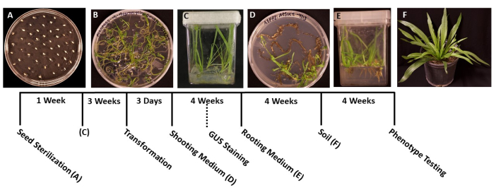
Figure 1: Timeline of Plantago lanceolata transformation. Representative images of each stage of the protocol. (A) Ungerminated seeds plated on an MS plate. (B) Seeds germinated after 1 week, ready to be transferred into magenta boxes. (C) Plants in MS boxes after 3 weeks of growth. Roots are green and healthy, at the ideal stage for transformation. (D) Shoots in shoot induction media after 4 weeks are ready to be transferred into the rooting medium. At this stage, β-glucuronidase (GUS) histochemical staining can be conducted, if applicable. (E) Plants in boxes with root induction media, where roots have formed after 4 weeks of growth. (F) Transgenic plants are grown to full length after 4 weeks of growth in soil. Please click here to view a larger version of this figure.
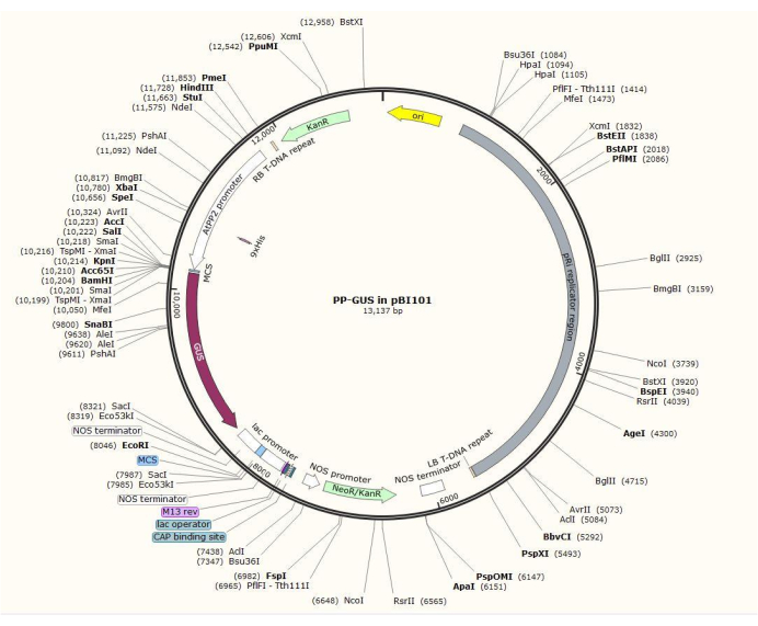
Figure 2: Diagram of the binary vector plasmid pBI101 + β-glucuronidase (GUS) with the inserted phloem-specific promoter AtPP2. Please click here to view a larger version of this figure.
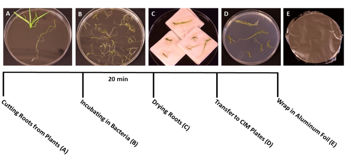
Figure 3: Steps of transformation. Representative images of each step of transformation. (A) Separating roots from shoots during transformation. (B) Soaking roots in bacteria/SS suspension. (C) Drying roots on paper towels to remove excess bacteria. (D) Roots plated on co-culture medium. (E) SS plates wrapped in aluminum foil. Plants were incubated for 2-3 days before being transferred to the shooting medium. Please click here to view a larger version of this figure.

Figure 4: GUS-staining. β-glucuronidase (GUS) staining results of narrowleaf plantain leaf segments. (A) Wild type. (B) Narrowleaf plantain transformed with the plasmid that harbors the AtPP2 promoter (empty vector). (C) Narrowleaf plantain transformed with the plasmid that harbors the AtPP2 promoter and the β-glucuronidase (GUS) gene. Each leaf was stained using the GUS histochemical staining protocol, then imaged with a microscopic camera. Images (B) and (C) show no staining pattern due to the absence of the GUS gene. The right image shows a clear blue staining pattern in the veins, confirming that the plants are transgenic. The bar represents 1 mm, with each leaf segment measuring approximately 1 cm in length. Please click here to view a larger version of this figure.
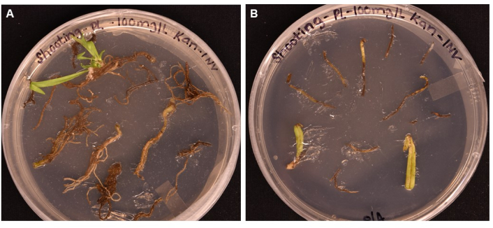
Figure 5: Comparison of transformation efficiency of different tissue types after >1 month incubation on shooting media. (A) Root tissues after over 1 month of growth. Roots have experienced expanded callus, and shoot initials have emerged. Non-transformed callus has begun to die in response to antibiotic selection. (B) Leaf and petiole tissues after over 1 month of growth. Tissues experienced some callus expansion but soon died in response to the antibiotic. No shoots emerged from either tissue. Please click here to view a larger version of this figure.

Figure 6: Emergence of callus and shoots on transformed tissue. Representative images of tissues placed on shooting medium after different lengths of incubation. (A) Root tissues just after being plated on the shooting medium. (B) Root tissues after 1 week on shooting medium. Callus expansion can be observed, and (B1) the first shoot initials have begun to emerge. (C) Root tissues after 3 weeks on shooting medium. More shoot initials have emerged. (C1) The shoot that emerged from the B1 shoot initial. (D) Root tissues after 4 weeks of incubation. Non-transformed tissue has begun to turn black/brown and die, and emerging shoots continue to grow. At this stage, shoots are ready to be moved to the rooting medium. Please click here to view a larger version of this figure.
Table 1: Media preparation recipes. A description of how to prepare mediums for transformation. The quantity of vitamins added is calculated based on the indicated stock solution concentration. See Table 2 for vitamin stock solution preparation. For all mediums, add reagents to 900 mL of double-distilled H2O, pH to the indicated level, and then add water to a final volume of 1,000 mL. * = add after sterilization. ** = pH with 1 M KOH. *** = pH with 1 M NaOH. Please click here to download this Table.
Table 2: Vitamin stocks for Plantago mediums. All vitamins must be filter sterilized and labeled accurately before storage. Where indicated, dissolve the powders first in 1 N NaOH, then make up the desired volume with double distilled H2O. Please click here to download this Table.
Discussion
The lack of a transformation protocol for plants in the genus Plantago limits the use of these plants as models, particularly when researchers are interested in exploring gene functions. P. lanceolata was chosen to develop a genetic transformation protocol because it is the most commonly studied plant of its genus16. The protocol that has been developed will likely be used as a tool to further advance studies related to vascular biology, ecology, plant-insect interactions, and abiotic stress physiology.
The protocol presented clearly outlines steps that allow a user to obtain transgenic plants. Besides the ability of P. lanceolata to thrive in a tissue culture environment, multiple factors contributed to the success of our transformation method. First, the importance of using high-quality, sterile plant root tissue for transformation was observed. Roots had the highest transformation rates when they were taken from 3-4-week-old plants, and appeared green or pale white. Roots taken from boxes with any amount of bacterial or fungal contamination often resulted in contaminated shooting cultures, and older roots that appeared brown did not result in successful transformation. Root tissue was the most efficient tissue type for transformation using the current method, as leaf and petiole tissue were unsuccessful at developing shoots.
Another important observation was that the optimal method for collecting root tissue for transformation was to place freshly cut root material in sterile water. This step effectively allowed root material to remain hydrated while the remainder of the tissue was collected, as roots tend to dry out quickly when being removed from their growth containers. This step also helped to increase the success rate of the transformation, because it allowed more roots to be incubated in the bacteria at one time.
This protocol could be modified by decreasing the time that the root tissue incubates in the co-culture media to 2 days. It was observed that a 2 or 3 day incubation period is sufficient to allow infection that results in shoot initials. However, longer incubation times are not recommended, since it was observed that the absence of an antibiotic inhibitor in the media often results in A. tumefaciens overgrowth, which can kill the emerging tissue.
A limitation of this study is the lack of available data on the performance of other methods or species of A. tumefaciens in P. lanceolata transformation for comparison. To our knowledge, this protocol is novel. During the initial trials, a high transformation efficiency was noted with A. tumefaciens GV3101, and we focused on refining the technique using this strain instead of experimenting with other strains. Our transformation efficiency of 20% is relatively high for plant transformation-many conventional methods consider anything >1% to be successful26,27,28. However, using another strain of A. tumefaciens, such as A. rhizogenes, known for its use in root transformation in multiple species29,30,31, may result in an even higher success rate. Further experimentation would be needed to assess the impact of using other strains to promote increased transformation efficiency in P. lanceolata.
The successful transformation of P. lanceolata will likely benefit many fields of study. The high transformation efficiency, and the rapid growth of the plant in tissue culture media, make P. lanceolata a feasible candidate for gene function studies15.
Disclosures
The authors have nothing to disclose.
Acknowledgements
This work was supported by the National Science Foundation (EDGE IOS-1923557 to C.Z. and Y.Z.).
Materials
| Name | Company | Catalog Number | Comments |
|---|---|---|---|
| 14 mL Round Bottom TubeA4A2:A34 | ThermoFisher Scientific | 150268 | |
| 1-Naphthylacetic acid | Gold Biotechnology | N-780 | |
| 3M Micropore Surgical Paper Tape | ThermoFisher Scientific | 19-027761 | |
| 50 mL Centrifuge Tubes | Research Products International Corp. | 163227LC | |
| 600 Watt High Pressure Sodium Lights | Plantmax | PX-LU600 | |
| 6-Benzylaminopurine (6-BAP) | Gold Biotechnology | B-110 | |
| Aluminum Foil | ThermoFisher Scientific | 01-213-100 | |
| Bacto Agar | Thermofisher Scientific | 214010 | |
| Binary Plasmid pBI101 | Clontech, USA | 632522 | |
| Cool White Grow Light Sylvania LLC | Home Depot | 315952205 | |
| D-biotin | ThermoFisher Scientific | BP232-1 | |
| ddH2O | |||
| DH5a E. coli | Invitrogen, USA | 18258012 | |
| Disposable Petri Dishes, Sterile 150 x 16 mm | ThermoFisher Scientific | FB0875712 | |
| Disposable Petri Dishes, Sterile 95 x 15 mm | ThermoFisher Scientific | FB0875714G | |
| Dissecting Scissors | Leica Biosystems | 38DI12044 | |
| Ethanol 200 Proof | Decon Labs | 2705 | |
| Folic Acid | Fisher Scientific | BP2519-5 | |
| Forceps | Leica Biosystems | 38DI18031 | |
| Gelrite | Research Products International Corp. | G35020-1000 | |
| Glycerol | ThermoFisher Scientific | 17904 | |
| Glycine | Sigma | 241261 | |
| Incubated Tabletop Orbital Shaker | ThermoFisher Scientific | SHKE420HP | |
| Indole-3-Acetic Acid (IAA) | Gold Biotechnology | I-110 | |
| Indole-3-Butyric Acid (IBA) | Gold Biotechnology | I-180 | |
| Kanamycin Monosulfate | Gold Biotechnology | K-120 | |
| Macrocentrifuge | ThermoFisher Scientific | 75007210 | |
| Magenta Boxes | ThermoFisher Scientific | 50255176 | |
| Micro Pipet Tips 1000 µL | Corning | 4140 | |
| Micro Pipet Tips 200 µL | Corning | 4138 | |
| Micro Pipette Tips 10 µL | Corning | 4135 | |
| Microcentrifuge | ThermoFisher Scientific | 75002410 | |
| Micropipettor 0.5-10 µL | Corning | 4071 | |
| Micropipettor 100-1000 µL | Corning | 4075 | |
| Micropipettor 20-200 µL | Corning | 4074 | |
| Micropipettor 2-20 µL | Corning | 4072 | |
| Murashige & Skooge Basal Medium with Vitamins | PhytoTech | M519 | |
| Murashige & Skooge Basal Salt Mixture | PhytoTech | M524 | |
| myo-Inositol | Gold Biotechnology | I-25 | |
| Nicotinic acid | Sigma | N0761-100g | |
| Parafilm (paraffin film) | ThermoFisher Scientific | S37440 | |
| Potassium Hydroxide (KOH) | Research Products International Corp. | P44000 | |
| Pyridoxine HCl | Sigma | P6280-10g | |
| Scalpel Blade Handle | Leica Biosystems | 38DI36419 | |
| Scalpel Blades | Leica Biosystems | 3802181 | |
| Sodium Chloride, Crystal (NaCl) | Mallinckrodt Chemicals | 7581-06 | |
| Sodium Hydroxide (NaOH) | Research Products International Corp. | S24000 | |
| Sodium Hypochlorite | Walmart | 23263068401 | |
| Soil- Bark Mix | Berger, USA | BM7 | |
| Square Pots (3.5 inches squared) | Greenhouse Megastore | CN-TRK-1835 | |
| Sucrose | Research Products International Corp. | S24060 | |
| Thermocycler | ThermoFisher Scientific | A24811 | |
| Thiamine HCl | Sigma | T4625-5G | |
| Timentin Ticarcillin/Clavulanate (15/1) (Timentin) | Gold Biotechnology | T-104 | |
| trans-Zeatin Riboside (ZR) | Gold Biotechnology | Z-100 | |
| Tryptone | Thermofisher Scientific | 211705 | |
| Wild Type Plantago lanceolata seeds | Outsidepride Seed Source, OR, USA | F1296 | Outsidepride.com |
| Yeast Extract Granulated | Research Products International Corp. | Y20025-1000 |
References
- Koornneef, M., Meinke, D. The development of Arabidopsis as a model plant. The Plant Journal. 61 (6), 909-921 (2010).
- Bragg, J. N., Anderton, A., Nieu, R., Vogel, J. P. Brachypodium distachyon. Agrobacterium Protocols. , Springer. New York, NY. 17-33 (2015).
- Chang, C., Bowman, J. L., Meyerowitz, E. M. Field guide to plant model systems. Cell. 167 (2), 325-339 (2016).
- Müller, B., Grossniklaus, U. Model organisms-a historical perspective. Journal of Proteomics. 73 (11), 2054-2063 (2010).
- Ding, X., et al. Different fruit-specific promoters drive AtMYB12 expression to improve phenylpropanoid accumulation in tomato. Molecules. 27 (1), 317(2022).
- Niedbała, G., Niazian, M., Sabbatini, P. Modeling agrobacterium-mediated gene transformation of tobacco (Nicotiana tabacum)-a model plant for gene transformation studies. Frontiers in Plant Science. 12, 695110(2021).
- Kumari, D., Prasad, B., Dwivedi, P. Genome Editing Platforms in Rice (Oryza sativa L.): Basic methodology and troubleshooting. , (2022).
- Zhang, B. Agrobacterium-mediated transformation of cotton. Transgenic Cotton. , Humana Press. Totowa, NJ. 31-45 (2013).
- Tiwari, R., Singh, A. K., Rajam, M. V. Improved and reliable plant regeneration and Agrobacterium-mediated genetic transformation in soybean (Glycine max L). Journal of Crop Science and Biotechnology. , 1-10 (2022).
- Bakhsh, A. Development of efficient, reproducible and stable Agrobacterium-mediated genetic transformation of five potato cultivars. Food Technology and Biotechnology. 58 (1), 57-63 (2020).
- Bates, R., Craze, M., Wallington, E. J. Agrobacterium-mediated transformation of oilseed rape (Brassica napus). Current Protocols in Plant Biology. 2 (4), 287-298 (2017).
- Hopp, H. E., Spangenberg, G., Herrera-Estrella, L. Plant transformation. Frontiers in Plant Science. 13, 876671(2022).
- Abdelmuhsin, A. A., Alghamdi, A. A., Ibrahim, N. A. Assessing the phenotypic and genotypic variations of Plantago ciliata in Ha'il region, Saudi Arabia. Entomology and Applied Science Letters. (1), 14-22 (2021).
- Coleman, N. Plantago Lanceolata. Botanical Bulletin. 1 (11), 45(1876).
- Huang, J., et al. Tissue-specific transcriptomic profiling of Plantago major provides insights for the involvement of vasculature in phosphate deficiency responses. Molecular Genetics and Genomics. 294 (1), 159-175 (2019).
- Penczykowski, R. M., Sieg, R. D. Plantago spp. as models for studying the ecology and evolution of species interactions across environmental gradients. The American Naturalist. 198 (1), 158-176 (2021).
- Pommerrenig, B., et al. Common plantain. A collection of expressed sequence tags from vascular tissue and a simple and efficient transformation method. Plant Physiology. 142 (4), 1427-1441 (2006).
- Bergero, R., Levsen, N., Wolff, K., Charlesworth, D. Arms races with mitochondrial genome soft sweeps in a gynodioecious plant, Plantago lanceolata. Molecular Ecology. 28 (11), 2772-2785 (2019).
- Aalto, E. A., Koelewijn, H. P., Savolainen, O. Cytoplasmic male sterility contributes to hybrid incompatibility between subspecies of Arabidopsis lyrata. G3: Genes, Genomes, Genetics. 3 (10), 1727-1740 (2013).
- Bloch, K. D., Grossmann, B. Digestion of DNA with restriction endonucleases. Current Protocols in Molecular Biology. 3 (1), (1995).
- Du, C., et al. Prokaryotic expression, purification, physicochemical properties and antifungal activity analysis of phloem protein PP2-A1 from cucumber. International Journal of Biological Macromolecules. 194, 395-401 (2022).
- Deshmukh, S. 22, Kulkarni, R., Bose, K. Transformation and protein expression. Textbook on Cloning, Expression and Purification of Recombinant Proteins. , Springer. Singapore. 83-114 (2022).
- Bergkessel, M., Guthrie, C. Colony PCR. Methods in Enzymology. 529, Academic Press. 299-309 (2013).
- Broehan, G., Kroeger, T., Lorenzen, M., Merzendorfer, H. Functional analysis of the ATP-binding cassette (ABC) transporter gene family of Tribolium castaneum. BMC Genomics. 14, 6(2013).
- Dong, Z., et al. Stable transformation of fluorescent proteins into Nosema bombycis by electroporation. Parasites & Vectors. 15 (1), 141(2022).
- Li, S., et al. Optimization of Agrobacterium-mediated transformation in soybean. Frontiers in Plant Science. 8, 246(2017).
- Manimaran, P., et al. Infection of early and young callus tissues of indica rice BPT 5204 enhances regeneration and transformation efficiency. Rice Science. 20 (6), 415-426 (2013).
- Wang, J., Nie, J., Pattanaik, S., Yuan, L. Efficient Agrobacterium-mediated transformation of Artemisia annua L. using young inflorescence. In Vitro Cellular & Developmental Biology-Plant. 52 (2), 204-211 (2016).
- Niazian, M., Belzile, F., Torkamaneh, D. CRISPR/Cas9 in planta hairy root transformation: a powerful platform for functional analysis of root traits in soybean. Plants. 11 (8), 1044(2022).
- Geng, S., et al. An efficient root transformation system for CRISPR/Cas9-based analyses of shoot-root communication in cucurbit crops. Horticulture Research. 9, (2022).
- Xiao, Y., et al. Efficient Agrobacterium-mediated genetic transformation using cotyledons, hypocotyls and roots of 'Duli'(Pyrus betulifolia Bunge). Scientia Horticulturae. 296, 110906(2022).
Reprints and Permissions
